Tag: immunotherapy

Promising Results for Immunotherapy Drug Nivolumab in Advanced Skin Cancer

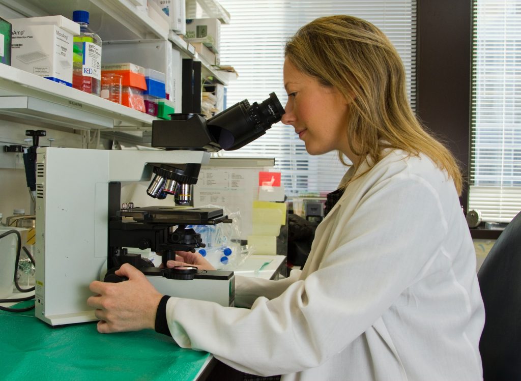
Female scientist in laboratory
Photo by Gustavo Fring on Pexels

A phase II clinical trial has demonstrated that patients with advanced cutaneous squamous cell carcinoma can benefit from the immune checkpoint inhibitor nivolumab. The findings were published in the journal CANCER

Two other immune checkpoint inhibitors, cemiplimab and pembrolizumab, have been approved by the US Food and Drug Administration for the treatment of advanced cutaneous squamous cell carcinoma in recent years. This new study is the first to report clinical trial results for nivolumab. 

The single-arm trial included 24 patients who received nivolumab at 3mg/kg every two weeks until they experienced cancer progression, developed unacceptable toxicity, or had received 12 months of treatment.  

During the trial, 14 patients (58.3%) benefited from the treatment, with their cancers demonstrating a response. Treatment-related adverse events of any grade occurred in 21 patients (87.5%) and, for and grade ≥ 3, in six patients (25%). One patient discontinued nivolumab due to toxicities. Prior radiotherapy exposure was associated with a worse response. 

“This is the first study to investigate nivolumab in this patient population, and it provides further evidence supporting the use of immune checkpoint blockers as standard therapies in cutaneous squamous cell carcinoma,” said lead author Rodrigo R. Munhoz, MD, of the Hospital Sírio-Libanês, in Brazil. 

An accompanying editorial notes that although the trial was small, its results were similar to those reported with pembrolizumab and cemiplimab. “In addition to providing more assurance to the clinical activity of different [immune checkpoint] inhibitors in this disease, this replicated data may permit a more widespread utilisation of these agents in managing a common disease with global implications,” the authors wrote. 

Source: Wiley

Why Lung Cancer Doesn’t Respond Well to Immunotherapy

A new MIT study explains why dendritic cells (green) in lymph nodes that drain from the lungs fail to stimulate killer T cells (white) to attack lung tumours.
Credits: MIT/ Courtesy of the researchers

Immunotherapy works well against some types of cancer, but it has shown mixed success against lung cancer. A new study from MIT helps to shed light on why the immune system mounts such a lacklustre response to lung cancer, even after treatment with immunotherapy drugs. In a study of mice, the researchers found that bacteria naturally found in the lungs help to create an environment that suppresses T-cell activation in the lymph nodes near the lungs.

The researchers did not find that kind of immune-suppressive environment in lymph nodes near tumours growing near the skin of mice. They hope that their findings could help lead to the development of new ways to rev up the immune response to lung tumours.

“There is a functional difference between the T-cell responses that are mounted in the different lymph nodes. We’re hoping to identify a way to counteract that suppressive response, so that we can reactivate the lung-tumour-targeting T cells,” says Stefani Spranger, the Howard S. and Linda B. Stern Career Development Assistant Professor of Biology, a member of MIT’s Koch Institute for Integrative Cancer Research, and the senior author of the new study.

MIT graduate student Maria Zagorulya is the lead author of the paper, which appears today in the journal Immunity.

Failure to attack

For many years, scientists have known that cancer cells can send out immunosuppressive signals, which leads to a phenomenon known as T cell exhaustion. The goal of cancer immunotherapy is to rejuvenate those T cells so they can begin attacking tumours again.

One type of drug commonly used for immunotherapy involves checkpoint inhibitors, which remove the brakes on exhausted T cells and help reactivate them. This approach has worked well with cancers such as melanoma, but not as well with lung cancer.

Spranger’s recent work has offered one possible explanation for this: She found that some T cells stop working even before they reach a tumour, because of a failure to become activated early in their development. In a 2021 paper, she identified populations of dysfunctional T cells that can be distinguished from normal T cells by a pattern of gene expression that prevents them from attacking cancer cells when they enter a tumour.

“Despite the fact that these T cells are proliferating, and they’re infiltrating the tumour, they were never licensed to kill,” Spranger says.

In the new study, her team delved further into this activation failure, which occurs in the lymph nodes, which filter fluids that drain from nearby tissues. The lymph nodes are where ‘killer T cells’ encounter dendritic cells, which present antigens (tumour proteins) and help to activate the T cells.

To explore why some killer T cells fail to be properly activated, Spranger’s team studied mice that had tumours implanted either in the lungs or in the flank. All of the tumours were genetically identical.

The researchers found that T cells in lymph nodes that drain from the lung tumours did encounter dendritic cells and recognise the tumour antigens displayed by those cells. However, these T cells failed to become fully activated, as a result of inhibition by another population of T cells called regulatory T cells.

These regulatory T cells became strongly activated in lymph nodes that drain from the lungs, but not in lymph nodes near tumours located in the flank, the researchers found. Regulatory T cells are normally responsible for making sure that the immune system doesn’t attack the body’s own cells. However, the researchers found that these T cells also interfere with dendritic cells’ ability to activate killer T cells that target lung tumours.

The researchers also discovered how these regulatory T cells suppress dendritic cells: by removing stimulatory proteins from the surface of dendritic cells, which prevents them from being able to turn on killer T cell activity.

Microbial influence

Further studies revealed that the activation of regulatory T cells is driven by high levels of interferon gamma in the lymph nodes that drain from the lungs. This signalling molecule is produced in response to the presence of commensal bacterial – bacteria that normally live in the lungs without causing infection.

The researchers have not yet identified the types of bacteria that induce this response or the cells that produce the interferon gamma, but they showed that when they treated mice with an antibody that blocks interferon gamma, they could restore killer T cells’ activity.

Interferon gamma has a variety of effects on immune signalling, and blocking it can dampen the overall immune response against a tumour, so using it to stimulate killer T cells would not be a good strategy to use in patients, Spranger says. Her lab is now exploring other ways to help stimulate the killer T cell response, such as inhibiting the regulatory T cells that suppress the killer T cell response or blocking the signals from the commensal bacteria, once the researchers identify them.

Reprinted with permission of MIT News

New Immunotherapy Sees 73% Success Rate in Multiple Myeloma Patients

Woman using lab equipment
Source: NCI on Unsplash

A new immunotherapy to kill bone marrow cancer cells was successful in as many as 73% of patients in two clinical trials, according to Mount Sinai researchers. The new bispecific antibody therapy binds to both T cells and multiple myeloma cells and directs the T cells to kill multiple myeloma cells. The researchers described this strategy as “bringing your army right to the enemy.”

The success of the off-the-shelf immunotherapy, called talquetamab, was even seen in patients whose cancer resisted previous therapies. It uses a different target than other approved therapies: a receptor expressed on the surface of cancer cells known as GPRC5D.

Talquetamab was tested in phase 1 and phase 2 trials. The phase 1 trial, reported in NEJM, established two recommended doses that were tested in the phase 2 trial. The results of the phase 2 trial were reported at the American Society of Hematology annual meeting. The study participants had all received at least three different therapies without achieving lasting remission, suggesting talquetamab could offer new hope for patients with hard-to-treat multiple myeloma.

“This means that almost three-quarters of these patients are looking at a new lease on life,” said lead author of the studies, Ajai Chari, MD, Director of Clinical Research in the Multiple Myeloma Program at The Tisch Cancer Institute. “Talquetamab induced a substantial response among patients with heavily pretreated, relapsed, or refractory multiple myeloma, the second-most-common blood cancer. It is the first bispecific agent targeting the protein GPRC5d in multiple myeloma patients.”

Nearly all patients with myeloma who receive standard therapies continually relapse. Patients who relapse or become resistant to all approved multiple myeloma therapies have a poor prognosis, so additional treatments are urgently needed. This study, while an early-phase trial designed to detect tolerability and find a safe dose, is an important step in meeting that need.

This international Phase 1 clinical trial enrolled 232 patients between January 2018 and November 2021. Patients received a variety of doses of the therapy either intravenously or subcutaneously; future studies will focus on doses only administered subcutaneously either weekly or every other week

The efficacy and safety findings in the phase 1 study were validated in the phase 2 trial presented at ASH. The phase 2 trial included 143 patients treated on a weekly dose and 145 patients treated at a higher biweekly dose.

The overall response rate in these two groups was about 73%, Dr Chari said. The response rate was maintained throughout various subgroups examined, with the exception of patients with a rare form of multiple myeloma that also extends to organs and soft tissues. More than 30% of patients in both groups had a complete response (no detection of myeloma-specific markers) or better, and nearly 60% had a “very good partial response” or better (indicating the cancer was substantially reduced but not necessarily down to zero).

The median time to a measurable response was approximately 1.2 months in both dosing groups and the median duration of response to date is 9.3 months with weekly dosing. Researchers are continuing to collect data on the duration of response in the group receiving 0.8 mg/kg every other week and for patients in both dosing groups who had a complete response or better.

Side effects were relatively frequent, but typically mild. About three-quarters of patients experienced a common side effect of immunotherapy – cytokine release syndrome, a constellation of symptoms including fever. About 60% experienced skin-related side effects such as rash, about half reported taste changes, and about half reported nail disorders. Only 5–6% of patients stopped treatment due to side effects.

The response rate observed in the study, which Dr Chari explained is higher than that for most currently accessible therapies, suggests talquetamab could offer a viable option for patients whose myeloma has stopped responding to most available therapies, offering a chance to extend life and benefit from other new and future therapies as they are developed.

Source: Mount Sinai Hospital

A Shot of Vitamin C Gives Dendritic Cells a Potent Cancer-fighting Boost

Vitamin C pills and orange
Photo by Diana Polekhina on Unsplash

New research published in Nucleic Acids Research has shown that vitamin C improves the immunogenic properties of dendritic cells, activating genes involved in the immune response. This discovery could help the development of potent new dendritic cell-based immunotherapies.

Since the onset of anticancer cell therapies, many types of immune cells have been used. The best-known of these cell therapies use lymphocytes, as in the highly successful CAR-T therapies. Recently, researchers have to turned to dendritic cells, known as the ‘master regulators of the immune system‘, for their ability to uptake and present antigens to the T-lymphocytes and induce an antigen-specific potent immune activation. This approach entails loading dendritic cells with specific antigens to create immune memory to make dendritic cell (DC)-vaccines.

To study dendritic cells in the lab, researchers differentiate them from monocytes using a particular set of molecular signalling. This differentiation is accomplished through a complex set of gene activation processes in the nucleus, mostly thanks to the activity of the chromatin remodelling machinery spearheaded by the TET family of demethylases, proteins that act upon the DNA epigenetic marks.

Vitamin C was already known to interact with several TET proteins to enhance its activity, but the specific mechanism was still poorly understood in human cells. In this study, a team lead by Dr Esteban Ballestar hypothesised that treating monocytes in vitro while differentiating into dendritic cells, would help the resulting cells be more mature and active.

The results obtained show that vitamin C treatment triggers an extensive demethylation at NF- kB/p65 binding sites compared with non-treated cells, promoting the activity of genes involved in antigen presentation and immune response activation. Vitamin C was also found to increase the communication of the resulting dendritic cells with other components of the immune system and stimulates the proliferation of antigen-specific T cells.

The researchers proved that vitamin C-stimulated dendritic cells loaded with antigens specific for the SARS-CoV-2 virus were able to activate T cells in vitro more efficiently than non-treated cells.

Overall, these new findings support the hypothesis that treating monocyte-derived dendritic cells with vitamin C may help generate more effective DC-vaccines. After consolidating these results in preclinical models and, hopefully, in clinical trials, a new generation of cell therapies based on dendritic cells may be used in the clinic to fight cancer more efficiently.

Source: Josep Carreras Leukaemia Research Institute

‘Striking’ Colon Cancer Trial Data gets Standing Ovation

Woman using lab equipment
Source: NCI on Unsplash

In a clinical trial, nearly every one of the 112 patients with mismatch repair-deficient (dMMR) colon cancer achieved a pathologic response with just two cycles of neoadjuvant immunotherapy, prompting a presentation panellist to describe it as “striking data” – though a note of caution was given.

Patients in the NICHE-2 single-arm study received PD-1 plus CTLA-4 blockade – nivolumab plus ipilimumab, and successfully underwent surgical resection, 98% on time, meeting the study’s primary safety endpoint, reported Myriam Chalabi, MD, at the at the European Society for Medical Oncology (ESMO) annual congress.

And with a median follow-up of 13.1 months, the disease-free survival (DFS) rate was 100%, said Dr Chalabi of the Netherlands Cancer Institute in Amsterdam. She pointed out that, by this point, the expected rate for this patient population was about 15%. The primary efficacy endpoint for the trial is DFS at 3 years, with success defined as a rate of 93% (data are expected next year).

A standing ovation erupted when Dr Chalabi displayed the waterfall plot showing the depth of pathologic response with just four weeks of nivolumab plus ipilimumab.

Credit: NICHE-2 Study

Among the 107 patients evaluable for efficacy, all but one had a pathologic response, 95% had a major pathologic response (MPR), and 67% had a pathologic complete response (pCR), ie no residual viable tumour in both the primary tumour bed and lymph nodes.

“As you can appreciate, the pathologic regression observed was near-complete or complete in almost all patients,” she said.

In contrast, pathologic response rates in the range of 5% to 7% for this population have been shown in prior trials involving neoadjuvant chemotherapy, said Chalabi.

Some 10–15% of colon cancers are classified as dMMR, she explained, which are highly sensitive to immune checkpoint inhibitors, where a number of agents have been approved for the metastatic setting.

The first to comment in Q&A, Alexander Eggermont, MD, PhD, stressed the potential impact of the findings, saying that patients with dMMR tumours scheduled for resection “should be taken off the surgical program.”

“They should be sent to the medical oncologist for the first dose of ipi/nivo,” he said. “We will live the day that they will not undergo surgery anymore after these schedules – that’s the next step.”

A multicentre, single-arm study, NICHE-2 enrolled 112 patients with previously untreated non-metastatic dMMR colon cancer undergoing surgery. The first cycle of neoadjuvant treatment included ipilimumab (1mg/kg) and nivolumab (3mg/kg). The second cycle, given two weeks later, was limited to nivolumab alone. After the first dose, median time to surgery was 5.4 weeks.

The trial defined pathologic response as 50% or less residual viable tumour; MPR was defined as 10% or less residual viable tumour, and included patients with pCRs in the primary tumour but viable tumour in the lymph nodes.

The median age of patients was 60, and 58% were women. About three-fourths had high-risk stage III disease, 13% had low-risk stage III disease, and 13% had stage I/II disease. About half had radiologic high-risk disease (both T4 and N2), said Dr Chalabi, and abdominal wall involvement was common.

When asked whether randomised data would be needed to make this approach standard, Dr Chalabi pointed out the group with T4 tumours.

“I wouldn’t want to randomise those patients,” she said. “Surgeons usually would prefer to have some type of downstaging before continuing on to surgery in order to increase the chances of achieving tumour-free resection margins and also to limit the extent of surgery needed to achieve that.”

The case for randomisation is stronger in earlier disease, she said, but if recurrences can be prevented even in stages where recurrence is more rare, such as stage II tumours (about 10% at 3 years), “we’re curing 10% more patients.”

A little less than a third of patients had Lynch syndrome, and pCRs were more frequent in this subset (78% vs 58% in those with sporadic tumours). Immune-related adverse events (AEs) were reported in 61% of patients, with 4% being grade 3/4.

When the prospect of a NICHE-3 trial came up, Dr Chalabi said that it ideally would have been an international study to validate the approach. However, a subsequent trial is being developed and will likely involve nivolumab plus anti-LAG-3 relatlimab, “which is a shame,” she noted.

“If we do get similar responses with nivolumab and anti-LAG-3, then that may be an avenue to test organ-sparing approaches with that combination in this population,” she added.

Source: MedPage Today

Durvalumab Plus Radiotherapy Boosts NSCLC Survival Rate

Shown here is a pseudo-colored scanning electron micrograph of an oral squamous cancer cell (white) being attacked by two cytotoxic T cells (red), part of a natural immune response. Photo by National Cancer Institute on Unsplash

Durvalumab with radiotherapy for unresectable locally advanced non-small cell lung cancer (NSCLC) without chemotherapy achieve had a much greater 12-month progression-free survival rate than expected, with tolerable adverse reactions.  This phase II clinical trial was reported at the IASLC 2022 World Conference on Lung Cancer in Vienna.

Immunotherapy plays an important role in NSCLC and combination of radiotherapy and immunotherapy have been reported to have a synergistic effect. Durvalumab after concurrent chemoradiotherapy has been standard of care with unresectable locally advanced NSCLC (stage III/postoperative recurrent NSCLC). But some patients are unable to complete concurrent curative radiation therapy and cannot receive durvalumab.

Dr M. Tachihara of Kobe University Graduate School of Medicine and colleagues developed the DOLPHIN study – the first Phase II study of immunotherapy combined with curative radiotherapy for unresectable locally advanced non-small cell lung cancer. 

The team enrolled 35 adult patients with unresectable locally advanced non-small cell lung cancer with an ECOG performance status (PS) 0-1, PD-L1≥1% (SP263 clone). Of the 35 patients (median age, 72 years), 88.6% were male, 54.3% had ECOG PS 0, 96.1% had a history of smoking, 57.1% had non-squamous histology, and 25.7% were postoperative recurrence.  

Patients received curative radiation therapy (60Gy) plus durvalumab 10 mg/kg every two weeks simultaneously, followed by maintenance with durvalumab for up to 12 months until disease progression (PD) or unacceptable toxicity.

Thirty-four patients were evaluated for safety, and 33 patients for efficacy. The 12-month progression-free survival rate by ICR was 72.1% (90% CI, 59.1-85.1) after a median follow-up of 18.7 months. Confirmed overall response rate was 90.9% (95% CI, 75.7-98.1, ICR-assessed) with complete and partial response rates of 36.4% and 54.5%, respectively. Median progression-free survival was not reached by ICR-assessed, and 24.1 months (95% CI, 16.0-NR) by investigator-assessed. Thirteen patients (39.4%) discontinued durvalumab; six patients due to progression disease and seven due to adverse events (AE). Grade 3/4 adverse events occurred in 47.1%; the most common adverse event of grade 3/4 was lung infection (11.8%) and pneumonitis (11.8%). Grade 5 adverse events of any cause occurred in 2 patients (5.9%), one with lung infection and one with broncho-oesophageal fistula because of tumour progression during follow-up.

“This DOLPHIN study is the first report of immunotherapy combined with curative radiotherapy for unresectable LA-NSCLC. The primary endpoint of 12-months PFS rate was met and much higher than expected value. It suggests that this treatment strategy is promising with tolerable adverse effects and appropriate as a study treatment for phase III trials,” said Dr. Tachihara.

Source: IASLC

‘Goldilocks’ Window for Immunotherapy Without Side Effects

Shown here is a pseudo-colored scanning electron micrograph of an oral squamous cancer cell (white) being attacked by two cytotoxic T cells (red), part of a natural immune response. Photo by National Cancer Institute on Unsplash

Researchers have developed a way to potentially reduce the toxic side-effects of CAR T cell immunotherapy, in findings that could overcome the pioneering treatment’s biggest limitation.

Their new study, reported in eLife, has come up with a way to identify a ‘goldilocks’ window that fine-tunes the cells used in the immunotherapy so that their activity is strong enough to eliminate the cancer but not so strong that they generate toxic side-effects.

Therapy provokes ‘perfect storm’

CAR T cell therapies involve collecting T cells from a cancer patient and supercharging the cells by individually re-engineering them in the laboratory. These enhanced cells are then put back into patients.

CAR T cell immunotherapy can be up to 90% effective in certain blood cancers, even curing some patients. But the treatment has harmful side-effects, with about 50% of patients experiencing dangerous complications.

The T cells are engineered to produce proteins on their surface called chimeric antigen receptors (CARs), which enable T cells to recognise and bind to specific proteins on the surface of cancer cells more efficiently.

Associate Professor Matthew Call said this synthetic sensor is what gives T cells the enhanced ability to attack and eliminate threats, like cancer cells.

“While putting these supercharged T cells into a patient with a high tumour burden can swiftly eradicate cancer cells, it also creates the perfect storm for an ongoing toxic response that can be harmful,” Associate Prof Call said.

There is currently no way of reliably predicting how strong CAR T cell therapy will be for a patient.

While previous studies have attempted to fine-tune T cells by targeting the end sections of the sensor, which either bind to the cancer cell or instruct the T cells to kill, the new research is the first to look at completely redesigning the middle part.

Researchers leveraged the computational expertise of the Weizmann Institute of Science to stitch together pieces of natural immune sensors with custom-designed synthetic elements, to generate new circuits that could be used to tune and assess variations of potency.

“Focusing on the connector fragment in the middle allows us to generate different versions of CARs that we know are stronger or weaker, enabling us to customise them to a patient’s potency requirements,” Associate Prof Call said.

“Being able to predictably tune this T cell activity significantly broadens our research, contrary to previous studies, because we are targeting something that exists in every immunotherapy scenario.

“For the first time, we can establish rules that will be applicable to any cancer where CAR T cell immunotherapy is being used.”

Enhanced treatment

Associate Prof Call said the ability to fine-tune T cells would dramatically reduce the number of patients experiencing severe side-effects from the treatment, which can include fever, high blood pressure and respiratory distress.

“CAR T cell therapy has proven effective in eradicating very advanced leukaemias and lymphomas, while also keeping the cancer at bay for many years – even after a patient has stopped taking cancer medication,” Associate Professor Call said.

“The therapy has incredible potential for cancer patients, but is currently used as a last resort due to these potentially severe side-effects.

“Our tools could lead to a fundamental rethink of the way CAR T cell therapy is offered by reducing a patient’s exposure risk to harmful side-effects. This would allow patients with a broad range of cancers to be given CAR T cell therapy far earlier in the treatment process.”

There are currently over 600 clinical trials of CAR T cell immunotherapy, with the treatment already being used for several blood cancers.

Researchers hope their new tool could be used to triage immunotherapy patients according to the potencies required in the early treatment phases, bringing the field closer to hitting that ‘goldilocks’ treatment window for many different cancers.

The next research phase will focus on progressing these findings into a clinical setting to see CAR T cell therapy used as a safer, first-line treatment.

Source: Walter and Eliza Hall Institute of Medical Research

SSRI Antidepressants Could be Used To Fight Cancer

Natural killer (NK) cells target a cancel cell for destruction. Credit: NCI

Long used to treat depression, selective serotonin reuptake inhibitors (SSRIs) could help improve modern cancer treatments.

In mouse experiments, they slowed the growth of pancreatic and colon cancers, and when combined with immunotherapy, they even halted cancer growth long-term. In some cases the tumours disappeared completely. The researchers’ findings will now be tested in human clinical trials.

The neurotransmitter serotonin, known as the happiness molecule, has many other functions and is mostly found outside the brain, stored in blood platelets. Serotonin reuptake inhibitors (SSRIs), which are used to treat depression, increase serotonin levels in the brain but reduce serotonin in platelets.

Serotonin was already known to be involved  in carcinogenesis. Until now, however, the underlying mechanisms had remained obscure. Now, researchers at the University of Zurich (UZH) and University Hospital Zurich (USZ) have shown that SSRIs or other drugs that lower peripheral serotonin levels can also slow cancer growth in mice.

Pierre-Alain Clavien, Director, Department of Surgery and Transplantation, University of Zurich, said: “Drugs that are already approved for clinical use as antidepressants could help improve treatment of hitherto incurable pancreatic and colorectal cancers.”

Although recent years have seen new, effective treatments such as targeted antibodies or immunotherapies, most patients with advanced-stage abdominal tumours such as colon or pancreatic cancer die within a few years of diagnosis. Tumour cells eventually become resistant to the drugs and are no longer recognised by the immune system. Now, the researchers have discovered the role serotonin plays in this tumour cell resistance mechanism.

Cancer cells use serotonin to boost production of an immunoinhibitory molecule, PD-L1, which binds to killer T cells, rendering them dysfunctional. The cancer cells thus escape destruction by the immune system. In mouse models, the researchers were able to show that SSRIs or peripheral serotonin synthesis inhibitors prevent this mechanism. “This class of antidepressants and other serotonin blockers cause immune cells to recognise and efficiently eliminate tumor cells again. This slowed the growth of colon and pancreatic cancers in the mice,” Clavien said.
PD-L1, via which serotonin exerts its effect, is also the target of modern immunotherapies, also called immune checkpoint inhibitors. The researchers then tested a dual treatment approach in mice: immunotherapy, which increases the activity of killer T cells, was combined with drugs that reduce peripheral serotonin. Cancer growth was suppressed in the animal models in the long term, and in some mice, the tumours disappeared completely.

“Our results provide hope for cancer patients, as the drugs used are already approved for clinical use. Testing such drug combinations on cancer patients in clinical trials can be fast-forwarded due to the known safety and efficacy of the drugs,” said Clavien.

Source: University of Zurich

Oxford-AstraZeneca Vaccine Tech Tapped to Treat Cancer

Source: National Cancer Institute on Unsplash

The Oxford-AstraZeneca vaccine’s success against SARS-CoV-2 has prompted scientists to develop a vaccine for cancer, using Oxford’s viral vector vaccine technology.

When tested in mouse tumour models, the two-dose therapeutic cancer vaccine increased the numbers of anti-tumour T cells infiltrating the tumours and improved the efficacy of cancer immunotherapy. Compared to immunotherapy alone, combination with the vaccine resulted in a greater reduction in tumour size and improved survival.

The study, which was done by Professor Benoit Van den Eynde’s group at the Ludwig Institute for Cancer Research, University of Oxford in collaboration with co-authors Professor Adrian Hill and Dr Irina Redchenko at the University’s Jenner Institute, has been published in the Journal for ImmunoTherapy of Cancer.

Cancer immunotherapy has improved outcomes for some cancer patients. Anti-PD-1 immunotherapy works by unleashing anti-tumour T cells to allow them to kill cancer cells. However, in the majority of cancer patients, anti-PD-1 therapy is still ineffective .

One reason for the poor efficacy of anti-PD-1 cancer therapy is that some patients have low levels of anti-tumour T cells. Oxford’s vaccine technology generates strong CD8+ T cell responses, which are necessary for strong anti-tumour effects.

The team developed a two-dose therapeutic cancer vaccine with different prime and boost viral vectors, one of which is the same as the vector in the Oxford-AstraZeneca COVID vaccine. In order to create a vaccine treatment that specifically targets cancer cells, the vaccine was designed to target two MAGE-type proteins found on the surface of many types of cancer cells.

Preclinical experiments in mouse tumour models demonstrated that the cancer vaccine increased the levels of tumour-infiltrating CD8+ T cells and enhanced the response to anti-PD-1 immunotherapy. The combined vaccine and anti-PD-1 treatment resulted in a greater reduction in tumour size and improved the survival of the mice compared to anti-PD-1 therapy alone.

Benoit Van den Eynde, Professor of Tumour Immunology at the University of Oxford, said: “We knew from our previous research that MAGE-type proteins act like red flags on the surface of cancer cells to attract immune cells that destroy tumours.

“MAGE proteins have an advantage over other cancer antigens as vaccine targets since they are present on a wide range of tumour types. This broadens the potential benefit of this approach to people with many different types of cancer.

“Importantly for target specificity, MAGE-type antigens are not present on the surface of normal tissues, which reduces the risk of side-effects caused by the immune system attacking healthy cells.”

Human trials in 80 patients with non-small cell lung cancer will be launched later this year.

Adrian Hill, Lakshmi Mittal and Family Professorship of Vaccinology and Director of the Jenner Institute, University of Oxford, said: “This new vaccine platform has the potential to revolutionise cancer treatment. The forthcoming trial in non-small cell lung cancer follows a Phase 2a trial of a similar cancer vaccine in prostate cancer undertaken by the University of Oxford that is showing promising results.

“Our cancer vaccines elicit strong CD8+ T cell responses that infiltrate tumours and show great potential in enhancing the efficacy of immune checkpoint blockade therapy and improving outcomes for patients with cancer.”

Source: Oxford University

Unleashing the Immune System to Attack Cancers

Shown here is a pseudo-colored scanning electron micrograph of an oral squamous cancer cell (white) being attacked by two cytotoxic T cells (red), part of a natural immune response. Photo by National Cancer Institute on Unsplash

A potential treatment has been identified, that could boost the immune system’s ability to find and destroy cancer cells, by impeding certain cells which regulate the immune system, which in turn can unleash other immune cells to attack tumours in cancer patients.

“A patient’s immune system is more than able to detect and remove cancer cells and immunotherapy has recently emerged as a novel therapy for many different types of cancers,” explained study leader Nullin Divecha, Professor of Cell Signalling at the University of Southampton. “However, cancer cells can generate a microenvironment within the tumour that stops the immune system from working thereby limiting the general use and success of immunotherapy,” he continued.

One of a number of types of T cells, Teffector cells (Teffs) carry out the task of detection and removal of cancer cells . How well Teff cells work in detecting and removing cancer cells is partly governed by other T cells called T-regulatory cells, or Tregs for short. Tregs physically interact with the Teff cells, producing molecules which dampen the functioning of the Teff cells.

Prof Divecha added, “Tregs carry out an important function in the human body because without them, the immune system can run out of control and attack normal cells of the body. However, in cancer patients we need to give the Teff cells more freedom to carry out their job.”

Molecules released by tumour cells exacerbate the problem by attracting and gathering Tregs, reducing the activity and function of Teff cells even further. Though there are mechanisms to inhibit Treg cells, since Treg and Teff cells are very similar, Teff cells are also generally inhibited.

In this new study, published in PNAS, scientists from the University of Southampton and the National Institute of Molecular Genetics in Milan showed that inhibition of a family of enzymes in cells called PIP4K could be the answer to how to restrict Tregs without affecting Teffs.

The research team isolated Tregs from healthy donors and used genetic technology to suppress the production of the PIP4K proteins. They saw that loss of PIP4Ks from Treg cells stopped their growth and response to immune signals, in turn stopping them from impeding Teff cell growth and function.

Importantly, the loss of the same enzymes in Teff cells did not limit their activity.

“This was surprising because PIP4Ks are in both types of T cells in similar concentrations but our study shows that they seem to have a more important function for Tregs than Teffectors,” said Dr. Alessandro Poli who carried out the experimental research.

Scientists must next develop molecules in order to inhibition of PIP4K as a potential therapy for patients. “Towards this end we show that treatment with a drug like inhibitor of PIP4K could enable the immune system to function more strongly and be better equipped to destroy tumour cells.”

Source: EurekAlert!