Tag: immune checkpoint inhibitors

Unique Genetic Pattern can Predict Severe Side Effects of Melanoma Immunotherapy

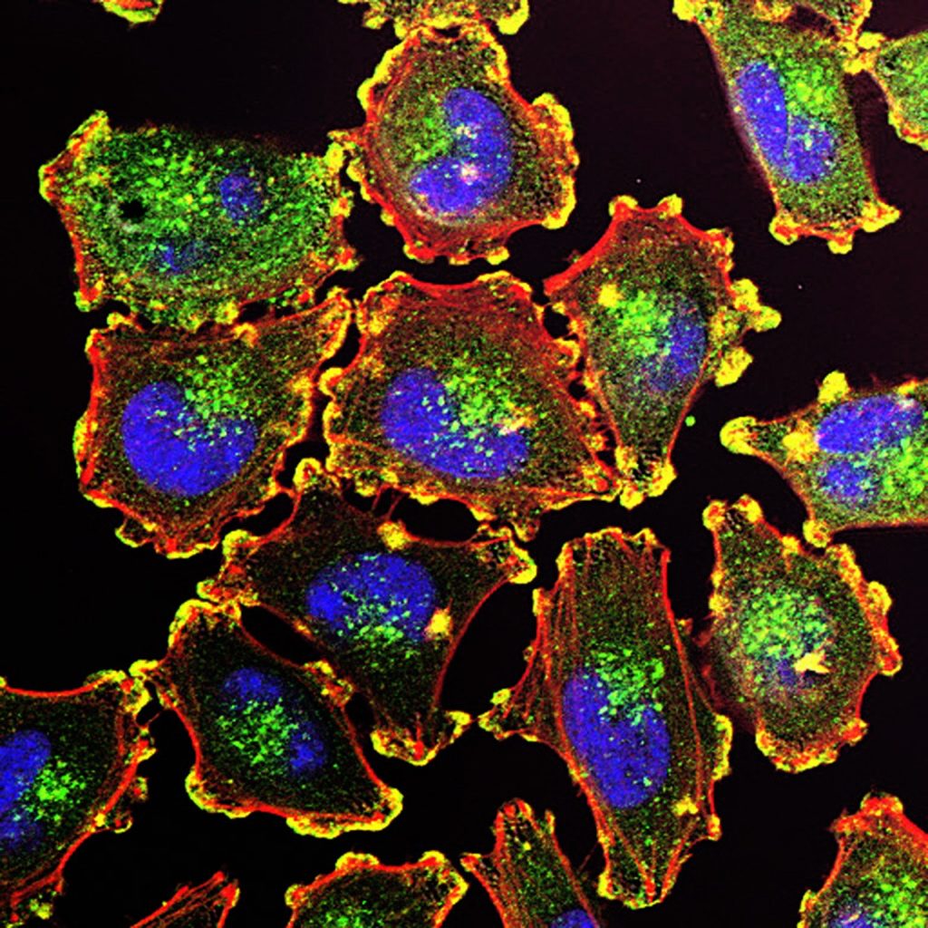
Melanoma Cells. Credit: National Cancer Institute

An activity pattern in certain genes responsible for building proteins known as spleen tyrosine kinases can predict which melanoma patients are likely to have severe side effects from immunotherapy designed to treat the most deadly skin cancer, as shown by a new study published in the journal Clinical Cancer Research.

Led by researchers at NYU Langone Health and its Perlmutter Cancer Center, the latest experiments focused on checkpoint inhibitors, drugs that have in the last decade become a mainstay of treating melanoma. This form of skin cancer kills nearly 10 000 Americans annually.

The drugs work by blocking molecules (checkpoints) that sit on the surface of immune T which the immune system uses to recognise and protect healthy cells. Cancer cells are able to hijack and turn off immune cell surveillance, evading detection. Immunotherapy drugs like nivolumab and ipilimumab are designed to block checkpoints, making cancer cells more “visible” again to T cells.

More than a third of melanoma patients given checkpoint inhibitors develop side effects so severe that they compromise their quality of life and ability to continue therapy. Side effects most often involve some form of inflammation, a sign of an overactive immune response. Patients may experience severe skin rashes, diarrhoea, or hyperthyroidism. More-severe side effects can include liver toxicity, colitis, and rheumatoid arthritis.

In the new study researchers found that even before treatment began in their test subjects, the activity of genes controlling the production of spleen tyrosine kinases predicted 83% of melanoma patients who eventually developed severe side effects from combined immunotherapy with nivolumab and ipilimumab.

Moreover, the researchers found that this heightened gene signature, as evidenced by the production of spleen tyrosine kinases, or the SYK pathway, did not interfere with the effectiveness of therapies in preventing recurrence of melanoma. The impact was connected only to side effects.

“Predictive information of this kind is critically important to oncologists and patients to help guide their immunotherapy decisions, to either minimize these side effects by taking additional precautions or to choose alternative immunotherapies,” said study co-senior investigator Tomas Kirchhoff, PhD.

“Our study results show that increased gene activity in the spleen tyrosine kinase pathway could be the basis of a possible blood test that identifies those melanoma patients most susceptible to having severe side effects from immunotherapy, and well before they start treatment,” said study co-lead investigator Kelsey Monson, PhD. 

For the study, researchers analysed immune system cell samples from 212 men and women with melanoma participating in the CheckMate-915 trial. The trial was designed to test whether combined therapy with nivolumab and ipilimumab worked better than single therapy with nivolumab in preventing postsurgical recurrence of melanoma. All immune cell samples were taken prior to the start of immunotherapy. Both drugs are manufactured by the pharmaceutical company Bristol Myers Squibb, which sponsored the CheckMate-915 trial, and provided the patient specimens and data used in the analysis.

When researchers looked at what genes were more active than others in patients who experienced side effects from their immunotherapy, they found a specific pattern among 24 genes tied to the production of spleen tyrosine kinases. Further statistical analyses showed that increased or decreased activity (transcription) of only five of these genes – CD22, PAG1, CD33, HNRNPU, and FCGR2C – along with patients’ age and the stage severity of their melanoma served as the best predictors of who would experience side effects from immunotherapy.

Study co-senior investigator Jeffrey S. Weber, MD, PhD, says that the SYK pathway has previously been linked to other autoimmune diseases, including lupus, rheumatoid arthritis, and colitis. He also points out that immunotherapy side effects were also most common in areas affected by these autoimmune diseases, including the skin, colon, and liver.

Dr Weber says the team next plans to investigate if an activated SYK pathway is predictive of side effects in patients treated with ipilimumab alone or with other combination immunotherapies.

“If our future research can explain how an activated spleen tyrosine kinase pathway leads to increased risk of side effects from immunotherapy, then it could also potentially help us to design better cancer immunotherapies and potentially other treatments for autoimmune diseases,” said Dr Kirchhoff.

Source: NYU Langone Health / NYU Grossman School of Medicine

Enhancing Checkpoint Inhibitor Therapy by Striking at the Right Time

Photo by Malvestida on Unsplash

A multidisciplinary research team at the University of California, Irvine has revealed that the circadian clock can be leveraged to enhance the efficacy of checkpoint inhibitor cancer therapy. Checkpoint inhibitors block different proteins from binding to tumour cells, allowing the immune system’s T cells to kill the tumour.

The study, published online today in the journal Nature Immunology, provides deeper insights into the intricate relationship among the circadian clock, immune regulation and tumour development and found that a therapeutic approach optimising time-of-day delivery based on an individual’s unique circadian patterns offers new avenues for prevention and treatment.

“Disruption of the internal biological pacemaker is an inherent aspect of modern society that may contribute to the rising incidence of many cancer types. We found that proper regulation of circadian rhythms is necessary to suppress inflammation and support peak immune function,” said corresponding author Selma Masri, UC Irvine associate professor of biological chemistry. “Understanding precisely how circadian disruption promotes disease progression could lead to behaviour modification to reduce cancer risk.”

Team members used an advanced single-cell RNA sequencing technique in a genetic model of colorectal cancer and identified clock-dependent changes controlling the number of myeloid-derived cells that suppress T cell activation. They discovered that disruption of the internal clock in the epithelial cells lining the intestine alters secretion of cytokine proteins, leading to heightened inflammation, increased numbers of immunosuppressive myeloid cells and cancer progression. These findings were leveraged to demonstrate that providing immunotherapy at the time of day when these immunosuppressive myeloid cells are most abundant significantly enhanced the efficacy of immune checkpoint blockades in solid tumours.

“As we enhance our understanding of the fundamental mechanism of circadian regulation of immunity, we will be able to harness the power of the body’s natural rhythms to fight cancer and develop more personalised and effective treatment strategies,” said lead author Bridget Fortin, a UC Irvine doctoral student in the Department of Biological Chemistry.

While this study represents a significant step forward in defining circadian control of anti-tumour immunity, the team believes future research should focus on exploring additional factors and cell types influencing time-of-day response to checkpoint inhibitor therapy.

Source: University of California – Irvine

Vitamin D may be Necessary for Effective Immunotherapy in Skin Cancer

3D structure of a melanoma cell derived by ion abrasion scanning electron microscopy. Credit: Sriram Subramaniam/ National Cancer Institute

Vitamin D has many effects on the body, including regulation of the immune system. New research indicates that for patients with advanced skin cancer, it may be important to maintain normal vitamin D levels when receiving immunotherapy in the form of immune checkpoint inhibitors. The findings are published in CANCER.

To see whether levels of vitamin D might impact the effectiveness of immune checkpoint inhibitors, investigators analysed the blood of 200 patients with advanced melanoma both before and every 12 weeks during immunotherapy treatment.

A favourable response rate to immune checkpoint inhibitors was observed in 56.0% of patients in the group with normal baseline vitamin D levels or normal levels obtained with vitamin D supplementation, compared with 36.2% in the group with low vitamin D levels without supplementation. Progression‐free survival in these groups was 11.25 and 5.75 months, respectively.

“Of course, vitamin D is not itself an anti-cancer drug, but its normal serum level is needed for the proper functioning of the immune system, including the response that anti-cancer drugs like immune checkpoint inhibitors affect,” said lead author Łukasz Galus, MD, of Poznan University of Medical Sciences, in Poland. “In our opinion, after appropriately randomised confirmation of our results, the assessment of vitamin D levels and its supplementation could be considered in the management of melanoma.”

Source: Wiley

New Way to Assess Immune Checkpoint Effectiveness

Chromosomes prepared from a malignant glioblastoma visualized by spectral karyotyping (SKY) reveal an enormous degree of chromosomal instability — a hallmark of cancer.
Credit: Thomas Ried, NCI Center for Cancer Research, National Cancer Institute, National Institutes of Health

Researchers have used a new machine learning and protein profiling system to identify vulnerabilities in glioblastomas and to assess immune checkpoint blockade treatment effectiveness.

Neoadjuvant immune checkpoint blockade (ICB) is a promising treatment for melanoma and other cancer types, and has recently been shown to provide a modest survival benefit for patients with recurrent glioblastoma. To improve the treatment efficacy, researchers are looking for vulnerabilities in surgically removed glioblastoma tissues, but this has been difficult due to the vast differences within the tumours and between patients.

To tackle this problem, researchers at Institute for Systems Biology (ISB) and their collaborators developed a new way to study tumours. The method builds mathematical models using machine learning-based image analysis and multiplex spatial protein profiling of microscopic compartments in the tumour.

The team used this approach to analyse and compare tumour tissues gathered from 13 patients with recurrent glioblastoma and 23 patients with high-risk melanoma. Both groups had been treated with neoadjuvant ICB. Using melanoma to guide the interpretation of glioblastoma analyses, they were able to identify the proteins that correlate with tumour-killing T cells, tumour growth, and immune cell-cell interactions.

Co-lead author Dr Yue Lu described the research : “This work reveals similarities shared between glioblastoma and melanoma, immunosuppressive factors that are unique to the glioblastoma microenvironment, and potential co-targets for enhancing the efficacy of neoadjuvant immune checkpoint blockade.”

“This framework can be used to uncover pathophysiological and molecular features that determine the effectiveness of immunotherapies,” added Dr Alphonsus Ng, co-lead author of the paper.

ISB, UCLA and MD Anderson collaborated on the study, the findings of which were published in Nature Communications. Brain cancer represents one of the toughest settings for immunotherapy success. Collaboration between scientists and clinicians provides a great opportunity for improving patient care and achieving a deep understanding of cancer immunotherapy.


“We believe that the integrated biological, clinical and methodological insights derived from comparing two classes of tumors widely seen as at the opposite ends of the spectrum with respect to immunotherapy treatments should be of interest to broad scientific and clinical audiences,” said corresponding author and ISB President, Dr Jim Heath.

Source: PRWeb

Journal information: Yue Lu et al, Resolution of tissue signatures of therapy response in patients with recurrent GBM treated with neoadjuvant anti-PD1, Nature Communications (2021). DOI: 10.1038/s41467-021-24293-4

No Survival Benefit Seen for PD-1 Inhibitor in Triple-negative Breast Cancer

Results of a large randomised trial showed no survival improvement in previously treated metastatic triple-negative breast cancer (TNBC) with single-agent pembrolizumab versus chemotherapy.

Eric Winer, MD, of Dana-Farber Cancer Institute in Boston, presented findings from his team’s randomised trial KEYNOTE-119, which compared pembrolizumab monotherapy versus single-agent chemotherapy as second or third-line therapy for metastatic TNBC. Investigators randomised 622 patients to the two treatment arms. The primary analysis in patients with a PD-L1 combined positive score (CPS) ≥10 showed a median overall survival (OS) of 12.7 months with pembrolizumab and 11.6 months with investigator’s choice of chemotherapy. No significant advantage for pembrolizumab was seen in analyses of patients with CPS ≥1 and the overall population. 

Pembrolizumab led to fewer grade 3/4 treatment-related adverse events (TRAEs). The most common grade 3/4 TRAEs all  more often with chemotherapy, with serious AEs occurring in 20% of each group.
After a median follow-up of 31 months, analysis of the CPS ≥10 subgroup showed the pembrolizumab arm had a non-significant 22% reduction in the survival hazard. The CPS ≥1 analysis yielded median OS values of 10.7 months for the pembrolizumab arm and 10.2 months for the chemotherapy arm. Analysis of the overall population showed a median OS of 9.9 months with pembrolizumab and 10.8 months with chemotherapy. There was some evidence from a post hoc exploratory analysis that pembrolizumab activity might increase with higher CPS values.

“These findings might inform future research of pembrolizumab monotherapy for selected subpopulations of patients, especially those with PD-L1-enriched tumours, and inform a combinatorial approach for the treatment of patients with metastatic triple-negative breast cancer,” the researchers concluded.

The findings are consistent with the history of single-arm anti-PD-1/L1 therapy for breast cancer, said Eitan Amir, MD, and David W Cescon, MD, PhD, both of Princess Margaret Cancer Center in Toronto. In all types of breast cancer, checkpoint inhibitors have produced low response rates, but this has been consistent.

“Given the low response rates observed in the overall population with pretreated triple-negative breast cancer in previous studies of anti-PD-1 or anti-PD-L1 monotherapy, the primary results of KEYNOTE-119 are unsurprising,” they wrote. “Since KEYNOTE-119 was launched, clinical development has focused principally on combinations of chemotherapy and immunotherapy in the first-line setting.”

Drs Amir and Ceson cautioned that the results of the post hoc analysis are intriguing but must be treated with caution.

“The finding that this higher PD-L1 expression threshold might be a predictor of pembrolizumab monotherapy benefit adds to previously observed associations with single-drug immunotherapy benefit, including de novo metastatic disease, absence of previous chemotherapy, normal lactate dehydrogenase, lung or nodal involvement, and absence of liver metastases […] . It would be intriguing to see if similar results can be validated in triple-negative breast cancer,” Amir and Cescon added.

Source: MedPage Today

‘Obesity Paradox’ in Kidney Cancer Continues to Mystify

Obese patients with metastatic renal cell carcinoma (RCC) were more likely to survive compared to their normal weight counterparts when receiving immune checkpoint inhibitors (ICI), a study has shown.

RCC is the most deadly of the urogenital cancers, and its incidence is increasing. Males are twice as likely as females to develop it.
A team of researchers including Toni Choueiri, MD, of Dana-Farber Cancer Institute in Boston, conducted an analysis of 735 metastatic RCC patients who received PD-1/L1 immunotherapies. 

Those with a BMI of 25 or greater had significantly longer overall survival (OS), with 1-year rates of 79% versus 66% for those with a BMI below that cutoff. This relationship was observed across tumour categories.

“These findings are consistent with the obesity paradox that was previously seen during the VEGF-targeted therapy era,” the team noted.

“Several hypotheses have attempted to explain this clinical observation in RCC,” Choueiri’s team wrote. “Low fatty acid synthase gene expression, which is inversely correlated with BMI, was associated with longer OS in VEGF-treated patients. Transcriptomic analysis suggests that patients with obesity have tumors with increased angiogenesis gene signatures and peritumoral adipose tissues with increased hypoxia, inflammation, and immune cell infiltration signatures.”

In 319 patients with next-generation sequencing technology, there was no difference between groups for tumour mutation burden, at an average 6.8 mutations per megabase for the low and high BMI groups. Genomic alteration frequency analysis also picked up no differences.

Limitations of the study authors included its retrospective nature, incomplete gene-expression profiling, and between-group imbalances. Patients with higher BMI had greater odds of having better performance status and being in more favourable risk groups, had greater odds of having clear cell histology, having had prior nephrectomy, and having received a checkpoint inhibitor as first-line therapy.

Source: MedPage Today

Journal information: AKA Lalani, et al “Assessment of immune checkpoint inhibitors and genomic alterations by body mass index in advanced renal cell carcinoma” JAMA Oncol 2021; DOI: 10.1001/jamaoncol.2021.0019.

New Biomarker Can Predict Response to Checkpoint Inhibitor Therapy

A team of researchers at Roswell Park Comprehensive Cancer Center have identified a biomarker that could be used to predict how well immune checkpoint inhibitors will be tolerated.

Immune checkpoint inhibitors (ICI) activate anti-tumour defences either through the disruption of inhibitory interactions between antigen-presenting cells and T cells at so-called checkpoints or else through the stimulation of activating checkpoints. Not all patients can tolerate ICI well; side effects can be severe, including colitis, which is one of the most common.

Pre-treatment biomarkers are of limited value in predicting response to ICI. Tumour biopsy shortly after ICI therapy is started can provide helpful information, but is invasive and difficult to do in some certain cancers.
Uncovering blood-based biomarkers that reflect the change of the tumour microenvironment and can predict a patient’s response to ICIs could improve current treatment regimens significantly, Dr. Ito notes. The team’s previous research indicates that T cells with varying levels of the chemokine receptor CX3CR1 responded differently to ICI therapy.

Based on those findings, the researchers sought to test CX3CR1 as a T cell biomarker in ICI therapy. They found that ICI therapy is linked to increased frequency and clonality of some CX3CR1-positive T cells; that the frequency of these CD8+ T cells stays high during ICI therapy; and that there are many genomic similarities between CD8+ tumour-infiltrating lymphocytes and this subset of CX3CR1-positive T cells.

Fumito Ito, MD, PhD, FACS, explained: “Although ICIs revolutionized the cancer treatment for significant numbers of people, many cancer patients do not respond to them, and some develop severe toxicity.”

“Currently, we are in need of a better biomarker to predict the response to immunotherapy, which is part of standard treatment in advanced and metastatic lung cancer,” said Hongbin Chen, MD, PhD. “This study sheds light on a promising blood-based biomarker that is potentially very useful in identifying which patients with lung cancer are most likely to benefit from immunotherapy. We look forward to investigating its utility in further clinical research.”

Source: News-Medical.Net

Journal information: Yamauchi, T., et al. (2021) T-cell CX3CR1 expression as a dynamic blood-based biomarker of response to immune checkpoint inhibitors. Nature Communications.doi.org/10.1038/s41467-021-21619-0.