Tag: immune cells

More Physical Activity Linked to Fewer Respiratory Infections in Children

Boys running
Photo by Margaret Weir on Unsplash

A study of 104 children wearing pedometers to monitor daily activity showed that higher levels of physical activity are associated with reduced susceptibility to upper respiratory tract infections such as the common cold. Reporting the findings in Pediatric Research, the researchers suggest reduced inflammatory cytokines and improved immune responses as a possible mechanism.

Wojciech Feleszko, Katarzyna Ostrzyżek-Przeździecka and colleagues measured the physical activity levels and symptoms of upper respiratory tract infections of children aged between four and seven years in the Warsaw city region between 2018 and 2019. Participants wore a pedometer armband 24 hours a day for 40 days to measure their activity levels and sleep duration. For 60 days, parents used daily questionnaires to report their children’s symptoms of upper respiratory tract infections, such as coughing or sneezing. On a second questionnaire, parents reported their children’s vaccinations, participation in sport, whether they had siblings, and their exposure to smoking and pet hair.

The authors found that as the average daily number of steps taken by children throughout the study period increased by 1000, the number of days that they experienced symptoms of upper respiratory tract infections decreased by an average of 4.1 days. Additionally, children participating in three or more hours of sport per week tended to experience fewer days with respiratory tract infection symptoms than those not regularly participating in sports.

Higher activity levels at the beginning of the study were associated with fewer days with respiratory tract infection symptoms during the following six weeks. Among 47 children, with 5668 average daily steps during the first two weeks of the study period, the combined number of days during the following six weeks that these children experienced upper respiratory tract infection symptoms was 947. However, among 47 children whose initial average daily steps numbered 9368, the combined number of days during the following six weeks that these children experienced respiratory symptoms for was 724. Upper respiratory tract infection symptoms were not associated with sleep duration, siblings, vaccinations, or exposure to pet hair or smoking.

The authors speculate that higher physical activity levels could help reduce infection risk in children by reducing levels of inflammatory cytokines and by promoting immune responses involving T-helper cells. They also suggest that skeletal muscles could release small extracellular vesicles that modulate immune responses following exercise. However, they caution that future research is needed to investigate these potential mechanisms in children. In addition, since this was an observational study, causality could not be established.

Source: EurekAlert!

Newly Discovered Subarachnoidal Layer Protects the Brain

Advances in neuro-imaging and molecular biology have unearthed a subtle, previously unknown layer in the brain. As described in the journal Science, the newly discovered layer forms a previously unknown component of brain anatomy that acts as both a protective barrier and platform from which immune cells monitor the brain for infection and inflammation.

“The discovery of a new anatomic structure that segregates and helps control the flow of cerebrospinal fluid (CSF) in and around the brain now provides us much greater appreciation of the sophisticated role that CSF plays not only in transporting and removing waste from the brain, but also in supporting its immune defenses,” said Maiken Nedergaard, co-director of the Center for Translational Neuromedicine at University of Rochester and the University of Copenhagen. Nedergaard and her colleagues have made significant findings in the field of neuroscience, including detailing the many critical functions of previously overlooked cells in the brain called glia and the brain’s unique process of waste removal, which the lab named the glymphatic system.

The study focuses on the series of membranes that encase the brain, creating a barrier from the rest of the body and keeping the brain bathed in CSF.  The traditional understanding of what is collectively called the meningeal layer identifies the three individual layers as dura, arachnoid, and pia matter.

 This new layer discovered by the international research team further divides the space between the arachnoid and pia layers, the subarachnoid space, into two compartments, separated by the newly described layer, which the researchers name SLYM (Subarachnoidal LYmphatic-like Membrane).  While the paper mostly describes the function of SLYM in mice, it also reports its presence in the adult human brain as well.

SLYM is a type of membrane that lines other organs in the body, including the lungs and heart, called mesothelium. These membranes typically surround and protect organs, and harbour immune cells.

The new membrane is very thin and delicate, consisting of only a few cells in thickness.  Yet SLYM is a tight barrier, allowing only very small molecules to transit and it also seems to separate “clean” and “dirty” CSF.  This last observation hints at the likely role played by SLYM in the glymphatic system, which requires a controlled flow and exchange of CSF, allowing the influx of fresh CSF while flushing the toxic proteins associated with Alzheimer’s and other neurological diseases from the central nervous system.  This discovery will help researchers more precisely understand the mechanics of the glymphatic system.

Central nervous system immune cells (indicated here expressing CD45) use SLYM as a platform close to the brain’s surface to monitor cerebrospinal fluid for signs of infection and inflammation.

The SLYM also appears important to the brain’s defences.  The central nervous system has its own native population of immune cells, and the membrane’s integrity prevents outside immune cells from entering.  In addition, the membrane appears to host its own population of central nervous system immune cells that use SLYM as an observation point close to the surface of the brain from which to scan passing CSF for signs of infection or inflammation. 

Discovery of the SLYM opens the door for further study of its role in brain disease.  For example, the researchers note that larger and more diverse concentrations of immune cells congregate on the membrane during inflammation and aging.  Furthermore, when the membrane was ruptured during traumatic brain injury, the resulting disruption in the flow of CSF impaired the glymphatic system and allowed non-central nervous system immune cells to enter the brain. 

These and similar observations suggest that diseases as diverse as multiple sclerosis, central nervous system infections, and Alzheimer’s might be triggered or worsened by abnormalities in SLYM function. They also suggest that the delivery of drugs and gene therapeutics to the brain may be impacted by SLYM, which will need to be considered as new generations of biologic therapies are being developed.

Source: University of Rochester Medical Center

Montelukast Can Block Harmful SARS-CoV-2 Protein and Protect Immune Cells

Targeting Nsp1 with montelukast helps prevent shutdown of host protein synthesis Credit: Mohammad Afsar

Montelukast can bind to and block a crucial protein produced by SARS-CoV-2, reducing viral replication in human immune cells, according to a new study by researchers at the Indian Institute of Science (IISc).

Montelukast has been around for more than 20 years and is usually prescribed to reduce inflammation caused by conditions like asthma, hay fever and hives.

In the study, published in eLife, the researchers showed that the drug binds strongly to the C-terminal, which is one end of a SARS-CoV-2 protein called Nsp1, which is one of the first viral proteins unleashed inside human cells. NSp1 can bind to ribosomes inside immune cells, shutting down production of vital proteins that the immune system needs, thereby weakening it. Nsp1 could therefore be a target to reduce the virus’s damage.

“The mutation rate in this protein, especially the C-terminal region, is very low compared to the rest of the viral proteins,” explained IISc’s Assistant Professor Tanweer Hussain, senior author of the study. Since Nsp1 is unlikely to change in future variants, targeting it with drugs is a viable strategy, he added.

The researchers screened more than 1600 FDA-approved drugs with computational modelling to find the ones that bound strongly to Nsp1, coming up with a shortlist of drugs including montelukast and saquinavir, an anti-HIV drug. “The molecular dynamic simulations generate a lot of data, in the range of terabytes, and help to figure out the stability of the drug-bound protein molecule. To analyse these and identify which drugs may work inside the cell was a challenge,” said Mohammad Afsar, first author of the study.

The researchers then cultured human cells which produced Nsp1, treated them with montelukast and saquinavir separately, and found that only montelukast was able to rescue the inhibition of protein synthesis by Nsp1.

“There are two aspects [to consider]: one is affinity and the other is stability,” explained Afsar. This means that the drug needs to not only bind to the viral protein strongly, but also stay bound for a sufficiently long time to prevent the protein from affecting the host cell, he adds. “The anti-HIV drug (saquinavir) showed good affinity, but not good stability.” Montelukast, on the other hand, was found to bind strongly and stably to Nsp1, allowing the host cells to resume normal protein synthesis.

The researchers then tested the effect of the drug on live viruses and found that the drug was able to reduce viral numbers in infected cells in the culture.

“Clinicians have tried using the drug … and there are reports that said that montelukast reduced hospitalisation in COVID patients,” said A/Prof Hussain, adding that the exact mechanisms behind it still need to be fully understood. His team plans to work with chemists to see if they can modify the structure of the drug to increase its potency, and also plan to continue the hunt for more drugs.

Source: Indian Institute of Science

HIV-infected Cells Use Sugars to Avoid Immune Destruction

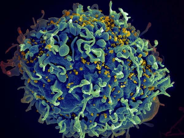
HIV invading a human cell
HIV invading a human cell: Credit NIH

A new study shows how key features on the surface of HIV-infected cells such as certain sugar molecules help the disease evade detection by the immune system, and how they can be disabled. The findings, published in PLOS Pathogens, represent a first step to eradicating this persistent virus in patients.

“We identified a glyco-immune checkpoint interaction as a novel mechanism that allows HIV-infected cells to evade immune surveillance,” said Mohamed Abdel-Mohsen, PhD, assistant professor in the Vaccine & Immunotherapy Center at The Wistar Institute and coauthor on the paper. “And we developed a novel approach that selectively targets these interactions on the surface of these infected cells.”

Existing treatments can reduce HIV to undetectable levels, but eradication remains elusive, with the disease typically returning quickly when treatment stops. And even when controlled, HIV increases risk for other health problems, including neurological disorders, cardiovascular disease, and cancer.

For the new study, researchers looked at a type of sugar molecule called sialic acid on the surface of HIV-infected cells. These sugars bind with receptors called siglecs on the surface of disease-fighting ‘natural killer’ immune cells. When activated, these receptors act as inhibitors, restraining the killer cells and causing them to stop killing. “We thought, ‘is it possible that these HIV-infected cells are using this interaction – covering themselves with these sugars to evade the natural killer immune surveillance?’” said Prof Abdel-Mohsen.

The researchers found that these infected cells can actually exploit this inhibitory connection to evade immune surveillance. They then investigated whether they could manipulate this connection to make the killer cells more effective at killing HIV-infected cells. Disabling the inhibitors from the killer cells was found to cause the immune cells to attack indiscriminately. The researchers turned to the HIV cells, using the enzyme sialidase to remove the sialic acid sugars that were activating the immune inhibitors but this affected all cells, again causing the killer cells to attack indiscriminately. Finally, they developed a sialidase conjugate linked to HIV antibodies, which only targeted sialic acid on HIV cells. With the sialic acid removed from these cells, the killer immune cells attacked and killed the HIV-infected cells, leaving healthy cells alone.

“The killer cells become a super killer for the HIV-infected cells and they now attack them in a selective manner,” said Prof Abdel-Mohsen. “The discovery could be a missing link in the “shock and kill” approach to HIV treatment that has been a focus of research for the past several years,” he added. This two-step process involves first “shocking” the HIV out of latency so it can be detected, and then stimulating the immune system to “kill” the virus once and for all. However, while effective methods have been discovered to reverse latency, scientists haven’t yet found a way to make HIV-infected cells more killable once reactivated. “We may have the shock, but we don’t yet have the kill,” Prof Abdel-Mohsen said. “Our method actually increases the susceptibility of HIV-infected cells to killing, which is one of the top unmet needs in the HIV field.”

First author Samson Adeniji, Ph.D., a postdoctoral fellow at Wistar, noted that the team’s approach could be tested in combination with broadly neutralizing antibody therapies currently being studied in clinical trials. “By combining approaches, we could turn these immune cells from a cop into a kind of Robocop,” he said.

The researchers also noted that, besides  HIV, the approach could be applied in infectious diseases that may evade the immune system, including hepatitis and COVID. In vivo tests with animals are the next step. They’re also investigating other sugar molecules on HIV that may play a similar role as sialic acid. “HIV-infected cells are likely evading immune surveillance through many potential glyco-immune checkpoints,” Abdel-Mohsen said. “We are investigating other mechanisms and how to break them.”

Source: Wistar Institute

Massage Heals Injured Muscle by Getting Immune Cells Out

Photo by Andrea Piacquadio from Pexels

While massage has been used to treat muscle pain and injury for thousands of years, it is only now that a study has scientifically confirmed that it improves recovery and strength gains. Intriguingly, the mechanism behind this is mechanically clearing out of immune cells from the injury site after they have done their job.

Using a custom-designed robotic system massage system for mice, the team found that this mechanical loading (ML) rapidly clears immune cells called neutrophils out of severely injured muscle tissue. This process also removed inflammatory cytokines released by neutrophils from the muscles, enhancing the process of muscle fiber regeneration. The research is published in Science Translational Medicine.

“Lots of people have been trying to study the beneficial effects of massage and other mechanotherapies on the body, but up to this point it hadn’t been done in a systematic, reproducible way. Our work shows a very clear connection between mechanical stimulation and immune function. This has promise for regenerating a wide variety of tissues including bone, tendon, hair, and skin, and can also be used in patients with diseases that prevent the use of drug-based interventions,” said first author Bo Ri Seo, PhD.

Dr Seo and her colleagues previously found in mouse studies that mechanical massage of injured muscles doubled the rate of muscle regeneration and reduced tissue scarring over the course of two weeks. With a new device inspired by soft robotics, the researchers sought to confirm these results. They found that the greater the force applied, the stronger the injured muscles became.

In vitro experiments suggested that neutrophil-secreted factors stimulate the growth of muscle cells, but the prolonged presence of those factors impairs the production of new muscle fibres. In vivo testing showed that stronger muscle fibre types predominated in treated, injured muscle types. Depleting neutrophils in mice after the third day resulted in greater strength recovery, indicating that they are important in the initial recovery period but removing them from the injury site early leads to improved muscle regeneration.

“The idea that mechanics influence cell and tissue function was ridiculed until the last few decades, and while scientists have made great strides in establishing acceptance of this fact, we still know very little about how that process actually works at the organ level. This research has revealed a previously unknown type of interplay between mechanobiology and immunology that is critical for muscle tissue healing, in addition to describing a new form of mechanotherapy that potentially could be as potent as chemical or gene therapies, but much simpler and less invasive,” said Don Ingber, MD, PhD, founding director of the Wyss Institute for Biologically Inspired Engineering at Harvard.

Source: Wyss Institute for Biologically Inspired Engineering at Harvard