Tag: hypoxia

Groundbreaking Study Discovers Differences in Oxygen Physiology in Down Syndrome

Source: Pixabay CC0

A groundbreaking new study published in Cell Reports by researchers from the University of Colorado Anschutz Medical Campus reports important differences in oxygen physiology and red blood cell function in individuals with Down syndrome. The study is part of the ongoing Human Trisome Project, a large and detailed cohort study of the population with Down syndrome, including deep annotation of clinical data, the largest biobank for the study of Down syndrome to date, and multi-omics datasets.

The Crnic Institute team first analysed hundreds of blood samples to identify physiological differences between research participants with Down syndrome versus controls from the general population. They observed that triplication of chromosome 21, or trisomy 21, the chromosomal abnormality that causes Down syndrome, leads to a physiological state reminiscent of hypoxia. They identified major changes in gene expression indicative of low oxygen availability, including induction of many hypoxia-inducible genes and proteins, as well as increased levels of factors involved in the synthesis of haeme, the molecule that transports oxygen inside red blood cells.

“These results reveal that hypoxia and hypoxic signalling should be front and centre when we talk about the health of people with Down syndrome,” says Dr Joaquín Espinosa, executive director of the Crnic Institute, professor of pharmacology, principal investigator of the Human Trisome Project, and one of the senior authors of the paper. “Given the critical role of oxygen physiology in health and disease, we need to understand the causes and consequences of hypoxia in Down syndrome, which could lead to effective interventions to improve oxygen availability in this deserving population.”

“The results are remarkable, it is safe to say that the blood of people with Down syndrome looks like that of someone who was quickly transported to a high altitude or who was injected with erythropoietin (EPO), the master regulator of erythropoiesis, the process of new red blood cell formation,” explains Dr Micah Donovan, lead author of the paper. “Although it has been known for many years that people with Down syndrome have fewer and bigger red blood cells, this is the first demonstration that they overproduce EPO and that they are undergoing stress erythropoiesis, a phenomenon whereby the liver and the spleen need to start producing red blood cells to supplement those arising from the bone marrow.”

The team discovered that these phenomena are also observed in a mouse model of Down syndrome, thus reinforcing the idea that these important physiological changes arise from triplication of genetic material and overexpression of specific genes.

“The fact that hypoxic signaling and stress erythropoiesis are conserved in the mouse model paves the way for mechanistic investigations that could identify the genes involved and reveal therapeutic interventions to improve oxygen physiology in Down syndrome,” explains Dr. Kelly Sullivan, associate professor of pediatrics, director of the Experimental Models Program at the Crnic Institute and co-author in the study.

The study team also investigated whether the elevated hypoxic signaling and associated stress erythropoiesis was tied to the heightened inflammatory state characteristic of Down syndrome. Although individuals with the stronger hypoxic signatures show more pronounced dysregulation of the immune system and elevated markers of inflammation, their results indicate that lowering inflammation does not suffice to reverse the hypoxic state.

“We will need a lot more data to understand what is causing the hypoxic state and its impacts on the health of people with Down syndrome,” says Dr Matthew Galbraith, assistant research professor of pharmacology, director of the Data Sciences Program at the Crnic Institute, and one of the senior authors of the paper. “The hypoxic state could be caused by obstructive sleep apnoea (which is common in Down syndrome), cardiopulmonary malfunction, or even perhaps defects in red blood cell function. We are very excited about several ongoing clinical trials funded by the NIH INCLUDE Project for obstructive sleep apnea in Down syndrome, which we believe will be very informative.”

The Crnic Institute study team is already planning several follow up studies, with the explicit goal of illuminating strategies to improve oxygen physiology in the population with Down syndrome.

Red Blood Cells Exposed to Oxygen Deficiency Protect against Myocardial Infarction

Source: Pixabay CC0

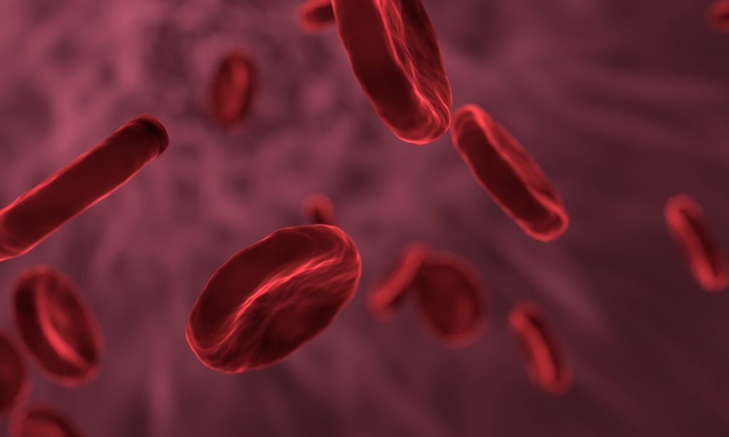
Red blood cells exposed to oxygen deficiency protect against myocardial infarction, according to a new study published in the Journal of Clinical Investigation. This study, conducted at Karolinska Institutet in collaboration with Karolinska University Hospital, also shows that that protection can be enhanced by a diet containing nitrate-rich vegetables, such as arugula and other green leafy vegetables.

“This effect was also shown in a clinical study in patients with high blood pressure who were randomly assigned to eat nitrate-rich vegetables or a diet low in nitrates,” says John Pernow, Professor of Cardiology at the Department of Medicine, Karolinska Institutet in Solna and senior physician at Karolinska University Hospital, and the study’s corresponding author together with Jon Lundberg, professor at the Department of Physiology and Pharmacology, Karolinska Institutet.

Part of the study was conducted through experiments with red blood cells from mice that were added to a myocardial infarction model with hearts from mice. Before the experiment, the red blood cells were exposed to low oxygen pressure, while nitrate was added to the drinking water.

In a clinical study, red blood cells were collected from patients with high blood pressure who were randomly assigned a nitrate-rich diet with green leafy vegetables or a diet with nitrate-poor vegetables. These red blood cells were given to the corresponding myocardial infarction model with hearts from rats.

“The results show both that the red blood cells convey protection against injury in the heart in the event of low oxygen levels, and how that protection can be enhanced through a simple dietary advice. This may be of great importance for patients at risk of myocardial infarction,” says the study’s first author Jiangning Yang, a researcher at the Department of Medicine, Solna, Karolinska Institutet.

The next step in the research is to develop additional drugs that can activate the protective signalling mechanism in red blood cells to provide protection to the body’s tissues and cells in the event of oxygen deficiency.

“In addition, we need to map how the blood cells transmit their protective signal to the heart muscle cells,” says John Pernow.

Source: Karolinska Institutet

Periods of Hypoglycaemia Worsen Progression of Diabetic Retinopathy

Credit: National Eye Institute

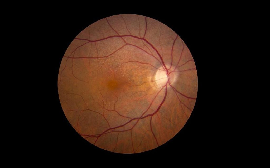
People with diabetes who experience periods of hypoglycaemia, a common event in those new to blood sugar management, are more likely to have worsening diabetic eye disease. Now, researchers say they have linked such low blood sugar levels with a molecular pathway that is activated in hypoxic cells in the eye.

The research, involving human and mouse eye cells and intact retinas grown in a low glucose environment in the laboratory, as well as mice with low glucose levels, was published in Cell Reports.

“Temporary episodes of low glucose happen once or twice a day in people with insulin-dependent diabetes and often among people newly diagnosed with the condition,” says Akrit Sodhi, MD, PhD, Johns Hopkins Medicine professor. Low glucose levels can also occur during sleep in people with non-insulin dependent diabetes. “Our results show that these periodic low glucose levels cause an increase in certain retinal cell proteins, resulting in an overgrowth of blood vessels and worsening diabetic eye disease,” adds Sodhi.

Up to a third of diabetic patients will develop diabetic retinopathy, which is characterised by the overgrowth of abnormal blood vessels in the retina.

Sodhi says the current study suggests that people with diabetic retinopathy may be particularly vulnerable to periods of low glucose, and keeping glucose levels stable should be an important part of glucose control.

For the study, the researchers analysed protein levels in human and mouse retinal cells and intact retinas grown in an environment of low glucose in the laboratory, as well as in mice that had occasional low blood sugar.

In human and mouse retinal cells, low glucose levels triggered a cascade of molecular changes that can lead to blood vessel overgrowth. First, the researchers saw that low glucose caused a decrease in retinal cells’ ability to break down glucose for energy.

When the researchers focused on Müller glial cells, which are supportive cells for neurons in the retina and rely primarily on glucose for energy production, they found that the cells increased the expression of the GLUT1 gene, which makes a protein that transports glucose into cells.

The researchers found that, in response to low glucose, the cells increased levels of a transcription factor, hypoxia-inducible factor (HIF)-1α. This turned on the cellular machinery, including GLUT1, needed to improve their ability to utilise available glucose, preserving the limited oxygen available for energy production by retinal neurons.

However, in hypoxic environments, as occurs in the retinas of patients with diabetic eye disease, this normal, physiologic response to low glucose triggered a flood of HIF-1α protein into the nucleus.

This resulted in an increase in the production of proteins such as VEGF and ANGPTL4, which cause the growth of abnormal, leaky blood vessels – the key culprit of vision loss in people with diabetic eye disease.

The researchers plan to study whether low glucose levels in people with diabetes may impact similar molecular pathways in other organs, such as the kidney and brain.

Sodhi says the HIF-1α pathway may serve as an effective target for developing new treatments for diabetic eye disease.

Source: Johns Hopkins Medicine

Delayed COVID Recovery could be a Protective Mechanism against Hypoxia

Photo by Andrea Piacquadio on Unsplash

COVID patients placed on ventilators can take a long time to regain consciousness. New research published the Proceedings of the National Academy of Sciences now shows that these delays may serve a purpose: protecting the brain from oxygen deprivation.

The existence of such a brain-preserving state could explain why some patients wake up days or even weeks after they stop receiving ventilation, and it suggests that physicians should take these lengthy recovery times into account when determining a patient’s prognosis.

In their study, investigators connect the pattern seen among those who have survived severe COVID with similar delays known to occur in a small fraction of cardiac arrest patients.

“The delayed recoveries in COVID patients are very much like the rare cases we’ve documented in previous research. In this new paper, we describe a mechanism to explain what we’re seeing in both types of patients,” said study co-senior author Dr Nicholas D. Schiff, a neurology professor at Weill Cornell Medicine.

He suggests that this mechanism is the brain protecting itself, pointing to animals, most notably painted turtles, that can tolerate extended periods without oxygen.

More than a decade ago, Dr Schiff and his colleagues first observed these delays among comatose cardiac arrest patients who received cooling therapy to reduce brain damage caused by a loss of blood flow. In one such case, a 71-year-old patient took 37 days to awaken, before ultimately making a near-complete recovery.

During the pandemic, Dr Schiff performed neurology consultations for COVID patients, and he soon began seeing similar, delayed awakenings occurring when patients were taken off ventilators and stopped receiving movement-limiting sedatives.

In a separate analysis of a large cohort of COVID patients from Weill Cornell Medicine and two other major U.S. medical centres, Dr Schiff and his colleagues, including co-author of the current paper, Dr Emery N. Brown, professor of anaesthesia at Harvard Medical School, found that a quarter of patients who survived ventilation took 10 days or longer to recover consciousness. The more oxygen deprivation they suffered while on the ventilator, the longer that delay.

In the prior study of cardiac patients, the researchers recorded a distinctive pattern in brain activity, one also seen in patients under deep anaesthesia. (Recordings from COVID patients are extremely limited.) Dr Schiff read that a similar pattern had been seen in the brains of painted turtles, which can withstand up to five months without oxygen under ice in the winter. To do so, they activate the same inhibitory system within the brain targeted by anaesthetics given to human cardiac and COVID patients but in novel ways developed by evolutionary specialisations.

Drs Schiff and Brown propose that, by chance, the same protective response emerges in the patients.

“It is our theory that oxygen deprivation as well as practices in the ICU, including commonly used anaesthetics, expose elements of strategies that animals use to survive in extreme conditions,” Dr Schiff said.

“These observations may offer new insights into the mechanisms of how certain anaesthetics produce unconsciousness and new approaches for ICU sedation and for fostering recovery from disorders of consciousness,” Dr Brown added.

When patients fail to regain consciousness for an extended time, physicians may recommend withdrawing life-supporting care. This threshold is typically set at 14 days or less for cardiac patients, while no such guidelines exist for COVID.

In light of this new research, however, so long as they lack brain injuries, physicians should avoid making negative projections about these patients’ potential to recover, note the researchers.

Source: Weill Cornell Medicine

Oxygen Deficiency in Newborns may Increase Later Cardiovascular Risk

Photo by Christian Bowen on Unsplash

A population-based observational study has shown that babies suffering oxygen-deficiency complications at birth are almost twice as likely to develop cardiovascular disease in childhood or early adulthood, though such conditions are rare in youth. The findings are published in the journal The Lancet Regional Health – Europe.

According to the Karolinska Institutet researchers, the study could be the first of its kind to examine how complications related to asphyxiation at birth, which affects four million babies annually, affects the risk of cardiovascular disease later in life. Previous research has mostly concentrated on the association between asphyxia in the neonatal period and brain development.

Despite the relatively high risk, the absolute number of babies who suffer from cardiovascular disease despite asphyxiation at birth is very low. After the 30-year follow-up period, only 0.3% of those with asphyxia-related complications had a cardiovascular diagnosis, compared with 0.15% of those without complications.

Since the study was observational, the researchers are unable to establish any causality or propose any underlying mechanisms.

Largest risk increase for stroke and heart failure

The study followed over 2.8 million individuals born in Sweden between 1988 and 2018, of whom 31 419 suffered asphyxia-related complications at birth. A total of 4165 cases of cardiovascular disease were identified during the follow-up period. The increase in risk was particularly salient for stroke and heart failure, as well as for atrial fibrillation. The researchers took into account potential confounders such as birth weight and maternal lifestyle.

“Even if the absolute risk of cardiovascular disease is low at a young age, our study shows that asphyxia-related complications at birth are associated with a higher risk of cardiovascular disease later in life,” says the study’s corresponding author Neda Razaz, assistant professor at the Department of MedicineSolna, Karolinska Institutet.

Source: Karolinska Institutet

Hypoxia can Trigger Immune System Reaction

Anatomical model of lungs
Photo by Robina Weermeijer on Unsplash

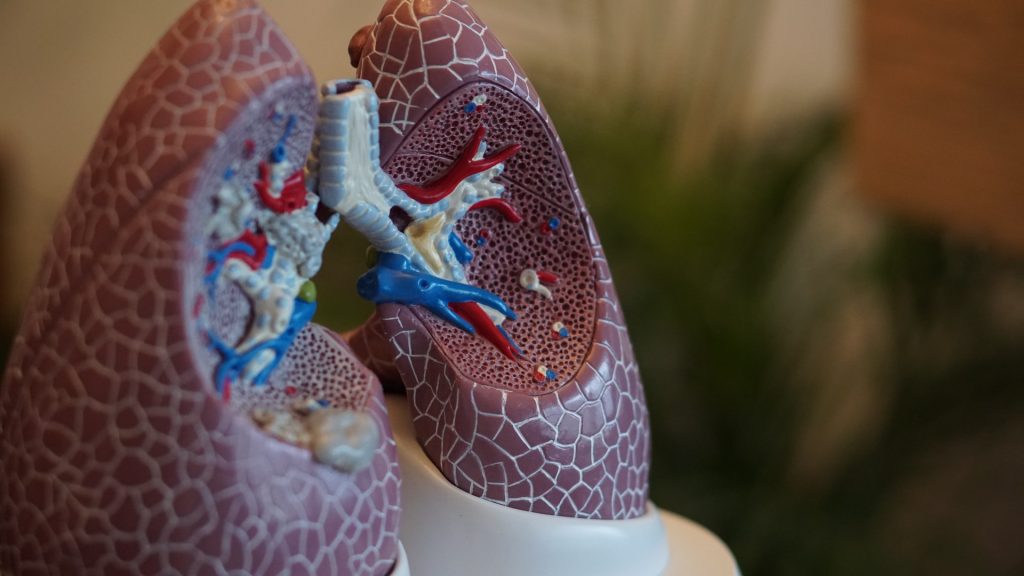
New research from scientists at La Jolla Institute for Immunology (LJI), shows that hypoxia can activate the same group of immune cells that cause inflammation during asthma attacks. During hypoxia, these cells flood the airways with lung-damaging molecules.

Hypoxia is a known trigger for developing and worsening lung conditions such as severe asthma, chronic obstructive pulmonary disease (COPD), and fibrosis. To treat and prevent these diseases, researchers need to understand why a lack of oxygen would affect the immune system.

“We show how lack of oxygen can be part of a feedback loop that can contribute to even worse inflammation,” says LJI Professor and Chief Scientific Officer Mitchell Kronenberg, PhD, a member of the LJI Center for Autoimmunity and Inflammation. “This work gives us insight into the causes of fibrosis of the lung and severe asthma.”

Prof Kronenberg and colleagues worked with a genetically altered mouse model to mimic the signals of hypoxia in the airway’s epithelial cells, which line the paths to the lungs. They discovered that combining the hypoxia signals with inflammatory signals stimulated the “innate,” or rapidly responding immunity, and an immune cell type called an ILC2.

An ILC2’s job is to make signaling molecules – cytokines – that quickly alert other immune cells to react to a pathogen. Unfortunately, ILC2s sometimes over-react and respond to harmless environmental allergens. In these cases, ILC2s churn out cytokines that drive mucus production and inflammation in the lungs. All this swelling and mucus leads to hypoxia.

As they report in Journal of Experimental Medicine, ILC2s respond to hypoxia as well, adding to the lung damage already caused during an asthma attack.

“That hypoxia may then contribute further to inflammation,” said Prof Kronenberg.

The next step was to figure out exactly how epithelial cells activate ILC2 during hypoxia. LJI Postdoctoral Fellow Jihye Han, PhD, led the work to uncover an unexpected culprit: adrenomedullin (ADM). ADM is known for its role in helping blood vessels dilate, but until now it had no known role in immune function.

Prof Kronenberg was surprised to see ADM involved — but not shocked. “We’re finding that many molecules with no previously known role in the immune system can also be important for immune function,” said Prof Kronenberg. “We need to understand that more generally.”

The researchers showed that human lung epithelial cells exposed to hypoxia also produced ADM. This means ADM or its receptor could be targets for treating inflammatory and allergic lung diseases.

The challenge is to find a balance between dampening the harmful immune response without leaving the body vulnerable to infections. Prof Kronenberg points out that the epithelial cell-ADM-ILC2 connection protected mice from hookworm infections, which damage the lungs and gut.

“ADM is a new target for lung diseases and has been implicated in bacterial pneumonia as well,” said Prof Kronenberg. “But blocking it would have to be done carefully.”

Source: La Jolla Institute for Immunology

Protecting Newborns’ Brains During Rewarming Stage of Cooling Therapy

Photo by William-Fortunato on Pexels

Oxygen-deprived newborns who undergo hypothermia therapy have a higher risk of seizures and brain damage during the rewarming period, according to a new study. The finding, published online in JAMA Neurology, could lead to better ways to protect these vulnerable patients during an often overlooked yet critical period of hypothermia therapy.

“A wealth of evidence has shown that cooling babies who don’t receive enough oxygen during birth can improve their neurodevelopmental outcomes, but few studies have looked at events that occur as they are rewarmed to a normal body temperature,” said study leader Lina Chalak, MD, MSCS, Professor at UT Southwestern. “We’re showing that there’s a significantly elevated risk of seizures during the rewarming period, which typically go unnoticed and can cause long-term harm.”

Millions of newborns around the world are affected by neonatal hypoxic-ischaemic encephalopathy (HIE), brain damage initially caused by hypoxia during birth. Although the World Health Organization estimates that birth asphyxia is responsible for nearly a quarter of all neonatal deaths, those babies that survive oxygen deprivation are often left with neurological injuries, Dr Chalak explained.

To help improve outcomes, babies diagnosed with HIE are treated with hypothermia, using a cooling blanket that brings the body temperature down to as low as 33.5°C, said Dr. Chalak.

Studies initially showed that during cooling, babies with HIE commonly have symptomless seizures, which are neurological events that can further damage the brain, prompting the addition of electroencephalographic (EEG) monitoring to the hypothermia protocol. However, Dr Chalak explained, babies typically haven’t been monitored during the rewarming period, in which the temperature of the blanket is increased by 0.5°C every hour.

To better understand seizure risk during rewarming, Dr. Chalak and colleagues studied 120 babies who were enrolled in another study that compared two different cooling protocols, one longer and colder than the other. The babies in the study were also monitored with EEG to check for seizures both during the cooling and the rewarming phases of hypothermia.

When the researchers compared data from the last 12 hours of cooling and the first 12 hours of rewarming, they found that rewarming roughly tripled the odds of seizures. Additionally, babies who had seizures during rewarming, there was twice the risk of mortality or neurological disability by age 2, compared with those who didn’t have seizures during this period. This finding held true even after adjusting for differences in medical centers and the newborns’ HIE severity.

While it is not known how to prevent seizures from occurring in babies with HIE, treating seizures when they do occur can help prevent further brain damage, Dr Chalak said. Thus, monitoring during both cooling and rewarming can help protect the babies’ brains from further insults while they heal.

“This study is telling us that there’s an untapped opportunity to improve care for these babies during rewarming by making monitoring a standard part of the protocol,” said Dr Chalak.

Source: EurekAlert!

The Effect of Hypoxia on Cancer Cells is a Matter of Timing

A new study from the University of Colorado School of Medicine shows that the effect of hypoxia on cancer cells varies in the short term versus the long term, opening new possibilities for cancer treatment.

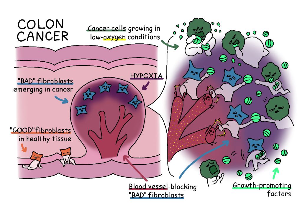
How cancer cells adapt to hypoxia, where insufficient oxygen reaches cells, is a key aspect of cancer biology.

“Most tumours cannot grow unless they figure out a way to induce formation of new blood vessels to supply them with oxygen and other nutrients,” explained Matthew Galbraith, PhD. “So, what happens inside of solid tumours is they undergo intermittent periods of low oxygen between rounds of new blood vessel formation.”

Previous research focussed on hypoxia in the long term, characterising it as oncogenic, or cancer promoting. However some studies showed that hypoxia-sensing factors, known as hypoxia inducible factors, or HIFs, can in some situations suppress tumour growth. To solve this, senior researcher Joaquin Espinosa, PhD and colleagues studied the immediate acute response to hypoxia.

“We employed a cutting-edge genomics technology that nobody had employed in this field before that allowed us to see what happens to cancer cells within minutes of depriving them of oxygen,” Dr Espinosa said.

Employing this technology, they identified hundreds of hypoxia-inducible genes activated shortly upon oxygen deprivation. Using computational biology approaches on large, publicly available datasets, they inferred the function of these genes on hundreds of lab-grown cancer cell lines and hundreds of tumour samples from cancer patients.

They found that when a cell is hypoxic, it reacts by ceasing growth to preserve its existing nutrients and oxygen. Thus, hypoxia causes a tumour-suppressive reaction at this point, mostly by preventing protein synthesis. Only after prolonged periods of hypoxia do cells metastasise and spread out in search of oxygen.

“There’s been a lot of debate about whether these hypoxia-inducible factors promote tumour growth or prevent tumour growth,” Dr Espinosa said. “The conclusion we came to is that everyone was right to a degree. Hypoxia-inducible factors can suppress tumour growth by preventing protein synthesis early on, but they can also advance tumour growth at later stages by promoting the ability of cancer cells to invade neighboring tissues. It depends on when you’re looking at it.”

The tumour suppression and promotion mechanisms elicited by HIFs can be exploited as drug targets. Tumour suppression is mediated by inhibition of an enzyme known as mTOR, which in turn can be inhibited by available drugs often used in cancer therapies. “mTOR inhibitors could mimic the tumour suppressive effects of HIFs,” Dr Galbraith explained.

When deprived of oxygen for a longer amount of time, the HIFs switch on a set of enzymes that can degrade the extracellular matrix that holds them in place, allowing the cancer cells to escape the oxygen-deprived tumour. The cancer cells can then enter the bloodstream and invade nearby tissues.

“These results emphasise the importance of developing inhibitors of hypoxia-inducible enzymes that degrade collagen and other components of the extracellular matrix,” Espinosa said.

Dr Espinosa and his team hope that their research will help new cancer treatments to be developed, which also target the cancer at the right times. 

“People have been trying to target the hypoxia-inducible factors with different therapeutics, but this research would suggest that you may want to exercise some caution about when you apply those therapeutics, given that the HIFs can be tumour suppressive in the early stages of hypoxia,” Dr Galbraith said.

“Since the hypoxic response can be tumour suppressive in some contexts and oncogenic in other contexts, it’s not a good idea to issue a blanket statement that we should always try to shut it down,” Dr Espinosa added. “Instead, we should be thinking about what aspect of the hypoxic response to target, and that’s the aspect where hypoxia drives invasion and metastasis.”

Hoping that other researchers would make use of the map his team developed, Dr Espinosa said, “I would say this is a definitive improvement in the mapping of the early events of hypoxia. And the beauty of that is that once you have a good map of the land, a lot of people can use it.”

Source:  Medical Xpress

Journal information: Zdenek Andrysik et al, Multi-omics analysis reveals contextual tumor suppressive and oncogenic gene modules within the acute hypoxic response, Nature Communications (2021). DOI: 10.1038/s41467-021-21687-2