Tag: Huntington's disease

A New Genetic Culprit in Huntington’s Disease

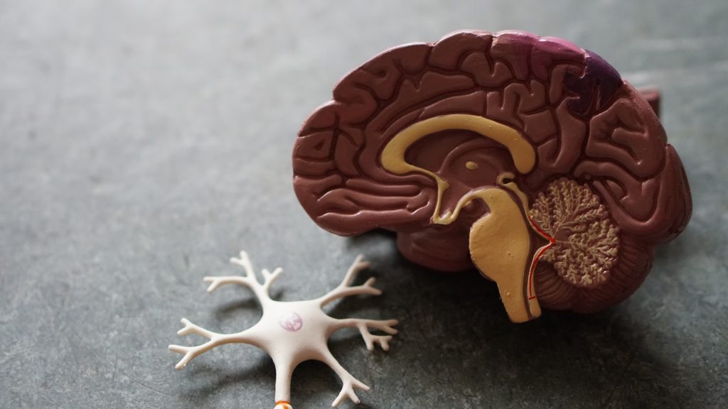
Photo by Sangharsh Lohakare on Unsplash

Researchers in Berlin and Düsseldorf have implicated a new gene in the progression of Huntington’s disease in a brain organoid model. The gene may contribute to brain abnormalities much earlier than previously thought. The study is out now in Nature Communications.

The researchers are the first to implicate the gene CHCHD2 in Huntington’s disease (HD) – an incurable genetic neurodegenerative disorder – and identified the gene as a potentially new therapeutic target. In a brain organoid model of the disease, the researchers found that mutations in the Huntington gene HTT also affect CHCHD2, which is involved in maintaining the normal function of mitochondria.

Six different labs at the Max Delbrück Center participated in the study, led by Dr Jakob Metzger of the “Quantitative Stem Cell Biology” lab at the and the “Stem Cell Metabolism” lab of Professor Alessandro Prigione at Heinrich Heine University Düsseldorf (HHU). Each lab contributed their unique expertise on Huntington’s disease, brain organoids, stem cell research and genome editing. “We were surprised to find that Huntington’s disease can impair early brain development through defects associated with mitochondrial dysfunction,” says Dr Pawel Lisowski, co-lead author in the Metzger lab at the Max Delbrück Center.

Moreover, “the organoid model suggests that HTT mutations damage brain development even before clinical symptoms appear, highlighting the importance of detecting the late-onset neurodegenerative disease early,” Selene Lickfett, co-lead author and a doctoral student in the Faculty of Mathematics and Natural Science in the lab of Prigione at HHU adds.

The unusual repetition of three letters

Huntington’s disease is caused when the nucleotides Cytosine, Adenine and Guanine are repeated an excessive number of times in the in the Huntington gene HTT. People with 35 or less repeats are generally not at risk of developing the disease, while carrying 36 or more repeats has been associated with disease. The greater the number of repeats, the earlier the disease symptoms are likely to appear, explains Metzger, a senior author of the study. The mutations cause nerve cells in the brain to progressively die. Those affected, steadily lose muscle control and develop psychiatric symptoms such as impulsiveness, delusions and hallucinations. Huntington’s disease affects approximately five to 10 in every 100 000 people worldwide. Existing therapies only treat the symptoms of the disease, they don’t slow its progression or cure it.

The challenge of HTT gene editing

To study how mutations in the HTT gene affect early brain development, Lisowski, first used variants of the Cas9 gene editing technology and manipulation of DNA repair pathways to modify healthy induced pluripotent stem cells such that they carry a large number of CAG repeats. This was technically challenging because gene editing tools are not efficient in gene regions that contain sequence repeats, such as the CAG repeats in HTT, says Lisowski.

The genetically modified stem cells were then grown into brain organoids – three-dimensional structures a few millimetres in size that resemble early-stage human brains. When the researchers analysed gene expression profiles of the organoids at different stages of development, they noticed that the CHCHD2 gene was consistently under expressed, which reduced metabolism of neuronal cells. CHCHD2 is involved in ensuring the health of mitochondria – the energy producing structures in cells. CHCHD2 has been implicated in Parkinson’s disease, but never before in Huntington’s.

They also found that when they restored the function of the CHCHD2 gene, they could reverse the effect on neuronal cells. “That was surprising,” says Selene Lickfett. “It suggests in principle that this gene could be a target for future therapies.”

Moreover, defects in neural progenitor cells and brain organoids occurred before potentially toxic aggregates of mutated Huntingtin protein had developed, adds Metzger, indicating that disease pathology in the brain may begin long before it is clinically evident.

“The prevalent view is that the disease progresses as a degeneration of mature neurons,” says Prigione. “But if changes in the brain already develop early in life, then therapeutic strategies may have to focus on much earlier time-points.”

Wide reaching implications

“Our genome editing strategies, in particular the removal of the CAG repeat region in the Huntington gene, showed great promise in reversing some of observed developmental defects. This suggests a potential gene therapy approach,” says Prigione. Another potential approach could be therapies to increase CHCHD2 gene expression, he adds.

The findings may also have broader applications for other neurodegenerative diseases, Prigione adds. “Early treatments that reverse the mitochondrial phenotypes shown here could be a promising avenue for counteracting age-related diseases like Huntington’s disease.”

Source: Max Delbrück Center for Molecular Medicine in the Helmholtz Association

Study Suggests Leprosy Drug may be Effective in Huntington’s Disease

Source: CC0

A preclinical study from Karolinska Institutet offers hope for treating severe neurodegenerative diseases with an existing drug: clofazimine, which is used to treat leprosy, may be effective in the treatment of Huntington’s disease.

The research group examined whether existing drugs could reduce the toxicity of so-called polyQ proteins. These proteins are found in patients with certain hereditary neurodegenerative diseases, including Huntington’s disease, for which there is no cure. 

Screening hundreds of drugs, they found that the leprosy drug clofazimine reduces the toxicity of polyQ proteins and restores mitochondrial function in zebrafish and worms. The finding, published in eBioMedicine, supports the previous hypothesis that polyQ diseases are associated with the dysfunction of mitochondria, the organelles in charge of producing energy within cells.

“Our work not only suggests the interest of a specific drug for the treatment of polyQ neurodegenerative diseases, but also helps us to better understand what causes these diseases. It is possible to find new uses for old drugs, which reduces the time needed to find novel therapies”, says last author Oscar Fernandez-Capetillo, Professor and research group leader at the Department of Medical Biochemistry and Biophysics at Karolinska Institutet.

Clofazimine is not very efficient in entering the nervous system, however. The research group are now trying to figure out solutions to this limitation, by testing the efficacy of clofazimine in mammalian models of neurodegenerative disease. 

“We hope that our discovery can be developed into a new medicine, but there are still some hurdles that need to be overcome,” says Oscar Fernandez-Capetillo.

The researchers are also conducting similar drug screens in other age-related pathologies such as cancer and other neurodegenerative disorders.

Source: Karolinska Institutet

Huntington’s Disease Impacts Neuron Types Differently

A healthy neuron.
A healthy neuron. Credit: NIH

In patients with Huntington’s disease, neurons in a part of the brain called the striatum are some of the worst affected. Degeneration of these neurons contributes to patients’ loss of motor control, which is one of the major hallmarks of the disease.

Neuroscientists at MIT have now shown that two distinct cell populations in the striatum are affected differently by Huntington’s disease. Reporting their results in Nature Communication, they believe that neurodegeneration of one of these populations leads to motor impairments, while damage to the other population, located in structures called striosomes, may explain the mood disorders that are often see in the early stages of the disease.

“As many as 10 years ahead of the motor diagnosis, Huntington’s patients can experience mood disorders, and one possibility is that the striosomes might be involved in these,” says Ann Graybiel, an MIT Institute Professor and one of the senior authors of the study.

Using single-cell RNA sequencing to analyse the genes expressed in mouse models of Huntington’s disease and postmortem brain samples from Huntington’s patients, the researchers found that cells of the striosomes and another structure, the matrix, begin to lose their distinguishing features as the disease progresses. The researchers hope that their mapping of the striatum and how it is affected by Huntington’s could help lead to new treatments that target specific cells within the brain.

This kind of analysis could also shed light on other brain disorders that affect the striatum, such as Parkinson’s disease and autism spectrum disorder, the researchers say.

Neuron vulnerability

Huntington’s disease leads to degeneration of brain structures called the basal ganglia, which are responsible for control of movement and also play roles in other behaviors, as well as emotions. For many years, Graybiel has been studying the striatum, a part of the basal ganglia that is involved in making decisions that require evaluating the outcomes of a particular action.

Many years ago, Graybiel discovered that the striatum is divided into striosomes, which are clusters of neurons, and the matrix, which surrounds the striosomes. She has also shown that striosomes are necessary for making decisions that require an anxiety-provoking cost-benefit analysis.

In a 2007 study, Richard Faull of the University of Auckland discovered that in postmortem brain tissue from Huntington’s patients, the striosomes showed a great deal of degeneration. Faull also found that while those patients were alive, many of them had shown signs of mood disorders such as depression before their motor symptoms developed.

To further explore the connections between the striatum and the mood and motor effects of Huntington’s, Graybiel teamed up with Kellis and Heiman to study the gene expression patterns of striosomal and matrix cells. To do that, the researchers used single-cell RNA sequencing to analyze human brain samples and brain tissue from two mouse models of Huntington’s disease.

Within the striatum, neurons can be classified as either D1 or D2 neurons. D1 neurons are involved in the “go” pathway, which initiates an action, and D2 neurons are part of the “no-go” pathway, which suppresses an action. D1 and D2 neurons can both be found within either the striosomes and the matrix.

The analysis of RNA expression in each of these types of cells revealed that striosomal neurons are harder hit by Huntington’s than matrix neurons. Furthermore, within the striosomes, D2 neurons are more vulnerable than D1.

The researchers also found that these four major cell types begin to lose their identifying molecular identities and become more difficult to distinguish from one another in Huntington’s disease. “Overall, the distinction between striosomes and matrix becomes really blurry,” Graybiel says.

Striosomal disorders

The findings suggest that damage to the striosomes, which are known to be involved in regulating mood, may be responsible for the mood disorders that strike Huntington’s patients in the early stages of the disease. Later on, degeneration of the matrix neurons likely contributes to the decline of motor function, the researchers say.

In future work, the researchers hope to explore how degeneration or abnormal gene expression in the striosomes may contribute to other brain disorders.

Previous research has shown that overactivity of striosomes can lead to the development of repetitive behaviors such as those seen in autism, obsessive compulsive disorder, and Tourette’s syndrome. In this study, at least one of the genes that the researchers discovered was overexpressed in the striosomes of Huntington’s brains is also linked to autism.

Additionally, many striosome neurons project to the part of the brain that is most affected by Parkinson’s disease (the substantia nigra, which produces most of the brain’s dopamine).

“There are many, many disorders that probably involve the striatum, and now, partly through transcriptomics, we’re working to understand how all of this could fit together,” Graybiel says.

Source: Massachusetts Institute of Technology

Fixing The Protein Behind Huntington Disease

Genetics
Image source: Pixabay

An international research effort has developed a new strategy to treat Huntington disease, which demonstrated that converting the disease-causing protein to its disease-free form results in it still retaining its original function. This discovery, published in the Journal of Clinical Investigation Insight, provides new avenues to approach Huntington disease.

Huntington disease is a rare neurodegenerative disorder with a worldwide prevalence of 2.7 per 100 000. Huntington’s disease is a dominantly inherited neurodegenerative disease and is caused by a mutation in a protein called ‘huntingtin’, which adds a distinctive feature of an expanded stretch of glutamine amino acids called polyglutamine to the protein. The patients would suffer a decade of regression before death, and, thus far, there is no known cure for the disease.

The cleavage near the stretched polyglutamine in mutated huntingtin is known to be the cause of the Huntington disease. However, as huntingtin protein is required for the development and normal function of the brain, it is critical to specifically eliminate the disease-causing protein while maintaining the ones that are still normally functioning. The research team showed that huntingtin delta 12 – the converted form of huntingtin that is resistant to developing cleavages at the ends of the protein, known to be the cause of Huntington disease – alleviated the disease’s symptoms while maintaining the functions of normal huntingtin.

Source: The Korea Advanced Institute of Science and Technology (KAIST)