Tag: heart failure

Is it Time to Stop Recommending Strict Salt Restriction in Heart Failure?

Credit: Pixabay CC0

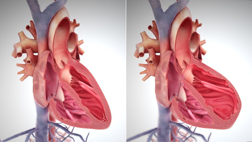
For decades, it’s been thought that people with heart failure should drastically reduce their dietary salt intake, but some studies have suggested that salt restriction could be harmful for these patients. A recent review in the European Journal of Clinical Investigation that assessed all relevant studies published between 2000 and 2023 has concluded that there is no proven clinical benefit to this strategy for patients with heart failure.

Most relevant randomised trials were small, and a single large, randomised clinical trial was stopped early due to futility. Although moderate to strict salt restriction was linked with better quality of life and functional status, it did not affect mortality and hospitalisation rates among patients with heart failure.

“Doctors often resist making changes to age-old tenets that have no true scientific basis; however, when new good evidence surfaces, we should make an effort to embrace it,” said author Paolo Raggi MD, PhD, of the University of Alberta.

Source: Wiley

Inflammatory Bowel Disease may Increase Risk of Heart Failure

Irritable bowel syndrome. Credit: Scientific Animations CC4.0

Inflammatory bowel disease (IBD) is associated with a slightly increased risk of heart failure up to 20 years after diagnosis, according to a comprehensive registry study from Karolinska Institutet published in the European Heart Journal.

The researchers analysed the risk of heart failure in over 80 000 patients with inflammatory bowel disease, that is, Crohn’s disease, ulcerative colitis or unclassified IBD, compared with 400 000 people from the general population, as part of the ESPRESSO study.

The results show that people with IBD have a 19% increased risk of developing heart failure up to 20 years after diagnosis. This corresponds to one extra heart failure case per 130 IBD patients in those 20 years, and the risk increase was seen regardless of the type of IBD. The highest risk of heart failure was seen in older patients, people with lower education and people with pre-existing cardiovascular-related disease at IBD diagnosis.

Contribute to new guidelines

“Both healthcare providers and patients should be aware of this increased risk, and it’s important that cardiovascular health is properly monitored,” says the study’s first author Jiangwei Sun, researcher at the Department of Medical Epidemiology and Biostatistics, Karolinska Institutet. “We hope the results will raise the awareness of health workers as to the increased risk of heart failure in individuals with IBD and contribute to new guidelines for cardiovascular disease management in IBD patients.”

Comparing siblings with and without ABD, the risk increase was slightly lower, 10%, suggesting that genetics and early environmental factors shared within families may play a role. 

“We don’t know if there is a causal relationship, but we will continue to explore genetic factors and the role of IBD medications and disease activities on the risk of heart failure,” says the study’s senior author Professor Jonas F. Ludvigsson from the Department of Medical Epidemiology and Biostatistics, Karolinska Institutet.

Source: Karolinska Institutet

New Advance against Heart Failure Caused by Y Chromosome Loss

Chromosomes. Credit: NIH

Researchers have discovered a gene on the Y chromosome that contributes to the greater incidence of heart failure in men when the Y chromosome is lost to ageing.

Y chromosome loss in men occurs progressively throughout life and can be detected in approximately 40% of 70-year-old men. In 2022, Kenneth Walsh, PhD, at University of Virginia discovered that this loss can contribute to heart muscle scarring and lead to heart failure. (That finding was the first to directly link Y chromosome loss to a specific harm to men’s health; Y chromosome loss is increasingly thought to play a role in diseases ranging from Alzheimer’s to cancer.)

In an important follow-up finding published in Nature Cardiovascular Research, Walsh and his team have discovered how Y chromosome loss triggers changes in heart immune cells that make the cells more likely to cause scarring and heart failure.

Further, the researchers found they could reverse the harmful heart changes by giving lab mice a drug that targets the process of fibrosis that leads to the heart scarring, which could lead to a similar treatment for men.

“Our previous work identified that it was loss of the entire Y chromosome that contributed to heart disease in men,” said Walsh, the director of UVA’s Hematovascular Biology Center. “This new work identified a single gene on the Y chromosome that can account for the disease-promoting effects of Y chromosome loss.”

About Y chromosome loss

Unlike women, who have two X chromosomes, men have an X and a Y. For a long time, the genes found on the Y chromosome were not thought to play important roles in disease. Sex hormones, scientists thought, explained the differences in certain diseases in men and women. But Walsh’s groundbreaking work has helped change that perception. It also suggested an explanation for why heart failure is more common in men than women. (Cardiovascular disease, which includes heart failure, is the leading cause of death worldwide.)

Y chromosome loss occurs in only a small percentage of affected men’s cells. This results in what is called “mosaicism,” where genetically different cells occur within one individual. Researchers aren’t entirely sure why this partial Y chromosome loss occurs, but predominantly it strikes elderly men and men who smoke compared to those who don’t.

To better understand the effects of Y chromosome loss, Walsh and his team examined genes found on the Y chromosome to determine which might be important to heart scarring. One gene they looked at, Uty, helps control the operating instructions for immune cells called macrophages and monocytes, the scientists determined. When the Uty gene was disrupted, either individually or through Y chromosome loss, that triggered changes in the immune cells in lab mice. Suddenly, the macrophages were much more “pro-fibrotic,” or prone to scarring. This accelerated heart failure as well, the scientists found.

“The identification of a single gene on the Y chromosome provides information about a new druggable target to treat fibrotic diseases,” said Walsh, of UVA’s Division of Cardiovascular Medicine and Robert M. Berne Cardiovascular Research Center.

Walsh and his team were able to prevent the harmful changes in the mice’s macrophages by giving them a specially designed monoclonal antibody. This halted the harmful changes in the heart, suggesting the approach might, with further research, lead to a way to treat or avoid heart failure and other fibrotic diseases in men with Y chromosome loss.

“Currently, we are working with our clinician colleagues in the Division of Cardiovascular Medicine at UVA to assess whether loss of the Y chromosome in men is associated with greater scarring in the heart,” Walsh said. “This research will provide new avenues for understanding the causes of heart disease.”

Based on their findings, Walsh and his team believe that a small group of genes found on the Y chromosome may have big effects on a wide array of diseases. Their new work identifies mechanisms that may lead to this, and they are hopeful that further research will provide a much better understanding of unknown causes of sickness and death in men.

“This research further documents the utility of studying the genetics of mutations that are acquired after conception and accumulate throughout life,” Walsh said. “These mutations appear to be as important to health and lifespan as the mutations that are inherited from one’s parents. The study of these age-acquired mutations represents a new field of human genetics.”

Source: University of Virginia Health System

No-aspirin Regimen Benefits Heart Failure Patients with LVADs

Photo by cottonbro studio

A recent clinical trial published in JAMA found that excluding aspirin for advanced heart failure (HF) patients with a ventricular assist device saw a reduction in bleeding events while maintaining their survival rates.

The ARIES-HM3 Randomised Clinical Trial assessed the safety and efficacy of excluding aspirin from the antithrombotic regimen in patients with advanced HF who have undergone implantation of a fully magnetically levitated left ventricular assist device (LVAD).

“We can now safely say that not giving aspirin is not only safe from a thromboembolic risk profile but results in improved adverse event rate by a significant reduction in non-surgical bleeding which is a well-known complication related to LVAD therapy,” said Mirnela Byku, MD, P.D, MBA, co-author of the study and director of the UNC Durable Mechanical Circulatory Device Program at the UNC School of Medicine.

“Improving not only longevity but also reducing morbidity and improving quality of life is a big focus in the field of MCS.”

Until this study, there had been no consensus in the field about use of or dose of aspirin in the LVAD population.

The international clinical trial followed a randomised, double-blind, placebo-controlled design and involved 628 patients across 51 centres in 9 countries.

The patients were divided into two groups: one receiving aspirin (100mg/d) and the other receiving a placebo in addition to vitamin K antagonist (VKA) therapy.

A focus was to determine if the likelihood a patient experiences major nonsurgical haemocompatibility-related adverse events (such as stroke, pump thrombosis, major bleeding, or arterial peripheral thromboembolism) within 12 months differed between the two groups.

The results showed that not giving aspirin to patients with advanced HF, treated with a fully magnetically levitated LVAD who are receiving VKAs, did not make their survival worse. Furthermore, aspirin avoidance was associated with a significant reduction (34%) in major nonsurgical bleeding events.

Source: University of North Carolina Health Care

Yoga Therapy Improves Quality of Life and Cardiovascular Function

Photo by Surface on Unsplash

Yoga therapy and lifestyle modifications have been shown to improve heart failure (HF) patients’ quality of life and enhance their cardiovascular function. A new study, presented at the American College of Cardiology Asia 2023 conference, examines the long-term outcomes of yoga therapy to determine the benefit of adding yoga therapy as a complementary treatment in the management of HF. After 12 months, participants with HF receiving yoga therapy continued to show improvement in left ventricular systolic function as well as quality of life.

The study included 75 heart failure patients (aged 30–70 years old) at a tertiary care centre in South India, who underwent coronary intervention, revascularisation or device therapy within in the previous six to 12 months. All of the patients included in the study were less than or equal to New York Heart Association (NYHA) Class III and had been on optimised medical therapy for at least 6 months to 12 months, and had a left ventricular ejection fraction (LVEF) of < 45%.

The interventional group included 35 participants (31 men and 4 women) and 40 (30 men and 10 women) were in the non-interventional (control) group. The interventional group received yoga therapy and guideline-directed medical therapy, while the control group only continued with standard guideline-directed medical therapy. Echocardiographic parameters were compared at various follow-ups to see the impact of yoga therapy on heart failure patients.

“Yoga is a combination of mind-body techniques, which is a set of physical exercises [asana] with breathing techniques [pranayama], relaxation and meditation that can be effectively used to stimulate physical and mental well-being,” said lead author Ajit Singh, PhD, research scientist for the Indian Council for Medical Research at Kasturba Medical College & Hospital, Manipal Academy of Heart Education in Manipal, India. “Our patients observed improvement in systolic blood pressure and heart rate compared to patients who were on medication without yoga.”

Participants in the yoga group were taken to the Department of Yoga at the hospital and an experienced yoga therapist taught selected yoga therapy like pranayama, meditation and relaxation techniques. Each session lasted around 60 minutes and participants were supervised for one week at the training centre before being asked to continue self-administered yoga at home. Those in the yoga group were advised to perform yoga at least five days a week for 12 months. At the training centre all the participants were taught together to perform the same steps, but individual support was available.

Researchers measured quality of life improvements using the World Health Organization Quality of Life questionnaire, which uses 26 questions to evaluate quality of life in four aspects: physical, psychological, social and environmental health. The participants completed the questionnaire at enrolment, as well as at 24 weeks and 48 weeks of follow-up. According to the researchers, the study showed participants in the yoga group had improvement in endurance, strength, balance, symptom stability and quality of life. They also observed that while patients improved physically and psychologically, there was no improvement in social and environmental health.

Echocardiographic parameters did not show any significant differences between the two groups at baseline. At both the six- and 12-month follow-up, improved biventricular systolic function was seen in the interventional (yoga) group compared to the control group. The interventional group also showed substantial improvement in functional outcomes as assessed by NHYA classification.

“This study proves that the addition of yoga therapy to standard medical management of heart failure leads to an improvement in left ventricular systolic function and quality of life in heart failure patients,” Singh said. “Hence, yoga therapy may improve physical well-being and left ventricular function among heart failure patients on guideline-directed optimal medical therapy.”

Source: American College of Cardiology

MRI Scan Combination Could Detect Hypertrophic Cardiomyopathy Early

Credit: Pixabay CC0

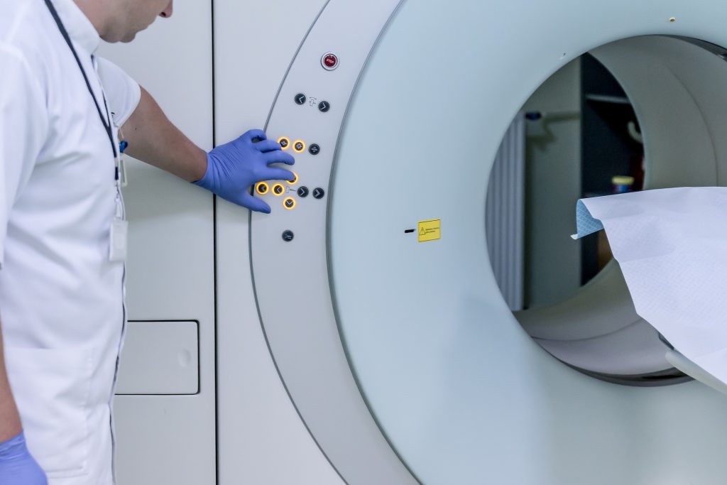
Combining two types of heart scan techniques could help detect hypertrophic cardiomyopathy (HCM) before symptoms and signs on conventional tests appear, according to a new study led by UCL researchers. To do this, they used two cutting-edge heart scanning techniques: cardiac diffusion tensor imaging (cDTI), which shows the heart’s microstructure and cardiac MRI perfusion (perfusion CMR), which reveals microvascular disease. Their findings, published in Circulation, will help doctors select appropriate treatments.

HCM is a disease which affects around 1 in 500 in the UK, causing thickening of heart muscle and can lead to heart failure and cardiac arrest.

Researchers studied the hearts of three groups: healthy people, people who already had HCM, and people with an HCM-causing genetic mutation but no overt signs of disease.

The scans showed that people with overt signs of HCM have very abnormal organisation of their heart muscle cells and a high rate and severity of microvascular disease compared to healthy volunteers, helping doctors more accurately spot the early signs of HCM.

Crucially, the scans were also able to identify abnormal microstructure and microvascular disease in the people who had a problematic gene but no symptoms or muscle thickening. They found that 28% had defects in their blood supply, compared to healthy volunteers. This meant that doctors were able to more accurately spot the early signs of HCM developing in patient’s hearts.

The first drug to slow HCM progression, mavacamten, has recently been approved for use in Europe and will allow doctors to reduce the severity of the disease once symptoms and muscle thickening have appeared. Genetic therapies are also in development which could prevent symptoms entirely by intercepting HCM development at an early stage.

Perfusion CMR is already being used in some clinics to help differentiate people with HCM from other causes of muscle thickening. The researchers think that these revolutionary new therapies, combined with cDTI and perfusion CMR scans, give doctors the best ever chance of treating people at risk of HCM early enough that the condition never develops.

Dr George Joy, who led the research with Professor James Moon and Dr Luis Lopes (all UCL Institute of Cardiovascular Science), said: “The ability to detect early signs of HCM could be crucial in trials testing treatments aimed at preventing early disease from progressing or correcting genetic mutations. The scans could also enable treatment to start earlier than we previously thought possible.

“We now want to see if we can use the scans to identify which patients without symptoms or heart muscle thickening are most at risk of developing severe HCM and its life-changing complications. The information provided from scans could therefore help doctors make better decisions on how best to care for each patient.”

Dr Luis Lopes (UCL Institute of Cardiovascular Science), senior author of the study, said: “By linking advanced imaging to our cohort of HCM patients (and relatives) with extensive genetic testing, this study detected microstructural abnormalities in vivo in mutation carriers for the first time and was the first to compare these parameters in HCM patients with and without a causal mutation.

“The findings allow us to understand more about the early subclinical manifestations of this serious condition but also provide additional clinical tools for screening, monitoring and hopefully in the near future for therapeutic decision-making.”

Source: University College London

SGLT-2 Inhibitors Reduce HF Hospitalisation Risk in Type 2 Diabetes

A study published in Annals of Internal Medicine has suggested that the new sodium-glucose co-transporter 2 inhibitors (SGLT-2i) may be viable as a first-line treatment in patients with type 2 diabetes (T2D), with reduced odds of hospitalisation for heart failure compared to those receiving metformin.

In cardiovascular outcome trials among adults with T2D, SGLT-2i have shown therapeutic promise, including reduced risk of hospitalisation for heart failure compared to placebo. However, SGLT-2i have mainly been evaluated as a second-line treatment, as metformin is generally given as a first-line, antidiabetic treatment.

In a new study, researchers from the Brigham compared cardiovascular outcomes among adults with T2D who initiated first-line treatment with either metformin or SGLT-2i. For the study, 8613 patients treated with SGLT-2i were matched to 17 226 patients treated with metformin. The authors found that patients receiving SGLT-2i showed a similar risk for myocardial infarction, stroke, and all-cause mortality, and a lower risk for hospitalization for heart failure compared with patients who received metformin. The risk for adverse events was similar except for an increased risk for genital infections compared with those receiving metformin.

“Our results suggest that SGLT-2i may be considered as first-line treatment for patients with T2D and cardiovascular disease or who are at increased risk for cardiovascular events,” said lead author HoJin Shin, BPharm, PhD, of the Division of Pharmacoepidemiology and Pharmacoeconomics. “However, more evidence from randomised clinical trials or observational studies will help us to identify patients who would benefit most from using SGLT-2i as first-line type 2 diabetes treatment.”

Source: EurekAlert!

Lessons Learnt From First Human Transplant of Genetically Modified Pig Heart

Photo by Natanael Melchor on Unsplash

A new study published in The Lancet has revealed the most extensive analysis to date on what led to the eventual heart failure in the world’s first successful transplant of a genetically modified pig heart into a human patient. This groundbreaking procedure was conducted by University of Maryland School of Medicine (UMSOM) physician-scientists in January 2022.

The patient, 57-year-old David Bennett, was treated at the University of Maryland Medical Center. He experienced strong cardiac function with no obvious signs of acute rejection for nearly seven weeks after the surgery. A sudden onset of heart failure led to his death two months after the transplant. Since then, the transplant team has been extensively studying the physiologic processes that led to the heart failure to identify what needs to be prevented in future transplants to improve the odds of success.

“Our paper provides crucial insight into how a multitude of factors likely played a role in the functional decline of the transplanted heart,” said study lead author Muhammad M. Mohiuddin, MD, Professor of Surgery and Scientific/Program Director of the Cardiac Xenotransplantation Program at UMSOM. “Our goal is to continue moving this field forward as we prepare for clinical trials of xenotransplants involving pig organs.”

Mr. Bennett, who was in end-stage heart failure and nearing the end of his life, did not qualify for a traditional heart transplant, but the experimental procedure was authorised by the US Food and Drug Administration under compassionate use.

“We were determined to shed light on what led to the heart transplant dysfunction in Mr. Bennett, who performed a heroic act by volunteering to be the first in the world,” said study co-author Bartley Griffith, MD, Professor of Surgery and The Thomas E. and Alice Marie Hales Distinguished Professor in Transplantation at UMSOM. “We want our next patient to not only survive longer with a xenotransplant but to return to normal life and thrive for months or even years.”

To better understand the processes that led to dysfunction of the pig heart transplant, the research team performed extensive testing on the limited available tissues in the patient. They carefully mapped out the sequence of events that led to the heart failure demonstrating that the heart functioned well on imaging tests like echocardiography until day 47 after surgery.

The new study confirms that no signs of acute rejection occurred during the first several weeks after the transplant. Likely, several overlapping factors led to heart failure in Mr. Bennett, including his poor state of health prior to the transplant that led him to become severely immunocompromised. This limited the use of an effective anti-rejection regimen used in preclinical studies for xenotransplantation. As a result, the researchers found, the patient was likely more vulnerable to rejection of the organ from antibodies made by the immune system. The researchers found indirect evidence of antibody-mediated rejection based on histology, immunohistochemical staining and single cell RNA analysis.

The use of an intravenous immunoglobulin, IVIG, a drug that contains antibodies, may also have contributed to damage to the heart muscle cells. It was given to the patient twice during the second month after the transplant to help prevent infection, likely also triggering an anti-pig immune response. The research team found evidence of immunoglobulin antibodies targeting the pig vascular endothelium layer of the heart.

Lastly, the new study investigated the presence of a latent virus, called porcine cytomegalovirus (PCMV), in the pig heart, which may have contributed to the dysfunction of the transplant. Activation of the virus may have occurred after the patient’s anti-viral treatment regimen was reduced to address other health issues. This may have initiated an inflammatory response causing cell damage. However, there is no evidence that the virus infected the patient or spread to organs beyond the heart. Improved PCMV testing protocols have been developed for sensitive detection and exclusion of latent viruses for future xenotransplants.

“Valuable lessons can be learned from this groundbreaking surgery and the courageous first patient, Mr. Bennett, that will better inform us for future xenotransplants,” said UMSOM Dean Mark T. Gladwin, MD, Vice President for Medical Affairs, University of Maryland, Baltimore, and the John Z. and Akiko K. Bowers Distinguished Professor. “In the future, our team of surgeon-scientists will utilise newly designed immune cell assays to monitor the patient more precisely in the days, weeks, and months following the xenotransplant. This will provide stricter control of the earliest signs of rejection and the promise of a truly lifesaving innovation.”

Source:

Autistic Adults Have Higher Risks of Injury and Disease

Photo by Andrew Neel on Unsplash

A large-scale registry study found that older autistic adults have a significantly higher risk of injury, especially self-inflicted, and physical conditions such as type 2 diabetes, anaemia, heart failure and COPD. The findings were published in The Lancet Healthy Longevity.

“We found an increased disease burden in middle-aged and older autistic adults, both men and women, irrespective of the presence of intellectual disability,” says Shengxin Liu, doctoral student at Karolinska Institutet. “Our findings point up the need to improve the support and care of older autistic adults.”

In the population-based study, the KI researchers linked different national registers and compared the risk for five types of injury and 39 age-related physical conditions in people over the age of 45. Of the four million-plus people born between 1932 and 1967, 1930 women and 3361 men had an autism diagnosis. For each physical condition, they evaluated the 25-year cumulative incidence and the relative risk in autistic people compared with non-autistic people of the same sex and age.

Seven-fold risk of self-harm

Autistic people had a higher risk of four of five studied injures, for which self-harm accounted for the greatest risk increase, followed by poisoning, falls and other physical injuries.

“The risk of self-harm was worryingly high, a full seven times higher than in non-autistic people,” says Liu. “Reasons behind this remain largely unknown. One possible contributing factor could be mental health conditions that commonly co-occur with autism, such as anxiety and depression.”

The researchers also found a risk increase for 15 physical conditions. For example, autistic people had three times the risk of anaemia and glucose dysregulation and almost double the risk of heart failure, type 2 diabetes, and COPD (chronic obstructive pulmonary disease).

Multiple contributory factors

“We now need to find out the cause of these associations and how they are affected by factors such as biology, age when diagnosed with autism, psychotropic treatment and psychosocial environment,” says the study’s last author Mark Taylor, senior researcher at the Department of Medical Epidemiology and Biostatistics, Karolinska Institutet. “But most importantly, researchers, health services and policymakers need to cooperate to make sure that older autistic adults have a better quality of life.”

Since this was an observational study, no causal relationships can be ascertained, and the researchers were not able to take into account variables such as socioeconomic status. Furthermore, given that the study used Swedish registers, it is difficult to make generalisations to other countries.

Source: Karolinska Institutet

Strong Legs Reduce Risk of Heart Failure after Heart Attack

Photo by Henry Xu on Unsplash

People with strong legs are less likely to develop heart failure after a heart attack, according to new research. Myocardial infarction is the most common cause of heart failure, with around 6–9% of heart attack patients going on to develop the condition. Previous research has shown that having strong quadriceps is associated with a lower risk of death in patients with coronary artery disease.

Presented at Heart Failure 2023, a scientific congress of the European Society of Cardiology (ESC), this study tested the hypothesis that leg strength is associated with a lower risk of developing heart failure after acute myocardial infarction. The study included 932 patients hospitalised in 2007 to 2020 with acute myocardial infarction who did not have heart failure prior to the admission and did not develop heart failure complications during their hospital stay. The median age was 66 years and 753 participants (81%) were men.

Maximal quadriceps strength was measured as an indicator of leg strength. Patients sat on a chair and contracted the quadriceps muscles as hard as possible for five seconds. A handheld dynamometer attached to the ankle recorded the maximum value in kg. The measurement was performed on each leg and the researchers used the average of both values. Strength was expressed relative to body weight, meaning that quadriceps strength in kg was divided by body weight in kg and multiplied by 100 for a % body weight value. Patients were classified as ‘high’ or ‘low’ strength according to whether their value was above or below the median for their se

The median value for women was 33% body weight and the median value for men was 52% body weight. A total of 451 patients had low quadriceps strength and 481 had high strength. During an average follow-up of 4.5 years, 67 patients (7.2%) developed heart failure. The incidence of heart failure was 10.2 per 1000 person-years in patients with high quadriceps strength and 22.9 per 1000 person-years in those with low strength.

The researchers analysed the association between quadriceps strength (low vs. high) and the risk of developing heart failure. The analysis was adjusted for factors known to be associated with the development of heart failure after myocardial infarction including age, sex, body mass index, prior myocardial infarction or angina pectoris, diabetes, atrial fibrillation, chronic obstructive pulmonary disease, peripheral arterial disease and kidney function. Compared with low quadriceps strength, a high strength level was associated with a 41% lower risk of developing heart failure (hazard ratio [HR]: 0.59; 95% confidence interval [CI] 0.35–1.00; p=0.048).

The investigators also analysed the association between quadriceps strength as a continuous variable and the risk of developing heart failure. Each 5% body weight increment in quadriceps strength was associated with an 11% lower likelihood of heart failure (HR 0.89; 95% CI 0.81–0.98; p=0.014).

Study author Mr. Kensuke Ueno, a physical therapist at the Kitasato University Graduate School of Medical Sciences, Sagamihara, Japan said: “Quadriceps strength is easy and simple to measure accurately in clinical practice. Our study indicates that quadriceps strength could help to identify patients at a higher risk of developing heart failure after myocardial infarction who could then receive more intense surveillance. The findings need to be replicated in other studies, but they do suggest that strength training involving the quadriceps muscles should be recommended for patients who have experienced a heart attack to prevent heart failure.”

Source: European Society of Cardiology