Tag: gut microbiota

Surprisingly, Benefits of Dietary Fibre Vary Significantly between Individuals

Photo by Mariana Kurnyk

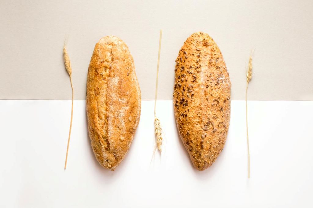
Nutritionists generally advise everyone to eat more dietary fibre, but a new study suggests that its effects on health can vary, suggesting that recommendations should be tailored to each individual’s gut microbiome. The study, published in Gut Microbes, focused on resistant starch, a category of dietary fibre found in such foods as bread, cereals, green bananas, whole-grain pasta, brown rice and potatoes.

The researchers identified the gut microbe species that change in response to two different types of resistant starch. They found evidence that each individual may have a unique response to eating a resistant starch, with some people benefiting and others experiencing little or no effect. The reason for the variation appears tied to the level of diversity and composition of a person’s gut microbiome.

“Precision nutrition definitely has a use in determining what dietary fibre we should tell people to eat,” said Angela Poole, assistant professor of molecular nutrition and senior author of the study.

“This is critical because we’ve had public messaging advising people to eat more dietary fibre for decades,” Poole said. “At the same time, less than 10% of people eat the recommended intake. Since there are many different types of dietary fibre and carbohydrates, a better strategy would be to collect data on each person and tell them which dietary fibre they can eat to get the most bang for their buck.”

Resistant starch comes in five types, and resists degradation by human digestive enzymes until it reaches the gut. There, it acts as a substrate for certain gut microbes to produce short chain fatty acids, which are important in signaling pathways that regulate glucose and lipid metabolism. Multiple microbe species may work together to create the fatty acids.

In the study, Poole and colleagues tested three dietary treatments on 59 participants over seven weeks.

The team had three different types of crackers manufactured. Two crackers had the same ingredients, except one contained resistant starch type 2, which occurs naturally, and the other contained resistant starch type 4, which is human-made. A third control cracker was digestible by human enzymes, similar to white bread, and the researchers expected none of the bacteria to act on the control.

Subjects were then divided into two groups. The first group ate the resistant starch type 2 cracker first, followed by the control, and then resistant starch type 4. Each cracker type was eaten for 10 days, with five days of no cracker consumption between treatments. The second group reversed the order, also with the control in the middle.

They then sequenced the microbiomes of each participant before and after each treatment. For resistant starch type 2, more than 30 bacteria changed in abundance, including Ruminococcus bromii, which is considered a keystone resistant starch degrader in the human gut. For type 4, more than 20 bacteria changed. And for the control, nothing changed.

“For the resistant starch crackers, we could detect that 20 or 30 of them were changing, but how much they changed and whether they changed at all, for each of those bacteria, depended on the person,” Poole said.

Similarly, each resistant starch type changed different short chain fatty acids, with variable levels of fatty acid increases and decreases based on the individual. For resistant starch type 2, the researchers identified a subset of 13 bacteria that predicted change in amounts of propionate, a type of short chain fatty acid. Also for resistant starch 2, by knowing the diversity of an individual’s gut microbiome, the researchers could roughly predict if two types of short chain fatty acids (acetate and butyrate) were going to increase.

The most surprising result was that the control digestible cracker led to the greatest gains of short chain fatty acids. More work is needed to understand why, but Poole suspects that the order of cracker consumption was key to the result. Since many microbes are involved in making short chain fatty acids, she hypothesises that eating a resistant starch first primed the gut to produce the fatty acids when that person ate the digestible starch.

“That’s one of the major takeaways, maybe I can get away with eating a French baguette some of the time, and it may be better than just eating whole grain all the time,” Poole said. “But I have to test that, and it probably varies between people.”

Source: Cornell University

Gut Bacteria in Parkinson’s Disease Produce Fewer B Vitamins

In Parkinson’s disease, a reduction in the gut bacteria of genes responsible for synthesising the essential B vitamins B2 and B7 was found. Credit: Reiko Matsushita

A study led by Nagoya University in Japan has revealed a link between gut microbiota and Parkinson’s disease (PD). The researchers found that the gut bacteria genes responsible for synthesising vitamins B2 and B7 were reduced. This gene reduction was also linked to low levels of agents that help maintain the integrity of the intestinal barrier, which when weakened causes the inflammation seen in PD. Their findings, published in npj Parkinson’s Disease, suggest that treatment with B vitamins to address these deficiencies can be used to treat PD. 

PD is characterized by a variety of physical symptoms that hinder daily activities and mobility, such as shaking, slow movement, stiffness, and balance problems. While the frequency of PD may vary between different populations, it is estimated to affect approximately 1-2% of individuals aged 55 years or older. 

Various physiological processes are heavily influenced by the microorganisms found in the gut, which are collectively known as gut microbiota. In ideal conditions, gut microbiota produce SCFAs and polyamines, which maintain the intestinal barrier that prevents toxins entering the bloodstream. Toxins in the blood can be carried to the brain where they cause inflammation and affect neurotransmission processes that are critical for maintaining mental health.

To better understand the relationship between the microbial characteristics of the gut in PD, Hiroshi Nishiwaki and Jun Ueyama from the Nagoya University Graduate School of Medicine conducted a metanalysis of stool samples from patients with PD from Japan, the United States, Germany, China, and Taiwan. They used shotgun sequencing, a technique that sequences all genetic material in a sample. This is an invaluable tool because it offers researchers a better understanding of the microbial community and genetic makeup of the sample.

They observed a decrease in the bacterial genes responsible for the synthesising of riboflavin (vitamin B2) and biotin (vitamin B7) in patients diagnosed with PD. Riboflavin and biotin, derived from both food and gut microbiota, have anti-inflammatory properties, which may counteract the neuroinflammation seen in diseases like PD. 

B vitamins play crucial roles in the metabolic processes that influence the production and functions of short-chain fatty acids (SCFAs) and polyamines, two agents that help maintain the integrity of the intestinal barrier, preventing toxins entering the bloodstream. An examination of fecal metabolites revealed decreases of both in patients with PD. 

The findings indicate a potential explanation for the progression of PD. “Deficiencies in polyamines and SCFAs could lead to thinning of the intestinal mucus layer, increasing intestinal permeability, both of which have been observed in PD,” Nishiwaki explained. “This higher permeability exposes nerves to toxins, contributing to abnormal aggregation of alpha-synuclein, activating the immune cells in the brain, and leading to long-term inflammation.” 

He added, “Supplementation therapy targeting riboflavin and biotin holds promise as a potential therapeutic avenue for alleviating PD symptoms and slowing disease progression.”

The results of the study highlight the importance of understanding the complex relationship among gut microbiota, metabolic pathways, and neurodegeneration. In the coming years, customised therapy could potentially be based on patients’ unique microbiome profiles. By altering bacterial levels in the microbiome, doctors can potentially delay the onset of symptoms associated with diseases like PD.

“We could perform gut microbiota analysis on patients or conduct faecal metabolite analysis,” Nishiwaki said. “Using these findings, we could identify individuals with specific deficiencies and administer oral riboflavin and biotin supplements to those with decreased levels, potentially creating an effective treatment.”

Source: Nagoya University

The study, “Meta-analysis of shotgun sequencing of gut microbiota in Parkinson’s disease,” was published in npj Parkinson’s Disease on May 21, 2024, at DOI:10.1038/s41531-024-00724-z.

New Antibiotic Kills Pathogenic Bacteria but Spares Healthy Gut Microbes

Gut Microbiome. Credit Darryl Leja National Human Genome Research Institute National Institutes Of Health

Researchers have developed a new antibiotic that reduced or eliminated drug-resistant bacterial infections in mouse models of acute pneumonia and sepsis while sparing healthy microbes in the mouse gut. The drug, called lolamicin, also warded off secondary infections with Clostridioides difficile, and was effective against more than 130 multidrug-resistant bacterial strains in cell culture.

The findings are detailed in the journal Nature.

“People are starting to realise that the antibiotics we’ve all been taking – that are fighting infection and, in some instances, saving our lives – also are having these deleterious effects on us,” said University of Illinois Urbana-Champaign chemistry professor Paul Hergenrother, who led the study with former doctoral student Kristen Muñoz. “They’re killing our good bacteria as they treat the infection. We wanted to start thinking about the next generation of antibiotics that could be developed to kill the pathogenic bacteria and not the beneficial ones.”

“Most clinically approved antibiotics only kill gram-positive bacteria or kill both gram-positive and gram-negative bacteria,” Muñoz said.

The few drugs available to fight gram-negative bacteria, which are protected by their double cell walls, also kill other potentially beneficial gram-negative bacteria. For example, colistin, one of the few gram-negative-only antibiotics approved for clinical use, can cause C. difficile-associated diarrhoea and pseudomembranous colitis, a potentially life-threatening complication. The drug also has toxic effects on the liver and kidney, and “thus colistin is typically utilised only as an antibiotic of last resort,” the researchers wrote.

To tackle the many problems associated with indiscriminately targeting gram-negative bacteria, the team focused on a suite of drugs developed by the pharmaceutical company AstraZeneca. These drugs inhibit the Lol system, a lipoprotein-transport system that is exclusive to gram-negative bacteria and genetically different in pathogenic and beneficial microbes. These drugs were not effective against gram-negative infections unless the researchers first undermined key bacterial defenses in the laboratory. But because these antibiotics appeared to discriminate between beneficial and pathogenic gram-negative bacteria in cell culture experiments, they were promising candidates for further exploration, Hergenrother said.

In a series of experiments, Muñoz designed structural variations of the Lol inhibitors and evaluated their potential to fight gram-negative and gram-positive bacteria in cell culture. One of the new compounds, lolamicin, selectively targeted some “laboratory strains of gram-negative pathogens including Escherichia coliKlebsiella pneumoniae and Enterobacter cloacae,” the researchers found. Lolamicin had no detectable effect on gram-positive bacteria in cell culture. At higher doses, lolamicin killed up to 90% of multidrug-resistant E. coliK. pneumoniae and E. cloacae clinical isolates.

When given orally to mice with drug-resistant septicemia or pneumonia, lolamicin rescued 100% of the mice with septicemia and 70% of the mice with pneumonia, the team reported.

Extensive work was done to determine the effect of lolamicin on the gut microbiome.

“The mouse microbiome is a good tool for modeling human infections because human and mouse gut microbiomes are very similar,” Muñoz said. “Studies have shown that antibiotics that cause gut dysbiosis in mice have a similar effect in humans.”

Treatment with standard antibiotics amoxicillin and clindamycin caused dramatic shifts in the overall structure of bacterial populations in the mouse gut, diminishing the abundance several beneficial microbial groups, the team found.

“In contrast, lolamicin did not cause any drastic changes in taxonomic composition over the course of the three-day treatment or the following 28-day recovery,” the researchers wrote.

Many more years of research are needed to extend the findings, Hergenrother said. Lolamicin, or other similar compounds, must be tested against more bacterial strains and detailed toxicology studies must be conducted. Any new antibiotics also must be assessed to determine how quickly they induce drug resistance, a problem that arises sooner or later in bacteria treated with antibiotics.

The study is a proof-of-concept that antibiotics that kill a pathogenic microbe while sparing beneficial bacteria in the gut can be developed for gram-negative infections – some of the most challenging infections to treat, Hergenrother said.

Source: University of Illinois at Urbana-Champaign, News Bureau

New Study Reveals that Gut Microbes Have an Arsenal against Pathogens

Gut Microbiome. Credit Darryl Leja National Human Genome Research Institute National Institutes Of Health

A study conducted by researcher Juan Du’s research group at the Karolinska Institutet sheds light on the capabilities of our gut microbes and their metabolites. The findings reveal potent inhibitory effects on the growth of antibiotic-resistant bacteria and suggest interactions and signaling between gut microbes and pathogens.

The study, published in the journal Gut Microbes, focuses on identifying key microbes within the gut microbiome that inhibit the growth of pathogens, particularly antibiotic-resistant strains. 

Strains from Clostridium perfringens, Clostridium butyricum, and Enterobacter maltosivorans and their metabolites were found to directly inhibit the growth of pathogens, including multi-drug-resistant ones. The study also reveals novel dipeptide features, suggesting interactions and signaling between gut microbes and pathogens.

“Multidrug-resistant microorganisms pose a global threat, and understanding the role of gut microbiota is crucial. Metabolites derived from these microbial communities play a significant role in regulating biochemical processes in the human body. Despite this, only a limited number of gut microbes and their bioactive metabolites have been explored so far”, explains author Juan Du. She continues:

“We plan to expand our screening to include a broader collection of commensal bacteria from various body sites. We’ll conduct mechanism studies to understand how these compounds function on pathogens, especially antibiotic-resistant strains”, says Juan Du.

Source: Karolinska Instutet

Autism and ADHD are Linked to Gut Flora Disturbance in First Year of Life

Photo by Christian Bowen on Unsplash

Disturbed gut flora during the first years of life is associated with diagnoses such as autism and ADHD later in life. One explanation for this disturbance could be from antibiotic treatment. This is according to a study led by researchers at the University of Florida and Linköping University and published in the journal Cell.

The study is the first prospective study to examine gut flora composition and a large variety of other factors in infants, in relation to the development of the children’s nervous system. The researchers have found many biological markers that seem to be associated with future neurological development disorders, such as autism spectrum disorder, ADHD, communication disorder and intellectual disability.

“The remarkable aspect of the work is that these biomarkers are found at birth in cord blood or in the child’s stool at one year of age over a decade prior to the diagnosis,” says Eric W Triplett, professor at the Department of Microbiology and Cell Science at the University of Florida, USA, one of the study leaders.

Antibiotic treatment could be involved

The study is part of the ABIS (All Babies in Southeast Sweden) study led by Johnny Ludvigsson at Linköping University. More than 16 000 children born in 1997–1999, representing the general population, have been followed from birth into their twenties. Of these, 1197 children (7.3%), have been diagnosed with autism spectrum disorder, ADHD, communication disorder or intellectual disability. Many lifestyle and environmental factors have been identified through surveys conducted on several occasions during the children’s upbringing. For some of the children, the researchers have analysed substances in umbilical cord blood and bacteria in their stool at the age of one.

“We can see in the study that there are clear differences in the intestinal flora already during the first year of life between those who develop autism or ADHD and those who don’t. We’ve found associations with some factors that affect gut bacteria, such as antibiotic treatment during the child’s first year, which is linked to an increased risk of these diseases,” says Johnny Ludvigsson, senior professor at the Department of Biomedical and Clinical Sciences at Linköping University, who led the study together with Eric W. Triplett.

Children who had repeated ear infections before one year of age had a higher risk of a developmental neurological disorder diagnosis later in life. It is probably not the infection itself that is the culprit, but the researchers suspect a link to antibiotic treatment. They found that the presence of Citrobacter bacteria or the absence of Coprococcus bacteria increased the risk of future diagnosis. One possible explanation may be that antibiotic treatment has disturbed the composition of the gut flora in a way that contributes to neurodevelopmental disorders. The risk of antibiotic treatment damaging the gut flora and increasing the risk of diseases linked to the immune system, such as type 1 diabetes and childhood rheumatism, has been shown in previous studies.

Coprococcus and Akkermansia muciniphila have potential protective effects. These bacteria were correlated with important substances in the stool, such as vitamin B and precursors to neurotransmitters which play vital roles orchestrating signalling in the brain. Overall, we saw deficits in these bacteria in children who later received a developmental neurological diagnosis,” says study first author Angelica Ahrens, Assistant Scientist in Eric Triplett’s research group at the University of Florida.

The present study also confirms that the risk of developmental neurological diagnosis in the child increases if the parents smoke. Conversely, breastfeeding has a protective effect, according to the study.

Differences at birth

In cord blood taken at the birth of children, the researchers measured substances such as fatty acids and amino acids, as well as exogenous ones such as nicotine and environmental toxins. They compared substances in the umbilical cord blood of 27 children diagnosed with autism with the same number of children without a diagnosis.

It turned out that children who were later diagnosed had low levels of several important fats in the umbilical cord blood. One of these was linolenic acid, which is needed for the formation of omega 3 fatty acids with anti-inflammatory properties and other effects in the brain. The same group also had higher levels than the control group of a PFAS substance, used as flame retardants and shown to negatively affect the immune system in several different ways. PFAS substances can enter the body via drinking water, food and the air we breathe.

Opens up new possibilities

As the relationships found in the Swedish children may not be generalisable to other populations, studies in other populations are needed. Another question is whether gut flora imbalance is a triggering factor or whether it has occurred as a result of underlying factors, such as diet or antibiotics. Yet even accounting for risk factors that might affect the gut flora, they found that the link between future diagnosis remained for many of the bacteria.

The research is at an early stage and more studies are needed, but the discovery that many biomarkers for future developmental neurological disorders can be observed at an early age opens up the possibility of developing screening protocols and preventive measures in the long term.

Source: Linköping University

Bacteria Subtype Linked to Growth in up to 50% of Human Colorectal Cancers

Human colon cancer cells. Credit: National Cancer Institute

Researchers at Fred Hutchinson Cancer Center have found that a specific subtype of a microbe commonly found in the mouth is able to travel to the gut and grow within colorectal cancer tumours. This microbe is also a culprit for driving cancer progression and leads to poorer patient outcomes after cancer treatment.

The findings, published in Nature, could help improve therapeutic approaches and early screening methods for colorectal cancer, which is the second most common cause of cancer deaths in adults in the U.S. according to the American Cancer Society.

Examining colorectal cancer tumours removed from 200 patients, the Fred Hutch team measured levels of Fusobacterium nucleatum, a bacterium known to infect tumours. In about 50% of the cases, they found that only a specific subtype of the bacterium was elevated in the tumour tissue compared to healthy tissue.

The researchers also found this microbe in higher numbers within stool samples of colorectal cancer patients compared with stool samples from healthy people.

“We’ve consistently seen that patients with colorectal tumours containing Fusobacterium nucleatum have poor survival and poorer prognosis compared with patients without the microbe,” explained Susan Bullman, PhD, Fred Hutch cancer microbiome researcher and co-corresponding study author. “Now we’re finding that a specific subtype of this microbe is responsible for tumour growth. It suggests therapeutics and screening that target this subgroup within the microbiota would help people who are at a higher risk for more aggressive colorectal cancer.”

In the study, Bullman and co-corresponding author Christopher D. Johnston, PhD, Fred Hutch molecular microbiologist, along with the study’s first author Martha Zepeda-Rivera, PhD, a Washington Research Foundation Fellow and Staff Scientist in the Johnston Lab, wanted to discover how the microbe moves from its typical environment of the mouth to a distant site in the lower gut and how it contributes to cancer growth.

First they found a surprise that could be important for future treatments. The predominant group of Fusobacterium nucleatum in colorectal cancer tumours, thought to be a single subspecies, is actually composed of two distinct lineages known as “clades.”

“This discovery was similar to stumbling upon the Rosetta Stone in terms of genetics,” Johnston explained. “We have bacterial strains that are so phylogenetically close that we thought of them as the same thing, but now we see an enormous difference between their relative abundance in tumours versus the oral cavity.”

By separating out the genetic differences between these clades, the researchers found that the tumour-infiltrating Fna C2 type had acquired distinct genetic traits suggesting it could travel from the mouth through the stomach, withstand stomach acid and then grow in the lower gastrointestinal tract. The analysis revealed 195 genetic differences between the clades.

Then, comparing tumour tissue with healthy tissue from patients with colorectal cancer, the researchers found that only the subtype Fna C2 is significantly enriched in colorectal tumour tissue and is responsible for colorectal cancer growth.

Further molecular analyses of two patient cohorts, including over 200 colorectal tumours, revealed the presence of this Fna C2 lineage in approximately 50% of cases.

The researchers also found in hundreds of stool samples from people with and without colorectal cancer that Fna C2 levels were consistently higher in colorectal cancer.

“We have pinpointed the exact bacterial lineage that is associated with colorectal cancer, and that knowledge is critical for developing effective preventive and treatment methods,” Johnston said.

Source: Fred Hutchinson Cancer Center

Certain Gut Bacteria Linked to Reduced Cardiovascular Disease Risk

Gut Microbiome. Credit Darryl Leja National Human Genome Research Institute National Institutes Of Health

Changes in the gut microbiome have been implicated in a range of diseases including type 2 diabetes, obesity, and inflammatory bowel disease. Now, a team of researchers has found that microbes in the gut may affect cardiovascular disease as well. In a study published in Cell, the team has identified specific species of bacteria that consume cholesterol in the gut and may help lower cholesterol and heart disease risk in people.

Researchers at the Broad Institute of MIT and Harvard along with Massachusetts General Hospital analysed metabolites and microbial genomes from more than 1400 participants in the Framingham Heart Study, a decades-long project focused on risk factors for cardiovascular disease.

The team discovered that bacteria called Oscillibacter take up and metabolise cholesterol from their surroundings, and that people carrying higher levels of the microbe in their gut had lower levels of cholesterol. They also identified the mechanism the bacteria likely use to break down cholesterol. The results suggest that interventions that manipulate the microbiome in specific ways could one day help decrease cholesterol in people. The findings also lay the groundwork for more targeted investigations of how changes to the microbiome affect health and disease.

“Our research integrates findings from human subjects with experimental validation to ensure we achieve actionable mechanistic insight that will serve as starting points to improve cardiovascular health,” said Xavier, who is a core institute member and a professor at Harvard Medical School and Massachusetts General Hospital.

Postdoctoral researcher Chenhao Li and research scientist Martin Stražar, both in Xavier’s lab, were co-first authors on the study.

Cholesterol cues

In the past decade, other researchers have uncovered links between composition of the gut microbiome and elements of cardiovascular disease, such as a person’s triglycerides and blood sugar levels after a meal. But scientists haven’t been able to target those connections with therapies in part because they lack a complete understanding of metabolic pathways in the gut.

In the new study, the Broad team gained a more complete and detailed picture of the impact of gut microbes on metabolism. They combined shotgun metagenomic sequencing, which profiles all of the microbial DNA in a sample, with metabolomics, which measures the levels of hundreds of known and thousands of unknown metabolites. They used these tools to study stool samples from the Framingham Heart Study.

“The project outcomes underline the importance of high-quality, curated patient data,” Stražar said. “That allowed us to note effects that are really subtle and hard to measure and directly follow up on them.”

More than 16 000 associations between microbes and metabolic traits were found, one of them particularly strong: People with several species of bacteria from the Oscillibacter genus had lower cholesterol levels than those who lacked the bacteria. The researchers found that species in the Oscillibacter genus were surprisingly abundant in the gut, representing on average 1 in every 100 bacteria.

The researchers then wanted to figure out the biochemical pathway the microbes use to break down cholesterol. To do this, they first needed to grow the organism in the lab. Fortunately, the lab has spent years collecting bacteria from stool samples to create a unique library that also included Oscillibacter.

After successfully growing the bacteria, the team used mass spectrometry to identify the most likely byproducts of cholesterol metabolism in the bacteria. This allowed them to determine the pathways the bacteria uses to lower cholesterol levels. They found that the bacteria converted cholesterol into intermediate products that can then be broken down by other bacteria and excreted from the body. Next, the team used machine-learning models to identify the candidate enzymes responsible for this biochemical conversion, and then detected those enzymes and cholesterol breakdown products specifically in certain Oscillibacter in the lab.

The team found another gut bacterial species, Eubacterium coprostanoligenes, that also contributes to decreased cholesterol levels. This species carries a gene that the scientists had previously shown is involved in cholesterol metabolism. In the new work, the team discovered that Eubacterium might have a synergistic effect with Oscillibacter on cholesterol levels, which suggests that new experiments that study combinations of bacterial species could help shed light on how different microbial communities interact to affect human health.

Microbial messages

The human gut microbiome remains mostly unmapped, but the team believes they have paved the way for the discovery of other similar metabolic pathways impacted by gut microbes, which could be targeted therapeutically.

“There are many clinical studies trying to do faecal microbiome transfer studies without much understanding of how the microbes interact with each other and the gut,” Li said. “Hopefully stepping back by focusing on one particular bug or gene first, we’ll get a systematic understanding of gut ecology and come up with better therapeutic strategies like targeting one or a few bugs.”

“Because of the large number of genes of unknown function in the gut microbiome, there are gaps in our ability to predict metabolic functions,” Li added. “Our work highlights the possibility that additional sterol metabolism pathways may be modified by gut microbes. There are potentially a lot of new discoveries to be made that will bring us closer to a mechanistic understanding of how microbes interact with the host.”

Source: Broad Institute of MIT and Harvard

Metabolic Diseases may be Driven by Gut Microbiome, Loss of Ovarian Hormones

Photo by Ravi Patel on Unsplash

The gut microbiome interacts with the loss of female sex hormones to exacerbate metabolic disease, including weight gain, fat in the liver and the expression of genes linked with inflammation, researchers report in the journal Gut Microbes.

The findings, using rodent models, may shed light on why women are at significantly greater risk of metabolic diseases such as obesity and Type 2 diabetes after menopause, when ovarian production of female sex hormones diminishes.

“Collectively, the findings demonstrate that removal of the ovaries and female hormones led to increased permeability and inflammation of the gut and metabolic organs, and the high-fat diet exacerbated these conditions,” said Kelly S. Swanson, the director of the Division of Nutritional Sciences and a professor in nutrition at the University of Illinois Urbana-Champaign who is a corresponding author of the paper. “The results indicated that the gut microbiome responds to changes in female hormones and worsens metabolic dysfunction.”

“This is the first time it has been shown that the response of microbiome to the loss of ovarian hormone production can increase metabolic dysfunction,” said first author Tzu-Wen L. Cross, a professor of nutrition science and the director of the Gnotobiotic Animal Facility at Purdue University. Cross was a doctoral student at the U. of I. when she began the research.

“The gut microbiome is sensitive to sex hormone changes and can further impact the risk of disease development.”

Cross said early microbiome research, beginning around 2005, looked at how the microbiome contributes to obesity development, but most of those studies focused on males.

“Metabolic dysfunction that is driven by the loss of ovarian-function in menopausal women – and how much the gut microbiome contributes to that – has not been studied. The aetiology is clearly very complex, but those gut-microbiome related factors are certainly components that we speculated play a role,” she said.

The scientists created diet-induced obesity in female mice and simulated the loss of female sex hormones by removing the ovaries in half of the population to examine any metabolic and inflammatory changes, including those to enzymes in the gut. The diets for both groups of mice were identical except for the proportion of fat, which constituted 60% or 10% of calories for those in the high-fat and low-fat groups, respectively.

In the second leg of the study, faecal samples were harvested from mice with or without ovaries and implanted in germ-free mice to study the impact on weight gain and metabolic and inflammatory activity in the gut, liver and fat tissue.

“The mice that were recipients of the gut microbiome of ovariectomized mice gained more weight and fat mass, and they had greater expression of genes in the liver associated with inflammation, obesity, Type 2 diabetes, fatty liver disease and atherosclerosis compared with those in the control group,” Swanson said.

Assessing the severity of fatty tissue and triglyceride concentrations in the liver, the scientists found that the triglyceride levels were significantly higher and fatty deposits in the liver and groin were greater in the mice that consumed the high-fat diet compared with all other treatment groups.

Those on the high-fat diet and those without ovaries had significantly larger fat cells, which are associated with cell death and the infiltration of macrophages. Along with elevated expression of the genes associated with inflammation and macrophage markers, these mice had lower expression of genes that are involved with glucose and lipid metabolism.

In the donor mice without ovaries that consumed the low-fat diet, the scientists found increased levels of beta-glucuronidase, an enzyme produced by the colon and some intestinal bacteria that breaks down and recycles steroidal metabolites such as oestrogen and various toxins, including carcinogens.

The scientists also examined the expression of genes coding for tight-junction proteins, which affect cell membranes’ permeability. They found that the mice without ovaries and those fed the high-fat diet had lower levels of these proteins in the liver and colon, which suggested their gut barriers were more permeable, compromised by either their diet or the absence of female hormones.

In the livers of the recipient mice that received transplants from donors without ovaries, the scientists found elevated expression levels of the gene for arginase-1, which plays a critical role in the elimination of nitrogenous waste. High levels of this protein have been associated with cardiovascular problems such as hypertension and atherosclerosis.

Source: University of Illinois at Urbana-Champaign, News Bureau

Gut Microbiome Composition Affects Sensitivity to Respiratory Viruses

Gut Microbiome. Credit Darryl Leja National Human Genome Research Institute National Institutes Of Health

The composition of microbiota found in the gut influences how susceptible mice are to respiratory virus infections and the severity of these infections, according to Georgia State University researchers. The findings, published in the journal Cell Host & Microbe, report that segmented filamentous bacteria, a bacterial species found in the intestines, protected mice against influenza virus infection when these bacteria were either naturally acquired or administered.

This protection against infection also applied to respiratory syncytial virus (RSV) and severe acute SARS-CoV-2. To maintain this protection, the study noted that segmented filamentous bacteria required immune cells in the lungs called basally resident alveolar macrophages.

In this study, the researchers investigated how differences in specific microbial species can impact outcomes of respiratory virus infections and how they might do so, which hasn’t been well defined previously.

They studied mice with discrete microbiome differences and mice differing in only the presence or absence of segmented filamentous bacteria.

Viral titers in the lung were measured several days after infection and varied significantly depending on the nature of the microbiome of the different animal groups.

“These findings uncover complex interactions that mechanistically link the intestinal microbiota with the functionality of basally resident alveolar macrophages and severity of respiratory virus infection,” said Dr. Andrew Gewirtz, co-senior author of the study and Regents’ Professor in the Institute for Biomedical Sciences at Georgia State.

The study found that in segmented filamentous bacteria-negative mice, basally resident alveolar macrophages were quickly depleted as respiratory virus infection progressed.

However, in segmented filamentous bacteria-colonised mice, basally resident alveolar macrophages were altered to resist influenza virus infection depletion and inflammatory signaling.

The basally resident alveolar macrophages disabled influenza virus, in large part by activating a component of the immune system referred to as the complement system.

“We find it remarkable that the presence of a single common commensal bacterial species, amidst the thousands of different microbial species that inhabit the mouse gut, had such strong impacts in respiratory virus infection models and that such impacts were largely attributable to reprogramming of basally resident alveolar macrophages,” said D. Richard Plemper, co-senior author of the study, Regents’ Professor and director of the Center for Translational Antiviral Research at Georgia State.

“If applicable to human infections, these findings will have major implications for the future risk assessment of a patient to advance to severe disease.”

“We find it highly unlikely that segmented filamentous bacteria is the only gut microbe capable of impacting the phenotype of alveolar macrophages, and consequently, proneness to respiratory virus infection,” Gewirtz said.

“Rather, we hypothesize that gut microbiota composition broadly influences proneness to respiratory virus infection. Microbiota mediated programming of basally resident alveolar macrophages may not only influence the severity of acute respiratory virus infection, but may also be a long-term post-respiratory virus infection health determinant.”

Source: Georgia State University

Yet Another Impact of High-fat Diets: Immune Changes

Photo by Patrick Fore on Unsplash

A new study from UC Riverside has added more reasons to stick to New Year’s diet resolutions: it showed that that high-fat diets affect genes linked not only to obesity, colon cancer and irritable bowels, but also to the immune system, brain function, and potentially COVID risk.

While other studies have examined the effects of a high-fat diet, this one is unusual in its scope. UCR researchers fed mice three different diets over the course of 24 weeks where at least 40% of the calories came from fat. Then, they looked not only at the microbiome, but also at genetic changes in all four parts of the intestines.

One group of mice ate a diet based on saturated fat from coconut oil, another got a monounsaturated, modified soybean oil, a third got an unmodified soybean oil high in polyunsaturated fat. Compared to a low-fat control diet, all three groups experienced concerning changes in gene expression, the process that turns genetic information into a functional product, such as a protein.

Plant-based or not, high-fat is bad

“Word on the street is that plant-based diets are better for you, and in many cases that’s true. However, a diet high in fat, even from a plant, is one case where it’s just not true,” said Frances Sladek, a UCR cell biology professor and senior author of the new study.

The study, published in Scientific Reports, documents the many impacts of high-fat diets. Some of the intestinal changes did not surprise the researchers, such as major changes in genes related to fat metabolism and the composition of gut bacteria. For example, they observed an increase in pathogenic E. coli and a suppression of Bacteroides, which helps protect the body against pathogens.

Other observations were more surprising, such as changes in genes regulating susceptibility to infectious diseases. “We saw pattern recognition genes, ones that recognise infectious bacteria, take a hit. We saw cytokine signalling genes take a hit, which help the body control inflammation,” Sladek said. ‘So, it’s a double whammy. These diets impair immune system genes in the host, and they also create an environment in which harmful gut bacteria can thrive.”

The team’s previous work with soybean oil documents its link to obesity and diabetes, both major risk factors for COVID. This paper now shows that all three high-fat diets increase the expression of ACE2 and other host proteins that are used by COVID spike proteins to enter the body.

Additionally, the team observed that high-fat food increased signs of stem cells in the colon. “You’d think that would be a good thing, but actually they can be precursors to cancer,” Sladek said.

In terms of effects on gene expression, coconut oil showed the greatest number of changes, followed by the unmodified soybean oil. Differences between the two soybean oils suggest that polyunsaturated fatty acids in unmodified soybean oil, primarily linoleic acid, play a role in altering gene expression.

Negative changes to the microbiome in this study were more pronounced in mice fed the soybean oil diet. This was unsurprising, as the same research team previously documented other negative health effects of high soybean oil consumption.

Soybeans are fine, but watch the oil

In 2015, the team found that soybean oil induces obesity, diabetes, insulin resistance, and fatty liver in mice. In 2020, the researchers team demonstrated the oil could also affect genes in the brain related to conditions like autism, Alzheimer’s disease, anxiety, and depression.

Interestingly, in their current work they also found the expression of several neurotransmitter genes were changed by the high fat diets, reinforcing the notion of a gut-brain axis that can be impacted by diet.

The researchers have noted that these findings only apply to soybean oil, and not to other soy products, tofu, or soybeans themselves. “There are some really good things about soybeans. But too much of that oil is just not good for you,” said UCR microbiologist Poonamjot Deol, who was co-first author of the current study along with UCR postdoctoral researcher Jose Martinez-Lomeli.

Also, the studies were conducted using mice, and mouse studies do not always translate to the same results in humans. However, humans and mice share 97.5% of their working DNA. Therefore, the findings are concerning, as soybean oil is the most commonly consumed oil in the United States, and is increasingly being used in other countries, including Brazil, China, and India.

By some estimates, Americans tend to get nearly 40% of their calories from fat, which mirrors what the mice were fed in this study. “Some fat is necessary in the diet, perhaps 10 to 15%. Most people though, at least in this country, are getting at least three times the amount that they need,” Deol said.

Readers should not panic about a single meal. It is the long-term high-fat habit that caused the observed changes. Recall that the mice were fed these diets for 24 weeks. “In human terms, that is like starting from childhood and continuing until middle age. One night of indulgence is not what these mice ate. It’s more like a lifetime of the food,” Deol said.

That said, the researchers hope the study will cause people to closely examine their eating habits.

Source: University of California – Riverside