Tag: gut microbiota

How Antibiotics in Infancy may Increase Diabetes Risk
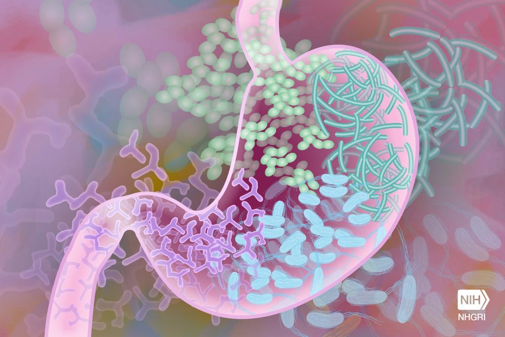
Gut Microbiome. Credit Darryl Leja National Human Genome Research Institute National Institutes Of Health

Exposure to antibiotics during a key developmental window in infancy can stunt the growth of insulin-producing cells in the pancreas and may boost risk of diabetes later in life, new research in mice suggests. The study, published this month in the journal Science, also pinpoints specific microorganisms that may help those critical cells proliferate in early life.

The findings are the latest to shine a light on the importance of the human infant microbiome—the constellation of bacteria and fungi living on and in us during our first few years. The research could lead to new approaches for addressing a host of metabolic diseases.

“We hope our study provides more awareness for how important the infant microbiome actually is for shaping development,” said first author Jennifer Hill, assistant professor in molecular, cellular and developmental biology at CU’s BioFrontiers Institute. “This work also provides important new evidence that microbe-based approaches could someday be used to not only prevent but also reverse diabetes.”

Something in the environment

More than 2 million U.S. adults live with Type 1 diabetes. The disease typically emerges in childhood, and genetics play a strong role. But scientists have found that, while identical twins share DNA that predisposes them to Type 1 diabetes, only one twin usually gets the disease.

“This tells you that there’s something about their environmental experiences that is changing their susceptibility,” said Hill.

For years, she has looked to microbes for answers.

Previous studies show that children who are breastfed or born vaginally, which can both promote a healthy infant microbiome, are less likely to develop Type 1 diabetes than others. Some research also shows that giving babies antibiotics early can inadvertently kill good bugs with bad and boost diabetes risk.

The lingering questions: What microbes are these infants missing out on?

“Our study identifies a critical window in early life when specific microbes are necessary to promote pancreatic cell development,” said Hill.

A key window of opportunity

She explained that human babies are born with a small amount of pancreatic “beta cells,” the only cells in the body that produce insulin. But some time in a baby’s first year, a once-in-a-lifetime surge in beta cell growth occurs.

“If, for whatever reason, we don’t undergo this event of expansion and proliferation, that can be a cause of diabetes,” Hill said.

She conducted the current study as a postdoctoral researcher at the University of Utah with senior author June Round, a professor of pathology.

They found that when they gave broad-spectrum antibiotics to mice during a specific window (the human equivalent of about 7 to 12 months of life), the mice developed fewer insulin producing cells, higher blood sugar levels, lower insulin levels and generally worse metabolic function in adulthood.

“This, to me, was shocking and a bit scary,” said Round. “It showed how important the microbiota is during this very short early period of development.”

Lessons in baby poop

In other experiments, the scientists gave specific microbes to mice, and found that several they increased their production of beta cells and boosted insulin levels in the blood. The most powerful was a fungus called Candida dubliniensis.

The team used faecal samples from The Environmental Determinants of Diabetes in the Young (TEDDY) study to make what Hill calls “poop slushies” and fed them to the mice.

When the researchers inoculated newborn mice with poop from healthy infants between 7 to 12 months in age, their beta cells began to grow. Poop from infants of other ages did not do the same. Notably, Candida dublineinsis was abundant in human babies only during this time period.

“This suggests that humans also have a narrow window of colonisation by these beta cell promoting microbes,” said Hill.

When male mice that were genetically predisposed to Type 1 diabetes were colonised with the fungus in infancy, they developed diabetes less than 15% of the time. Males that didn’t receive the fungus got diabetes 90% of the time.

Even more promising, when researchers gave the fungus to adult mice whose insulin-producing cells had been killed off, those cells regenerated.

Too early for treatments

Hill stresses that she is not “anti-antibiotics.” But she does imagine a day when doctors could give microbe-based drugs or supplements alongside antibiotics to replace the metabolism-supporting bugs they inadvertently kill.

Poop slushies (faecal microbiota transplants) have already been used experimentally to try to improve metabolic profiles of people with Type 2 diabetes, which can also damage pancreatic beta cells.

But such approaches can come with real risk, since many microbes that are beneficial in childhood can cause harm in adults. Instead, she hopes that scientists can someday harness the specific mechanisms the microbes use to develop novel treatments for healing a damaged pancreas—reversing diabetes.

She recently helped establish a state-of-the-art “germ-free” facility for studying the infant microbiome at CU Boulder. There, animals can be bred and raised entirely without microbes, and by re-introducing them one by one scientists can learn they work.

“Historically we have interpreted germs as something we want to avoid, but we probably have way more beneficial microbes than pathogens,” she said. “By harnessing their power, we can do a lot to benefit human health.”

Source: University of Colorado at Boulder

Gut Microbes may Play a Role Linking Sugary Drinks and Diabetes Risk

Photo by Breakingpic on Pexels

It is well known that consuming sugary drinks increases the risk of diabetes, but the mechanism behind this relationship is unclear. Now, in a paper published in the Cell Press journal Cell Metabolism, researchers show that metabolites produced by gut microbes might play a role.

In a long-term cohort of US Hispanic/Latino adults, the researchers identified differences in the gut microbiota and blood metabolites of individuals with a high intake of sugar-sweetened beverages. The altered metabolite profile seen in sugary beverage drinkers was associated with a higher risk of developing diabetes in the subsequent 10 years. Since some of these metabolites are produced by gut microbes, this suggests that the microbiome might mediate the association between sugary beverages and diabetes.

“Our study suggests a potential mechanism to explain why sugar-sweetened beverages are bad for your metabolism,” says senior author Qibin Qi, an epidemiologist at Albert Einstein College of Medicine. “Although our findings are observational, they provide insights for potential diabetes prevention or management strategies using the gut microbiome.”

Sugar-sweetened beverages are the main source of added sugar in the diets of US adults – in 2017 and 2018, US adults consumed an average of 34.8g of added sugar each day from sugary beverages such as soda and sweetened fruit juice. Compared to added sugars in solid foods, added sugar in beverages “might be more easily absorbed, and they have a really high energy density because they’re just sugar and water,” says Qi.

Previous studies in Europe and China have shown that sugar-sweetened beverages alter gut microbiome composition, but this is the first study to investigate whether this microbial change impacts host metabolism and diabetes risk. It’s also the first study to investigate the issue in US-based Hispanic/Latino population — a group that experiences high rates of diabetes and is known to consume high volumes of sugar-sweetened beverages.

The team used data from the ongoing Hispanic Community Health Study/Study of Latinos (HCHS/SOL), a large-scale cohort study with data from over 16 000 participants living in San Diego, Chicago, Miami, and the Bronx. At an initial visit, participants were asked to recall their diet from the past 24 hours and had blood drawn to characterise their serum metabolites. The researchers collected faecal samples and characterized the gut microbiomes of a subset of the participants (n = 3035) at a follow-up visit and used these data to identify association between sugar-sweetened beverage intake, gut microbiome composition, and serum metabolites.

They found that high sugary beverage intake, defined as two or more sugary beverages per day, was associated with changes in the abundance of nine species of bacteria. Four of these species are known to produce short-chain fatty acids: molecules that are produced when bacteria digest fibre and that are known to positively impact glucose metabolism. In general, bacterial species that were positively associated with sugary beverage intake correlated with worse metabolic traits. Interestingly, these bacteria were not associated with sugar ingested from non-beverage sources.

The researchers also found associations between sugary beverage consumption and 56 serum metabolites, including several metabolites that are produced by gut microbiota or are derivatives of gut-microbiota-produced metabolites. These sugar-associated metabolites were associated with worse metabolic traits, including higher levels of fasting blood glucose and insulin, higher BMIs and waist-to-hip ratios, and lower levels of high-density lipoprotein cholesterol (“good” cholesterol). Notably, individuals with higher levels of these metabolites had a higher likelihood of developing diabetes in the 10 years following their initial visit.

“We found that several microbiota-related metabolites are associated with the risk of diabetes,” says Qi. “In other words, these metabolites may predict future diabetes.”

Because gut microbiome samples were only collected from a subset of the participants, the researchers had an insufficient sample size to determine whether any species of gut microbes were directly associated with diabetes risk, but this is something they plan to study further.

“In the future, we want to test whether the bacteria and metabolites can mediate or at least partially mediate the association between sugar-sweetened beverages and risk of diabetes,” says Qi.

The team plans to validate their findings in other populations and to extend their analysis to investigate whether microbial metabolites are involved in other chronic health issues linked to sugar consumption, such as cardiovascular disease.

Source: Science Direct

Gut Microbes also Feed on Sugar to Produce Crucial Short-chain Fatty Acids

Source: CC0

Gut microbes that were thought to feed exclusively on dietary fibre also get fed sugar from our guts, from which they produce short-chain fatty acids that are crucial to many body functions. The Kobe University discovery of this symbiotic relationship also points the way to developing novel therapeutics.

Gut microbes produce many substances that our body needs but cannot produce itself. Among them are short-chain fatty acids that are the primary energy source for the cells lining our guts but have other important roles, too, and that are thought to be produced by bacteria who feed on undigested fibre. However, in a previous study, the Kobe University endocrinologist Ogawa Wataru found that people who take the diabetes drug metformin excrete the sugar glucose to the inside of their guts. He says: “If glucose is indeed excreted into the gut, it is conceivable that this could affect the symbiotic relationship between the gut microbiome and the host.”

Ogawa and his team set out to learn more about the details of the glucose excretion and its relationship with the gut microbiota. “We had to develop unprecedented bioimaging methods and establish novel analytical techniques for the products of the gut microbial metabolism,” he says. They used their new methods to not only see where and how much glucose enters the guts, but also used mouse experiments to find out how the sugar is transformed after that. In addition, they also checked how the diabetes drug metformin influences these results both in humans and in mice.

The Kobe University team now published their results in the journal Communications Medicine. They found that, first, glucose is excreted in the jejunum and is transported from there inside the gut to the large intestine and the rectum. “It was surprising to find that even individuals not taking metformin exhibited a certain level of glucose excretion into the intestine. This finding suggests that intestinal glucose excretion is a universal physiological phenomenon in animals, with metformin acting to enhance this process,” Ogawa explains. In both humans and mice, irrespective of whether they were diabetic or not, metformin increased the excretion by a factor of almost four.

And second, on the way down, the glucose gets transformed into short-chain fatty acids. Ogawa says: “The production of short-chain fatty acids from the excreted glucose is a huge discovery. While these compounds are traditionally thought to be produced through the fermentation of indigestible dietary fibres by gut microbiota, this newly identified mechanism highlights a novel symbiotic relationship between the host and its microbiota.”

Ogawa and his team are now conducting further studies with the aim of understanding how metformin and other diabetes drugs affect glucose excretion, the gut microbiome and their metabolic products. He says: “Intestinal glucose excretion represents a previously unrecognised physiological phenomenon. Understanding the underlying molecular mechanisms and how drugs interfere with this process could lead to the development of novel therapeutics aimed at the regulation of gut microbiota and their metabolites.”

Source: Kobe University

Gluten Free Diet Reduces Coeliac Symptoms – and ‘Good’ Gut Bacteria

Photo by Mariana Kurnyk: https://www.pexels.com/photo/two-baked-breads-1756062/

A research team led by the University of Nottingham has used magnetic resonance imaging (MRI) to better understand the impact a gluten free diet has on people with coeliac disease, which could be the first step towards finding new ways of treating the condition.

The MARCO study – MAgnetic Resonance Imaging in COliac disease is published in Clinical Gastroenterology and Hepatology (CGH) (link connects to BioRxiv copy).

Coeliac disease is a chronic condition affecting around one person in every 100 in the general population. When people with coeliac disease eat gluten, which is found in pasta and bread, their immune system produces an abnormal reaction that inflames and damages the gut tissue and causes symptoms such as abdominal pain and bloating.

The only treatment is a life- long commitment to a gluten free diet, which helps recovery of the gut tissue but still leaves many patients with gastrointestinal symptoms.

Luca Marciani, Professor of Gastrointestinal Imaging at the University, led the study. He said: “Despite being a common chronic condition, we still don’t precisely know how coeliac disease affects the basic physiological functioning of the gut and how the gluten free diet treatment may further change this.

“We launched the MARCO study to try and address this issue, by using MRI along with gut microbiome analysis to give us new insights into how a gluten-free diet affects people with coeliac disease.”

The team recruited 36 people who had just been diagnosed with coeliac disease and 36 healthy volunteers to participate in the study. Images were taken of their guts with MRI, along with blood and stool samples. The patients then followed a gluten free diet for one year and came back to repeat the study. The healthy participants came back one year later too and repeated the study, but they did not follow any diet treatment.

The study found that the newly diagnosed patients with coeliac disease had more gut symptoms, more fluid in the small bowel and that the transit of food in the bowel was slower than in the healthy controls.

The microbiota (the ‘bugs’ living in the colon) of the patients showed higher levels of ‘bad bugs’ such as E.coli. After one year of a gluten free diet, gut symptoms, bowel water and gut transit improved in the patients, but without returning to normal values. But the gluten free diet also reduced some of the ‘good bugs’ in the microbiota, such as Bifidobacteria associated with reduced intake of starch and wheat nutrients, due to the different diet.

The patient study was conducted by Radiographer Dr Carolyn Costigan, from Nottingham University Hospitals, as part of her PhD studies at the University of Nottingham.

It was particularly interesting to see how the imaging results on gut function correlated with changes in the ‘bugs’ in the colon microbiota. The findings increase our understanding of gut function and physiology in coeliac disease and open the possibility of developing prebiotic treatments to reverse the negative impact of the gluten free diet on the microbiome.”

Luca Marciani, Professor of Gastrointestinal Imaging

Dr Frederick Warren from the Quadram Institute, which contributed to the research, said: “This study is the result of an exciting and innovative research collaboration bringing together medical imaging technology and gut microbiome analysis. We provide important insights which pave the way for future studies which may identify novel approaches to alleviate long-term symptoms in coeliac patients.”

Source: University of Nottingham

Parkinson’s Drug Found to Promote Pathogenic Gut Bacteria

Fig. 1: Chemical imaging of active gut microbes. After brief incubation with heavy water, culture medium and a drug, various chemical bonds (here C-D and C-H) in the stool sample are shown in yellow and green, their ratio in yellow-purple (left). Selected microbes are detected in the same image section with fluorescence-labelled oligonucleotide probes in cyan. The activity of the detected microbes can be determined based on the amount of C-D bonds. C: Xiaowei Ge (Boston University)

An international team of scientists have revealed that the widely prescribed Parkinson’s disease drug entacapone significantly disrupts the human gut microbiome by inducing iron deficiency. This international study, provides new insights into the often-overlooked impact of human-targeted drugs on the microbial communities that play a critical role in human health. The findings, published in Nature Microbiology, suggest however that iron supplementation can help counteract these impacts.

While it is well established that antibiotics can significantly disrupt the human gut microbiome, emerging research shows that a wide range of human-targeted drugs – particularly those used to treat neurological conditions – can also profoundly affect the microbial communities living in our bodies. Despite their intended therapeutic effects on different organs, these drugs can inadvertently disrupt the balance of gut microbes, leading to potential health consequences. Until now, most studies investigating these interactions relied either on patient cohort analyses affected by many confounding factors or on experiments using isolated gut bacteria, which do not fully capture the complexity of the human microbiome.

Investigating drug–bug interactions

The team, which included some from the University of Vienna, used a novel experimental approach. The researchers studied the effects of two drugs – entacapone and loxapine, a medication for schizophrenia – on faecal samples from healthy human donors. They incubated the samples with therapeutic concentrations of these drugs, then analysed the impact on the microbial communities using advanced molecular and imaging techniques, including heavy water labelling combined with Stimulated Raman Spectroscopy (SRS). The team discovered that loxapine and even more so entacapone severely inhibited many microbiome members, while E. coli dramatically expanded in the presence of entacapone.

“The results were even more striking when we examined microbial activity, rather than just their abundance,” explained Fatima Pereira, lead author of the study and former Postdoctoral researcher at the University of Vienna. “The heavy water-SRS method allowed us to observe the subtle yet significant changes in the gut microbiome, which are often missed in traditional abundance-based measurements.”

Entacapone induces iron starvation, favours pathogenic microbes

The researchers hypothesised that entacapone might interfere with iron availability in the gut, a crucial resource for many microbes. Their experiments confirmed that adding iron to faecal samples containing entacapone counteracted the drug’s microbiome-altering effects. Further investigation revealed that E. coli, which thrived under these conditions, carried a highly efficient iron-uptake system (enterobactin siderophore). This system allowed the bacteria to overcome iron starvation and proliferate, even in the presence of the drug.

“By showing that entacapone induces iron deficiency, we have uncovered a new mechanism of drug-induced gut dysbiosis, in which the drug selects for E. coli and other potentially pathogenic microbes well adapted to iron limiting conditions,” said Michael Wagner, scientific director of the Excellence Cluster and vice-head of the Centre for Microbiology and Environmental Systems Science (CeMESS) at the University of Vienna.

Wider implications for drug–microbiome interactions

This discovery has broader implications for understanding how other human-targeted drugs might affect the gut microbiome. Several drugs, including entacapone, contain metal-binding catechol groups, suggesting that this mechanism could be a more common pathway for drug-induced microbiome alterations.

The findings also present an opportunity to mitigate the side effects of drugs like entacapone. By ensuring sufficient iron availability to the large intestine, it may be possible to reduce dysbiosis and the gastrointestinal issues that often accompany Parkinson’s disease treatment.

“The next step is to explore how we can modify drug treatments to better support the gut microbiome,” said Wagner. “We are looking at strategies to selectively deliver iron to the large intestine, where it can benefit the microbiome without interfering with drug absorption in the small intestine.”

Source: University of Vienna

Gut Health Signals could Transform Arthritis Treatment

Gut Microbiome. Credit Darryl Leja National Human Genome Research Institute National Institutes Of Health

Changes in the gut microbiome before rheumatoid arthritis is developed could provide a window of opportunity for preventative treatments, new research suggests.

Bacteria associated with inflammation is found in the gut in higher amounts roughly 10 months before patients develop clinical rheumatoid arthritis, according to a longitudinal study by researchers at the University of Leeds. 

This new research might give us a major opportunity to act sooner to prevent rheumatoid arthritis.

Dr Christopher Rooney, Leeds Institute of Medical Research

Previous research has linked rheumatoid arthritis to the gut microbiome, which is the ecosystem of microbes in your intestines. But this new study, published in the Annals of the Rheumatic Diseases, reveals a potential intervention point. 

Lead researcher Dr Christopher Rooney, NIHR Academic Clinical Lecturer at the University of Leeds and Leeds Teaching Hospitals NHS Trust, said: “Patients at risk of rheumatoid arthritis are already experiencing symptoms such as fatigue and joint pain, and they may know someone in their family who has developed the disease. As there is no known cure, at-risk patients often feel a sense of hopelessness, or even avoid getting tested.  

“This new research might give us a major opportunity to act sooner to prevent rheumatoid arthritis.” 

Major opportunity for treatment

Funded by Versus Arthritis, the longitudinal study was conducted on 19 patients at risk of rheumatoid arthritis, with samples taken five times during a 15-month period.  

Five of these patients progressed to clinical arthritis, and the research showed they had gut instability with higher amounts of bacteria including Prevotella, which is associated with rheumatoid arthritis, about ten months before progression. The remaining 14, whose disease didn’t progress, had largely stable amounts of bacteria in their gut. 

Potential treatments that the researchers want to test at the 10-month window include changes to diet like eating more fibre, taking prebiotics or probiotics, and improving dental hygiene to keep harmful bacteria from periodontal disease away from the gut. 

The exact relationship between gut inflammation and rheumatoid arthritis development remains unclear. In a small number of patients within the study, the gut changes occurred before there were any changes to the joints observed by a rheumatologist, but more research is needed to determine whether these influence each other. 

Although bacteria is associated with rheumatoid arthritis, the researchers want to make it clear that there is no evidence this is contagious. 

Lucy Donaldson, director for research and health intelligence at Versus Arthritis, said: “At Versus Arthritis, we welcome the findings of this study which could give the clinicians of the future a crucial window of opportunity to delay – or even prevent – the onset of rheumatoid arthritis. This success is testament to the dedication of UK researchers who are working to personalise treatment and prevent chronic conditions that have significant impacts on a person’s ability to work, raise families and live independently.” 

The study initially took data from 124 individuals who had high levels of CCP+, an antibody that attacks healthy cells in the blood, which indicates risk of developing rheumatoid arthritis. The researchers compared their samples to 22 healthy individuals and seven people who had a new rheumatoid arthritis diagnosis.  

The findings from this larger group showed that the gut microbiome was less diverse in the at-risk group, compared to the healthy control group. 

The longitudinal study, which took samples from 19 patients over 15 months, revealed the changes in bacteria at ten months before progression to rheumatoid arthritis. 

The Leeds research team will now carry out an analysis of treatments that have already been trialled, to inform future testing of treatments at this potential 10-month intervention point. 

Source: University of Leeds

The Gut Microbiome can Affect Symptoms of Hypopituitarism

Gut Microbiome. Credit Darryl Leja National Human Genome Research Institute National Institutes Of Health

In research published in PLOS Genetics, scientists have shown that the balance of bacteria in the gut can influence symptoms of hypopituitarism in mice. They also showed that aspirin was able to improve hormone deficiency symptoms in mice with this condition.

People with mutations in a gene called Sox3 develop hypopituitarism, where the pituitary gland doesn’t make enough hormones. It can result in growth problems, infertility and poor responses of the body to stress.

The scientists at the at the Francis Crick Institute removed Sox3 from mice, causing them to develop hypopituitarism around the time of weaning (starting to eat solid food).

They found that mutations in Sox3 largely affect the hypothalamus in the brain, which instructs the pituitary gland to release hormones. However, the gene is normally active in several brain cell types, so the first task was to ask which specific cells were most affected by its absence.

The scientists observed a reduced number of cells called NG2 glia, suggesting that these play a critical role in inducing the pituitary gland cells to mature around weaning, which was not known previously. This could explain the associated impact on hormone production.

The team then treated the mice with a low dose of aspirin for 21 days. This caused the number of NG2 glia in the hypothalamus to increase and reversed the symptoms of hypopituitarism in the mice.

Although it’s not yet clear how aspirin had this effect, the findings suggest that it could be explored as a potential treatment for people with Sox3 mutations or other situations where the NG2 glia are compromised.

An incidental discovery revealed the role of gut bacteria in hormone production

When the National Institute for Medical Research (NIMR) merged with the Crick in 2015, mouse embryos were transferred from the former building to the latter, and this included the mice with Sox3 mutations.

When these mice reached the weaning stage at the Crick, the researchers were surprised to find that they no longer had the expected hormonal deficiencies.

After exploring a number of possible causes, lead author Christophe Galichet compared the microbiome – bacteria, fungi and viruses that live in the gut – in the mice from the Crick and mice from the NIMR, observing several differences in its makeup and diversity. This could have been due to the change in diet, water environment, or other factors that accompanied the relocation.

He also examined the number of NG2 glia in the Crick mice, finding that these were also at normal levels, suggesting that the Crick-fed microbiome was somehow protective against hypopituitarism.

To confirm this theory, Christophe transplanted faecal matter retained from NIMR mice into Crick mice, observing that the Crick mice once again showed symptoms of hypopituitarism and had lower numbers of NG2 glia. 

Although the exact mechanism is unknown, the scientists conclude that the make-up of the gut microbiome is an example of an important environmental factor having a significant influence on the consequences of a genetic mutation, in this case influencing the function of the hypothalamus and pituitary gland.

Source: Francis Crick Institute

Breastfeeding Shapes the Gut Microbiome and Protects against Asthma

Photo by Wendy Wei

Human breast milk regulates a baby’s mix of microbes, known as the microbiome, during the infant’s first year of life, in turn lowers the child’s risk of developing asthma, according to a new study published in Cell.

Led by researchers at NYU Langone Health and the University of Manitoba, the study results showed that breastfeeding beyond three months supports the gradual maturation of the microbiome in the infant’s digestive system and nasal cavity, the upper part of the respiratory tract. Conversely, stopping breastfeeding earlier than three months disrupts the paced development of the microbiome and was linked to a higher risk of preschool asthma.

Some components in breast milk, such as complex sugars called human milk oligosaccharides, can only be broken down with the help of certain microbes. This provides a competitive advantage to microbes capable of digesting these sugars. By contrast, infants who are weaned earlier than three months from breast milk and who then rely solely on formula feeding, become home to a different set of microbes –ones that will help the infant to digest the components in formula. While many of these microbes that thrive on formula do eventually end up in all babies, the researchers showed that their early arrival is linked to an increased risk of asthma.

“Just as a pacemaker regulates the rhythm of the heart, breastfeeding and human milk set the pace and sequence for microbial colonisation in the infant’s gut and nasal cavity, ensuring that this process occurs in an orderly and timely manner,” said study co-senior investigator and computational biologist Liat Shenhav, PhD. “Healthy microbiome development is not only about having the right microbes. They also need to arrive in the right order at the right time,” said Dr Shenhav, an assistant professor at NYU Grossman School of Medicine, its Institute for Systems Genetics, and the school’s Department of Microbiology.

For the study, Dr Shenhav, who is also an assistant professor at NYU’s Courant Institute of Mathematical Sciences, worked in collaboration with study co-senior investigator Meghan Azad, PhD, director of the Manitoba Interdisciplinary Lactation Center, and a professor of paediatrics and child health, at the University of Manitoba.

Another key study finding was that the bacterium Ruminococcus gnavus appeared much sooner in the guts of children who were weaned early from breast milk than in those of children who were exclusively breastfed. The bacterium is known to be involved in the production of molecules called short-chain fatty acids, and the formation and breakdown of the amino acid tryptophan. Both tryptophan and its metabolites have been linked to immune system regulation and disruption in previous research, including an increased risk of asthma. The study authors noted that beyond aiding in digestion, an infant’s microbiome plays a crucial role in the immune system’s development.

The study tracked the ebb and flow of microbes in the guts and noses of infants during the first year of life, as well as details on breastfeeding and the composition of their mothers’ milk. All the children and their mothers were participating in the CHILD Cohort Study, a long-term research project that has been studying the same 3500 Canadian children at different stages of life from the womb well into adolescence.

The data provided by the CHILD Cohort Study enabled researchers to detangle the impact of breastfeeding on an infant’s microbiome from a range of other environmental factors, including prenatal smoke exposure, antibiotics, and the mother’s asthma history.

Even when these factors were accounted for, they found that breastfeeding duration remained a powerful determinant for the child’s microbial makeup over time. They also used these microbial dynamics and data on milk components to train a machine learning model that accurately predicted asthma years in advance. Finally, they created a statistical model to learn causal relationships, which showed that the primary way breastfeeding reduces asthma risk is through shaping the infant’s microbiome.

“The algorithms we developed provide valuable insights into microbial dynamics during an infant’s first year of life and how these microbes interacted with the infant,” said Dr Shenhav. “These insights allowed us to move beyond identifying associations, enhancing our ability to make predictions and explore causal relationships.

“Our research highlights the profound impact of breastfeeding on the infant microbiome and breastfeeding’s essential role in supporting respiratory health. By uncovering the mechanisms behind the protective effects of breast milk, as demonstrated in this study, we aim to inform national guidelines on breastfeeding and weaning from breast milk in a data-driven manner.

“With further research, our findings could also contribute to developing strategies to prevent asthma in children who cannot be breastfed for at least three months,” she added.

Source: NYU Langone Health / NYU Grossman School of Medicine

Ketogenic Diet Reduces Friendly Gut Bacteria and Raises Cholesterol Levels

Photo by Jose Ignacio Pompe on Unsplash

A study from the University of Bath reveals that ketogenic low-carbohydrate diets can increase cholesterol levels and reduce beneficial gut bacteria, specifically Bifidobacterium.

Published in Cell Reports Medicine, the research from the Centre for Nutrition, Exercise, and Metabolism involved 53 healthy adults for up to 12 weeks. Participants followed either a moderate sugar diet (control), a low-sugar diet (less than 5% of calories from sugar), or a ketogenic (keto) low-carbohydrate diet (less than 8% of calories from carbohydrates).

Key findings include:

•Increased Cholesterol: The keto diet raised cholesterol levels, particularly in small and medium sized LDL particles. The diet increased apolipoprotein B (apoB), which causes plaque buildup in arteries. In contrast, the low-sugar diet significantly reduced cholesterol in LDL particles.

•Reduced Favourable Gut Bacteria: The keto diet altered gut microbiome composition, notably decreasing Bifidobacteria, beneficial bacteria often found in probiotics. This bacteria has wide ranging benefits: producing b vitamins, inhibiting pathogens and harmful bacteria and lowering cholesterol. Sugar restriction did not significantly impact the gut microbiome composition.

•Glucose Tolerance: The keto diet reduced glucose tolerance, meaning the adults’ bodies became less efficient at handling carbohydrates.

•Both Diets Resulted In Fat Loss: Keto Diet resulted in an average of 2.9kg fat mass loss per person, whilst the sugar restricted diet followed with an average 2.1kg fat mass loss per person at 12 weeks.

•Metabolism: Researchers also noticed that the keto diet caused significant changes in lipid metabolism and muscle energy use, shifting the body’s fuel preference from glucose to fats.

•Physical Activity Levels: Both sugar restriction and keto diets achieved fat loss without changing physical activity levels. Previous studies from the Centre for Nutrition, Exercise and Metabolism have shown that skipping breakfast or intermittent fasting cause reductions in physical activity.

Lead researcher Dr. Aaron Hengist highlighted the concerning cholesterol findings:

“Despite reducing fat mass, the ketogenic diet increased the levels of unfavourable fats in the blood of our participants, which, if sustained over years, could have long-term health implications such as increased risk of heart disease and stroke.”

Dr. Russell Davies, who led the microbiome research, explained the impact on gut health:

“Dietary fibre is essential for the survival of beneficial gut bacteria like Bifidobacteria. The ketogenic diet reduced fibre intake to around 15 grams per day, half the NHS recommended intake. This reduction in Bifidobacteria might contribute to significant long-term health consequences such as an increased risk of digestive disorders like irritable bowel disease, increased risk of intestinal infection and a weakened immune function.”

Professor Javier Gonzalez, who oversaw the research, commented on the glucose findings:

“The ketogenic diet reduced fasting glucose levels but also reduced the body’s ability to handle carbs from a meal. By measuring proteins in muscle samples taken from participants’ legs, we think this is probably an adaptive response to eating less carbohydrates day-to-day and reflects insulin resistance to storing carbs in muscle. This insulin resistance is not necessarily a bad thing if people are following a ketogenic diet, but if these changes persist when people switch back to a higher carbohydrate diet it could increase the risk of developing type 2 diabetes in the long-term”

In light of this new research, the academics conclude that if you’re considering a diet, a low sugar one will be better for most people. More work is needed to understand how individuals may benefit from each type of diet. The government recommends that free sugars (those added to food or drink or found naturally in honey, syrups, fruit juices and smoothies) should be restricted to less than 5% of total energy intake. Professor Dylan Thompson, who also oversaw the work, said:

“The ketogenic diet is effective for fat loss, but it comes with varied metabolic and microbiome effects that may not suit everyone. In contrast, sugar restriction supports government guidelines for reducing free sugar intake, promoting fat loss without apparent negative health impacts.”

Source: University of Bath

Breast Cancer Chemo Disrupts Gut Microbiome and Impacts Cognition

Photo by Tima Miroshnichenko on Pexels

Chemotherapy is known to cause behavioural side effects, including cognitive decline. Notably, the gut microbiome communicates with the brain to affect behaviour, including cognition. 

“For the first time ever, our Intelligut Study found that the gut microbiome has been implicated in cognitive side effects of chemotherapy in humans,” said senior author Leah Pyter, associate professor of psychiatry and neuroscience at Ohio State University. “The potential connection between the gut and the brain would allow us to create treatments for the gut to treat the brain.”

Study findings are published in the journal Brain, Behavior, and Immunity.

This clinical longitudinal observational study explored whether chemotherapy-induced disruption of the gut microbiome relates to cognitive decline and circulating inflammatory signals. 

Faecal samples, blood and cognitive measures were collected from 77 patients with breast cancer before, during and after chemotherapy.

“We found that patients treated with chemotherapy who showed decreases in cognitive performance also had reductions in the diversity of their gut microbiome,” said Pyter, also a researcher with Ohio State’s Institute for Behavioral Medicine Research and member of the Cancer Control Research Program at The Ohio State University Comprehensive Cancer Center – Arthur G. James Cancer Hospital and Richard J. Solove Research Institute (OSUCCC – James)

This research builds on Pyter’s prior research in mouse models that found chemotherapy-induced shifts in the gut microbiome cause neurobiological changes and behavioural side effects.  The current study indicates that an association between gut microbiome and cognitive performance exists in humans as well. 

“Side effects of chemotherapy are common and may reduce quality of life, but these side effects can be dismissed as ‘part of chemotherapy’ and therefore overlooked and under-treated,” Pyter said. “We believe that gut microbiome-focused interventions, such as faecal microbial transplantation, may improve behavioural side effects of chemotherapy.” 

OSUCCC—James researchers are also conducting research studies on how the gut microbiome impacts cancer treatment effectiveness and its role in reducing or increasing cancer risk. 

“Chemotherapy is a very important tool for stopping many cancers and side effects should not deter patients who would benefit from this type of therapy from pursuing it, but we know the side effects of some treatment regimens can be quite challenging for patients to complete,” said David Cohn, MD, interim chief executive officer of the OSUCCC – James. “It’s a careful tightrope of walking between effective cancer control and side effect management – and our team is working every day, in the hospital clinics and the lab, to develop ways to manage the side effects of disease treatment with an eye toward quality of life.” 

Source: Ohio State University