Tag: glioma

Scars of Destroyed Brain Tumours are Fertile Grounds for Recurrence

Types of tumour cells. Credit: Scientific Animations CC4.0

A Ludwig Cancer Research study has discovered that recurrent tumours of the aggressive brain cancer glioblastoma multiforme (GBM) grow out of the fibrous scars of malignant predecessors destroyed by interventions such as radiotherapy, surgery and immunotherapy.

Led by Ludwig Lausanne’s Johanna Joyce, Spencer Watson and alumnus Anoek Zomer and published in the current issue of Cancer Cell, the study describes how these scars enable the regrowth of tumours and identifies drug targets to sabotage their malignant support. It also demonstrates the efficacy of such combination therapies in preclinical trials using mouse models of GBM.

“We’ve identified fibrotic scarring as a key source of GBM resurgence following therapy, showing how it creates a protective niche for the regrowth of the tumor,” said Joyce. “Our findings suggest that blocking the process of scarring in the brain by adding anti-fibrosis agents to current treatment strategies could help prevent glioblastoma from recurring and improve the outcomes of therapy.”

There is a great need for such interventions. GBM is the most common and aggressive form of brain cancer in adults. Despite considerable effort to develop effective therapies for the cancer, the average life expectancy of patients remains around 14 months following diagnosis.

The origins of the current study date back to 2016, when the Joyce lab reported in the journal Science its examination in mouse models of strategies to overcome resistance to a promising immunotherapy for the treatment of GBM. That experimental therapy, which inhibits signalling by the colony stimulating factor-1 receptor (CSF-1R) and currently in clinical trials, targets immune cells known as macrophages and their brain-resident versions, microglia, both of which are manipulated by GBM cells to support tumour growth and survival.

The Joyce lab has demonstrated that CSF-1R inhibition reprograms these immune cells into an anti-tumour state and so induces significant tumour regression. Yet, as the Science study showed, about half the mice show relapse following an initial response to the therapy. “What was most remarkable about that observation was that every single time a brain tumour recurred following immunotherapy, it regrew right next to a scar that had formed at the original site of a tumour,” said Joyce.

In the current study, Joyce, Watson, Zomer and their colleagues examined tumour samples obtained from patients undergoing GBM therapy and showed that fibrotic scarring occurs following therapy in humans as well – and that it is similarly associated with tumour recurrence. They also showed that the fibrotic scarring occurs in response to not only immunotherapy but also following the surgical and radiological removal of tumours.

To explore how fibrosis contributes to relapse, the researchers applied an integrated suite of advanced technologies to analyze the cellular and molecular geography of the scars and the microenvironment of resurgent tumors.

These technologies include the analysis of global gene expression in individual cells, the comprehensive analysis of proteins in the tissues as well a workflow and AI-powered suite of analytical methods for the spatial analysis of tissues named hyperplexed immunofluorescence imaging (HIFI). Recently developed by Watson and colleagues in the Joyce lab, HIFI permits the simultaneous visualisation of multiple molecular markers in and around cells across broad cross-sections of tissues, enabling the generation of granular maps of the tumour microenvironment.

“Applied together, these advanced methods allowed us to see exactly how fibrotic scars form,” said Watson. “They revealed that the fibrosis serves as a kind of protective cocoon for residual cancer cells and pushes them into a dormant state in which they are largely resistant to therapy. We found that it also shields them from surveillance and elimination by the immune system.”

Integrated analyses of the tissue microenvironment following therapy revealed that the descendants of cells associated with tumor-feeding blood vessels become functionally altered to resemble fibroblasts—fiber-producing cells commonly involved in wound-healing. These perivascular-derived fibroblast-like (PDFL) cells fan out across the region previously occupied by the regressing tumor, where they mediate the generation of fibrotic scars. These cells, the researchers found, are especially activated by neuroinflammation and immune factors known as cytokines, most notably one called transforming growth factor-β (TGF-β).

“To see if targeting fibrotic scarring could improve therapeutic outcomes for GBM, we devised a treatment regimen using existing drugs to block TGF-β signaling and suppress neuroinflammation in combination with CSF-1R inhibition and evaluated it in preclinical trials using mouse models of GBM,” said Joyce. “We also timed these additional treatments to coincide with the period of maximal PDFL activation identified by our studies. Our results show that the drug combination inhibited fibrotic scarring, diminished the numbers of surviving tumor cells and extended the survival of treated mice compared to controls.”

The researchers suggest that approaches to limit fibrotic scarring could significantly improve outcomes for GBM patients receiving surgical, radiation or macrophage-targeting therapies. Additional research, they note, will likely yield even better drug targets for such combination therapies.

Source: Ludwig Institute for Cancer Research

Glioma Cells can Also Fire off Electrical Signals in the Brain

Source: Pixabay

Researchers at Baylor College of Medicine and the Jan and Dan Duncan Neurological Research Institute at Texas Children’s Hospital have uncovered a new cell type in human brain cancers. Their study, published in Cancer Cell, reveals that a third of the cells in glioma, fire electrical impulses. Interestingly, the impulses, also called action potentials, originate from tumour cells that are part neuron and part glia, supporting the groundbreaking idea that neurons are not the only cells that can generate electric signals in the brain.

The scientists also discovered that cells with hybrid neuron-glia characteristics are present in the non-tumour human brain. The findings highlight the importance of further studying the role of these newly identified cells in both glioma and normal brain function.

“Previous studies have shown that patient survival outcomes are associated with tumour proliferation and invasiveness, which are influenced by tumour intrinsic and extrinsic factors, including communication between tumour cells and neurons that reside in the brain,” said Dr Benjamin Deneen, professor in the Department of Neurosurgery at Baylor.

Researchers have previously described that glioma and surrounding healthy neurons connect with each other and that neurons communicate with tumours in ways that drive tumour growth and invasiveness. 

“We have known for some time now that tumour cells and neurons interact directly,” said first author Dr Rachel N. Curry, postdoctoral fellow in paediatrics – neuro oncology at Baylor, who was responsible for conceptualising the project. “But one question that always lingered in my mind was, ‘Are cancer cells electrically active?’ To answer this question correctly, we required human samples directly from the operating room. This ensured the biology of the cells as they would exist in the brain was preserved as much as possible.”

To study the ability of glioma cells to spike electrical signals and identify the cells that produce the signals, the team used Patch-sequencing, a combination of techniques that integrates whole-cell electrophysiological recordings to measure spiking signals with single-cell RNA-sequencing and analysis of the cellular structure to identify the type of cells.

The electrophysiology experiments were conducted by research associate and co-first author Dr Qianqian Ma in the lab of co-corresponding author associate professor of neuroscience Dr Xiaolong Jiang. This innovative approach has not been used before to study human brain tumour cells. “We were truly surprised to find these tumour cells had a unique combination of morphological and electrophysiological properties,” Ma said. “We had never seen anything like this in the mammalian brain before.”

“We conducted all these analyses on single cells. We analysed their individual electrophysiological activity. We extracted each cell’s content and sequenced the RNA to identify the genes that were active in the cell, which tells us what type of cell it is,” Deneen said. “We also stained each cell with dyes that would visualise its structural features.”

Integrating this vast amount of individual data required the researchers to develop a novel way to analyse it.

“To define the spiking cells and determine their identity, we developed a computational tool – Single Cell Rule Association Mining (SCRAM) – to annotate each cell individually,” said co-corresponding author, Dr Akdes Serin Harmanci, assistant professor of neurosurgery at Baylor.

“Finding that so many glioma cells are electrically active was a surprise because it goes against a strongly held concept in neuroscience that states that, of all the different types of cells in the brain, neurons are the only ones that fire electric impulses,” Curry said. “Others have proposed that some glia cells known as oligodendrocyte precursor cells (OPCs) may fire electrical impulses in the rodent brain, but confirming this in humans had proven a difficult task. Our findings show that human cells other than neurons can fire electrical impulses. Since there is an estimated 100 million of these OPCs in the adult brain, the electrical contributions of these cells should be further studied.”

“Moreover, the comprehensive data analyses revealed that the spiking hybrid cells in glioma tumours had properties of both neurons and OPC cells,” Harmanci said. “Interestingly, we found non-tumour cells that are neuron-glia hybrids, suggesting that this hybrid population not only plays a role in glioma growth but also contributes to healthy brain function.”

“The findings also suggest that the proportion of spiking hybrid cells in glioma may have a prognostic value,” said co-corresponding author Dr Ganesh Rao, professor and chair of neurosurgery at Baylor. “The data shows that the more of these spiking hybrid glioma cells a patient has, the better the survival outcome. This information is of great value to patients and their doctors.”

“This work is the result of extensive equal collaboration across multiple disciplines – neurosurgery, bioinformatics, neuroscience and cancer modelling – disciplines strongly supported by state-of-the-art groups at Baylor,” Deneen said. “The results offer an enhanced understanding of glioma tumours and normal brain function, a sophisticated bioinformatics pipeline to analyse complex cellular populations and potential prognostic implications for patients with this devastating disease.”

Source: Baylor College of Medicine

New Drug Shows Promise for Treating Rare and Aggressive Gliomas

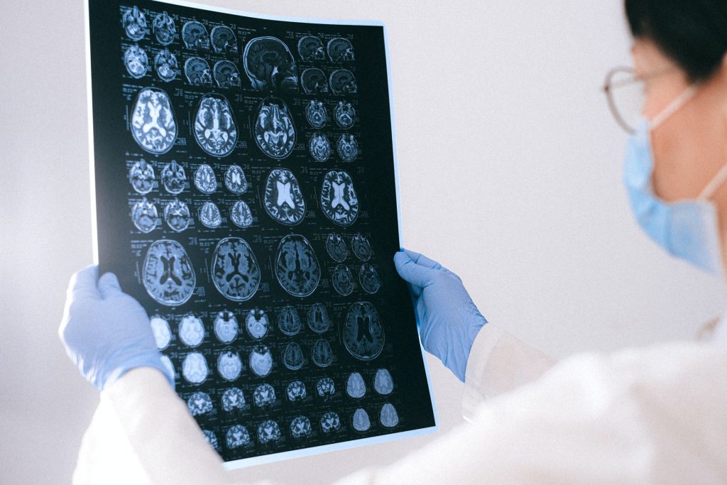
MRI scan showing brain cancer. Credit: Michelle Monje, MD, PhD, Stanford University

An experimental drug may provide a new treatment option for some patients with rare incurable brain tumours, according to an analysis published in the Journal of Clinical Oncology.

Diffuse midline gliomas are diagnosed in about 800 people per year in the U.S., according to the Centers for Disease Control and Prevention.

A subset of particularly aggressive diffuse midline gliomas are caused by a H3 K27M mutation and the only effective treatment is radiation, as the location of the tumour in the brain makes surgery difficult. Even with radiation, relapse is virtually inevitable and more than 70% of patients with this subtype of brain tumour die from the cancer, according to the National Institutes of Health.

In the study, investigators analysed the results of five previous clinical trials testing the effectiveness of dordaviprone, an experimental drug which works by blocking a certain protein in tumours with the mutation.

The study included results from 50 patients (including four children) with H3 K27M–mutant diffuse midline gliomas and found that 30% of patients responded well to the drug. The most common side effect reported was fatigue, according to the study.

Now, the researchers are launching a trial at Northwestern Medicine hospitals to investigate the drug’s effectiveness in newly diagnosed patients.

Source: Northwestern University

Scientists Evaluate Old Epilepsy Drug for Glioma Prevention

Photo by Anna Shvets on Pexels

A drug used to treat children with epilepsy prevents brain tumour formation and growth in two mouse models of neurofibromatosis type 1 (NF1), according to a study by researchers at Washington University School of Medicine in St. Louis. NF1 is a genetic condition that causes tumours to grow on nerves throughout the body.

The findings lay the groundwork for a clinical trial to assess whether the drug, lamotrigine, can prevent or delay brain tumours in children with NF1. The study is published online in the journal Neuro-Oncology.

“Based on these data, the Neurofibromatosis Clinical Trials Consortium is considering launching a first-of-its-kind prevention trial,” said senior author David H. Gutmann, MD, PhD, professor of neurology. “The plan is to enrol kids without symptoms, treat them for a limited time, and then see whether the number of children who develop tumours that require treatment goes down.

“This is a novel idea, so we took it to an NF1 patient focus group,” Gutmann continued. “They said, ‘This is exactly what we’re looking for.’ A short-term treatment with a drug that has been used safely for 30 years was acceptable to them if it reduced the chance their children would develop tumours and need chemotherapy that might have all kinds of side effects.”

Optic gliomas, tumours on the optic nerve are the most serious type that those with NF1 get. Such tumours typically appear between ages 3 to 7. Though rarely fatal, they cause vision loss in up to a third of patients as well as other symptoms, including early puberty. Standard chemotherapy for optic gliomas is only moderately effective at preventing further vision loss and can affect children’s developing brains, resulting in cognitive and behavioural problems.

In a previous study, Gutmann and Corina Anastasaki, PhD, an assistant professor of neurology and the first author on the new paper, showed that lamotrigine stopped optic glioma growth in NF1 mice by suppressing neuronal hyperactivity. Intrigued, the Neurofibromatosis Clinical Trial Consortium asked Gutmann and Anastasaki to clarify the connection between NF1 mutation, neuronal excitability and optic gliomas; assess whether lamotrigine was effective at the doses already proven safe in children with epilepsy; and conduct these studies in more than one strain of NF1 mice.

In humans, NF1 could be caused by any one of thousands of different mutations in the NF1 gene, with different mutations causing different medical problems. Repeating experiments in multiple strains of mice was a way of gauging whether lamotrigine was likely to work in people regardless of the underlying mutation.

Anastasaki and Gutmann not only showed that lamotrigine worked in two strains of NF1 mice, they also showed that the drug worked at lower doses than those used for epilepsy, meaning that it was probably safe. Even better, they found that a short course of the drug had lasting effects, both as a preventive and a treatment. Mice with tumours and that were treated for four weeks starting at 12 weeks of age saw their tumours stop growing and even showed no further damage to the retinas. Mice that received a four-week course of the drug starting at 4 weeks of age, before tumours typically emerge, showed no tumour growth even four months after treatment had ended.

These findings have led Gutmann to suggest that a one-year course of treatment for young children with NF1, maybe between the ages of 2 to 4, might be enough to reduce their risk of brain tumours.

“The idea that we might be able to change the prognosis for these kids by intervening within a short time window is so exciting,” Gutmann said. “If we could just get them past the age when these tumours typically form, past age 7, they may never need treatment. I’d love it if I never again had to discuss chemotherapy for kids who aren’t even in first grade yet.”

Source: Washington University School of Medicine

Approval for First-in Class Glioma Drug set to Change Practice

Photo by Anna Shvets on Pexels

A new drug for the treatment of a type of brain tumour that strikes young people could soon receive approval by the U.S. Food and Drug Administration. The drug, vorasidenib, could greatly extend the time before further therapy – and eventual resistance – is needed.

In an editorial in the New England Journal of Medicine, David Schiff, MD, the co-director of UVA Cancer Center’s Neuro-Oncology Center, outlines the potential significance of the drug vorasidenib for patients with most low-grade gliomas. The drug was fast-tracked by the FDA in August 2023 based on the strength of the findings, and filings for regulatory approval were made in February 2024. FDA approval is anticipated in the second half of 2024, and its approval in Europe will likely soon follow.

Adult-type diffuse gliomas represent approximately 81% of primary malignant brain tumours. Of those, approximately 20% harbour an isocitrate dehydrogenase (IDH) mutation, including 100% of grade 2 and grade 3 adult-type diffuse gliomas. Approximately 2500 Americans with a median age of only 40 are diagnosed with grade 2 IDH-mutant gliomas each year. The tumours cause steadily increasing disability, eventually become resistant to treatment options and typically prove fatal.

 Because of the limited treatment options available, doctors usually take a “watch and wait” approach to managing the brain tumours, holding off on treatment until after the tumour progresses.

In the randomised controlled INDIGO trial, 331 patients received either vorasidenib or placebo. The trial showed that the drug slowed tumour growth significantly and extended the average time until the tumour started growing from 11.1 months to more than 27 months. Vorasidenib also increased the time to next intervention (TTNI), the timeframe before patients need additional treatment such as radio- or chemotherapy. 

Schiff, in his editorial, describes the results as “striking.” Vorasidenib’s success could “put a nail in the coffin” of the watch-and-wait approach for such brain tumours, Schiff believes. 

“It used to be that we thought of all gliomas as being on a spectrum,” Schiff said. “We now understand that those with the IDH gene mutation have a markedly different biology, outcome and, as this study shows, vulnerabilities that new therapies can exploit.”

If the drug receives approval from the federal Food and Drug Administration, it would become the first targeted therapy for low-grade gliomas. But Schiff notes that there are also other recent advances that are improving our understanding of such gliomas.

“There are still many unanswered questions about how we can best utilise this new medication if and when it receives FDA approval,” Schiff said. “Nonetheless, considering that existing standard therapies for these tumours [radiation and chemotherapy] are tough on patients, with short- and long-term side effects, it will be wonderful to have a useful and very well-tolerated treatment option.”

Foundations Laid for Standardised PET Examination of Diffuse Gliomas

Photo by Mart Production on Pexels

Diffuse gliomas are malignant brain tumours that cannot be optimally examined by means of conventional MRI imaging. So-called amino acid PET (positron emission tomography) scans are better able to image the activity and spread of gliomas. An international team of researchers from the RANO Working Group have drawn up the first ever international criteria for the standardised imaging of gliomas using amino acid PET. It has published its results in the journal The Lancet Oncology.

PET uses a radioactive tracer to measure metabolic processes in the body. Amino acid PET is used in the diagnosis of diffuse gliomas, with tracers that work on a protein basis (amino acids) and accumulate in brain tumours.

The Response Assessment in Neuro-Oncology (RANO) Working Group is an international, multidisciplinary consortium founded to develop standardised new response criteria for clinical studies relating to brain tumours.

Under the joint leadership of nuclear physician Nathalie Albert from LMU and oncologist Professor Matthias Preusser from the Medical University of Vienna, the RANO group has developed new criteria for assessing the success of therapies for diffuse gliomas.

Nathalie Albert explains: “PET imaging with radioactively labelled amino acids has proven extremely valuable in neuro-oncology and permits reliable representation of the activity and extension of gliomas. Although amino acid PET has been used for years, it had not been evaluated in a structured manner before now. In contrast to MRI-based diagnostics, there have been no criteria for interpreting these PET images.” According to the researchers, the new criteria allow PET to be used in clinical studies and everyday clinical practice and create a foundation for future research and the comparison of treatments for improved therapies.

New criteria for PET examinations of brain tumours

Diffuse gliomas are malignant brain tumorus that cannot be optimally examined by means of conventional MRI imaging. So-called amino acid PET scans are better able to image the activity and spread of gliomas.

These malignant brain tumours develop out of glial cells and are generally aggressive and difficult to treat.

The RANO group has developed criteria that permit evaluation of the success of treatment using PET. Called PET RANO 1.0, these PET-based criteria open up new possibilities for the standardised assessment of diffuse gliomas.

Source: Ludwig-Maximilians-Universität München

First-in-human Treatment with Tumour Vaccine Shows Positive Results against Gliomas

Photo by Anna Shvets on Pexels

Tumour vaccines alert a cancer patient’s immune system to proteins that are carrying cancer-typical alterations, and may prove useful in gliomas, which are resistant to most forms of treatment including immune checkpoint inhibitors. Reporting in Nature Medicine, physicians and cancer researchers have now performed a first-in human treatment of eight adult patients with advanced midline gliomas with a peptide vaccine. The vaccine, which mimicked a histone protein mutation typical of this type of cancer, proved to be safe and induced the desired immune responses against the brain tumour, with one patient experiencing remission for more than 31 months.

Cancer vaccinations depend on distinct protein structures on cancer cells by which immune cells can differentiate them from healthy one. Mutations in the tumour genome often lead to protein structures that are altered in a way typical of cancer.

Diffuse midline gliomas are among the most aggressive brain tumours. They usually occur in children and young adults near the brain stem and are therefore difficult to access surgically. Chemotherapy or radiotherapy also have limited effectiveness. In this type of cancer, mutations characteristically occur in the gene encoding histone H3 (H3K27M), a packaging protein of DNA. The mutation gives rise to a novel protein structure (a neoepitope) that can be recognised as foreign by the patient’s immune system.

“Such mutations, which occur in identical form in many patients, are rare in cancer. They literally lend themselves to the development of tumor vaccines because they occur in all cancer cells, since the mutated histone is causative for the development of midline gliomas. This means that vaccination against the mutated protein gets to the root of the problem,” explains Michael Platten, Director of the Department of Neurology at the University Medical Center Mannheim and Head of Department at the German Cancer Research Center (DKFZ).

The researchers led by Katharina Sahm and Michael Platten synthetically reconstructed the section of the histone H3 protein with the characteristic mutation. Using this peptide, they were able to curb the growth of H3K27M-mutated tumours in a mouse model. Encouraged by the results, the team decided to test the mutation-specific vaccine produced at the University of Tübingen in patients in a phase I-trial*, which is still ongoing.

In parallel, the physicians, together with colleagues from Munich, Berlin, Bonn and Münster, treated eight adult patients with the peptide vaccine in time-limited individual curative trials. These patients, who could not be enrolled in the trial protocol, suffered from diffuse midline gliomas with H3K27M mutation that progressed after standard therapy. Some of the affected individuals received therapy with immune checkpoint inhibitors in addition to tumor vaccination.

No serious side effects were observed in any of the vaccinated patients. Five of the eight treated patients developed specific immune responses against the mutant protein, which were dominated by CD4 T-helper cells. In one of the patients who had shown a strong immune response, the tumour regressed completely and she remained tumour-free for 31 months.

The vaccine peptide, which is comparatively long at 27 amino acids, worked in patients with different HLA variants. HLA proteins are responsible for the presentation of the mutant peptide on the cell surface and differ from person to person depending on their genetic background. Supported by the HI-TRON Mainz — Helmholtz Institute of the DKFZ, the researchers also observed that immune responses decreased over time, so repeated administration of the vaccine could support a sustained effect.

“We cannot make any further statements about the efficacy of the vaccination based on these treatments. In any case, the current study has given us valuable information that will help us to further optimise the development of brain tumour vaccines in the future,” explains the study’s senior author Katharina Sahm, senior physician at the Neurological University Hospital Mannheim and DKFZ researcher. A phase I-trial is currently underway to test the vaccine against the H3K27M mutation in patients with newly diagnosed midline gliomas. Evaluation is expected to begin around 2025.

Source: German Cancer Research Center (Deutsches Krebsforschungszentrum, DKFZ)

Gliomas Have Folates Receptors That Could be Targeted for Imaging

Photo by Anna Shvets on Pexels

University of Turku researchers have discovered that gliomas contain increased amount of folate receptor expression relative to adjacent brain tissue. This discovery is a new and significant finding in the field, which could allow folate-based radiopharmaceuticals can be used in positron emission tomography (PET) imaging to detect folate receptors in gliomas.

This phenomenon, which is described in Frontiers in Immunology, has been observed in both experimental models and human tumour samples.

“Prior to this discovery, the presence of folate receptors and their increased presence in gliomas had not been recognised, and thus they have not yet been used for imaging nor treatment purposes,” summarises Doctoral Researcher Maxwell Miner from the Turku PET Centre at the University of Turku in Finland.

According to research group leader and InFLAMES PI Professor Anne Roivainen this presents an especially exciting target for potential future treatments.

“Our results show an average of 100-fold increase in folate-based radiopharmaceutical accumulation in glioma tissue versus that of adjacent healthy brain tissue,” says Professor Roivainen.

Urgent need for new chemotherapy treatments

Glioma brain tumours originate from the non-neuronal glial cells in the brain, which outnumber neurons in quantity. Gliomas comprise numerous subgroups, with even a high degree of morphological and receptor variability within a single cancerous lesion.

This exceptional cellular heterogeneity can make treatment difficult. There is an urgent need for new chemotherapy treatments particularly for the most malignant brain cancers as they often grow in an infiltrative web-like manner on their periphery making distinguishing the boundaries between glioma and non-glioma difficult. The researchers at the Turku PET Centre hope that this recent discovery will lead to further investigation into folate-targeted brain tumour detection and treatment.

Source: University of Turku

Vorasidenib Extends Progression-free Survival in Glioma Subtype

Photo by National Cancer Institute on Unsplash

In a study published in the New England Journal of Medicine, scientists report that a new targeted therapy drug can extend progression-free survival for a subtype of glioma. The finding suggests a possible new treatment option for people with the slow-growing but deadly brain tumour.

The team, co-led by UCLA, found the drug vorasidenib more than doubled progression-free survival in people with recurrent grade 2 glioma with IDH1 and IDH2 mutations. Compared with placebo, those who took vorasidenib went for nearly 17 more months without their cancer worsening, delaying the time before they needed to begin chemotherapy and radiation.

The type of glioma studied in the paper, recurrent grade 2 glioma with IDH1 and IDH2 mutations, tends to affect younger people, often those in their 30s. The current standard treatment, a combination of radiation and chemotherapy, can cause neurological deficits that make it hard for patients in an often challenging and busy stage of life.

UCLA professor of neuro-oncology Dr Timothy Cloughesy, co-senior author of the study, said that the availability of a treatment that enables patients to go for longer periods of time between chemotherapy and radiation treatments could have a major impact.

“We’re always concerned about the delayed effects of radiation,” said Cloughesy. “Having the ability to hold off on getting radiation therapy to the brain with an effective therapy is really critical and very meaningful to this population of patients.”

Vorasidenib is a dual inhibitor of mutant IDH1/2, meaning that it prevents the formation and accumulation of the onco-metabolite 2-Hydroxyglutarate, or 2-HG, that occurs when genetically altered versions of two enzymes, IDH1 and IDH2, are present in a tumour. 2-HG is thought to be responsible for the formation and maintenance of IDH-mutant gliomas.

The study is also the first clinical trial to analyse a targeted therapy drug specifically developed to treat brain cancer. Targeted therapies focus on specific molecules that are involved in cancer cell growth and metastasis. Unlike chemotherapy and other therapies that can affect both cancerous and healthy cells, targeted therapies only attack cancer cells with the mutated target while sparing normal cells.

While there has been great progress in using targeted therapies to treat many types of cancer, the difficulty of crossing the blood-brain barrier makes developing targeted therapies for brain tumours challenging. Vorasidenib is a brain-penetrant inhibitor, allowing it to cross the blood-brain barrier.

The study involved 331 people aged 12 and older who had been diagnosed with recurrent grade 2 glioma with the IDH1 and IDH2 mutations and who had undergone brain tumour surgery. From that group, 168 were randomised to vorasidenib and 163 to placebo.

Among those who received vorasidenib, the disease did not progress for an average of 27.7 months, significantly longer than the 11.1 months for those who received the placebo. And among those who received vorasidenib, 85.6% went for 18 months before their next treatment, while 83.4% went for 24 months between treatments.

The disease progressed in just 28% of people receiving v orasidenib, compared to 54% of those receiving placebos. And as of September 2022, which was 30 months after the study began, 72% of patients who were in the vorasidenib group were still taking the drug and their disease had not progressed.

For patients who were originally in the placebo group whose cancer began to progress during the study, doctors permitted a switch to vorasidenib. The researchers observed limited adverse side effects from vorasidenib. “This is the first targeted treatment that shows unequivocal efficacy in this population and is precedent-setting for this disease,” Cloughesy said.

Source: University of California – Los Angeles Health Sciences

A Link Between Head Injury and Increased Glioma Risk

Photo by John Simmons on Unsplash

Previous research has hinted at a possible link between head injury and increased rates of gliomas, rare but aggressive brain tumours. A University College London team has now identified a possible mechanism to explain this link, implicating genetic mutations acting in concert with brain tissue inflammation to change the behaviour of cells, making them more likely to become cancerous.

Publishing in Current Biology, the researchers have now identified a possible mechanism to explain this link, implicating genetic mutations acting in concert with brain tissue inflammation to change the behaviour of cells, making them more likely to become cancerous. Although this study was largely carried out in mice, it suggests that it would be important to explore the relevance of these findings to human gliomas.

The study was led by Professor Simona Parrinello (UCL Cancer Institute), Head of the Samantha Dickson Brain Cancer Unit and co-lead of the Cancer Research UK Brain Tumour Centre of Excellence. She said: “Our research suggests that a brain trauma may contribute to an increased risk of developing brain cancer in later life.”

Gliomas are brain tumours that often arise in neural stem cells. More mature types of brain cells, such as astrocytes, have been considered less likely to give rise to tumours. However, recent findings have demonstrated that after injury, astrocytes can exhibit stem cell behaviour again.

Professor Parrinello and her team therefore set out to investigate whether this property may make astrocytes able to form a tumour following brain trauma using a pre-clinical mouse model.

Young adult mice with brain injury were injected with a substance which permanently labelled astrocytes in red and knocked out the function of the p53 gene, known to have a vital role in suppressing many different cancers. A control group was treated the same way, but the p53 gene was left intact. A second group of mice was subjected to p53 inactivation in the absence of injury.

Professor Parrinello said: “Normally astrocytes are highly branched – they take their name from stars – but what we found was that without p53 and only after an injury the astrocytes had retracted their branches and become more rounded. They weren’t quite stem cell-like, but something had changed. So we let the mice age, then looked at the cells again and saw that they had completely reverted to a stem-like state with markers of early glioma cells that could divide.”

This suggested to Professor Parrinello and team that mutations in certain genes synergised with brain inflammation, which is induced by acute injury and then increases over time during the natural process of ageing to make astrocytes more likely to initiate a cancer. Indeed, this process of change to stem-cell like behaviour accelerated when they injected mice with a solution known to cause inflammation.

The team then looked for evidence to support their hypothesis in human populations. Working with Dr Alvina Lai in UCL’s Institute of Health Informatics, they consulted electronic medical records of over 20 000 people who had been diagnosed with head injuries, comparing the rate of brain cancer with a control group, matched for age, sex and socioeconomic status. They found that patients who experienced a head injury were nearly four times more likely to develop a brain cancer later in life, than those who had no head injury. It is important to keep in mind that the risk of developing a brain cancer is overall low, estimated at less than 1% over a lifetime, so even after an injury the risk remains modest.

Professor Parrinello said: “We know that normal tissues carry many mutations which seem to just sit there and not have any major effects. Our findings suggest that if on top of those mutations, an injury occurs, it creates a synergistic effect. In a young brain, basal inflammation is low so the mutations seem to be kept in check even after a serious brain injury. However, upon ageing, our mouse work suggests that inflammation increases throughout the brain but more intensely at the site of the earlier injury. This may reach a certain threshold after which the mutation now begins to manifest itself.”

Source: University College London