Tag: genetics

Study Reveals Mediaeval Plague Victims Buried With Care and Attention

Photo by Peter Kvetny on Unsplash

Mediaeval plague victims in the UK were mostly buried with care and attention, according to a new study from Cambridge University. 

In the mid-14th century, Europe was devastated by the Black Death which killed between 40 and 60 per cent of the population. For centuries afterward, waves of plague would continue to strike the region.

Due to the rapid onset of death in the absence of antibiotic treatment (less than a week for bubonic plague and under 48h for pneumonic plague), the disease leaves no visible evidence on the skeleton, so until now archaeologists have been unable to identify individuals who died of plague unless they were buried in mass graves.

Although it has been long believed that most plague victims in fact received an individual burial, this has been impossible to confirm until now.

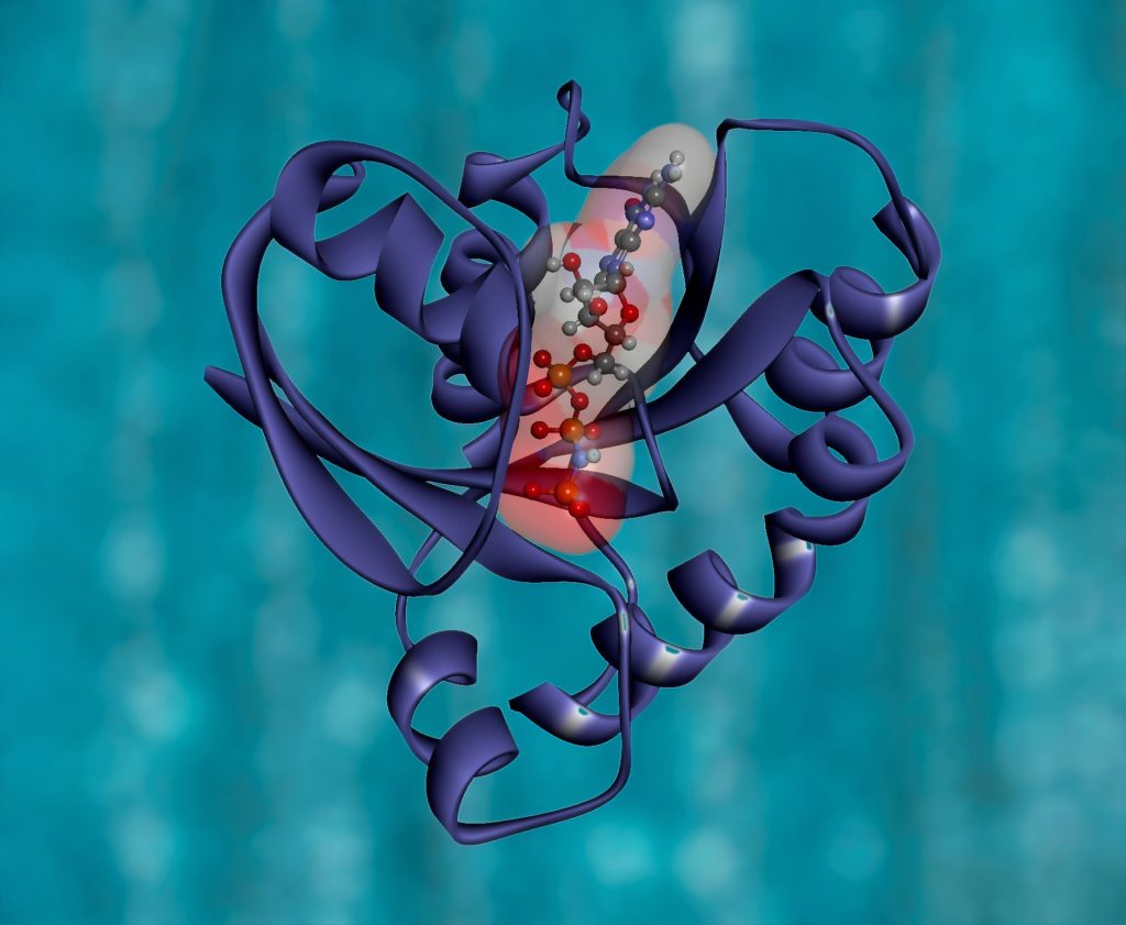
By studying DNA extracted from the teeth of individuals who died at this time, researchers from the Wellcome Trust-funded After the Plague project, based at the Department of Archaeology, University of Cambridge, have identified the presence of Yersinia Pestis, the bacterial pathogen that causes plague. The study is available to read online in the European Journal of Archaeology.

These include people who received normal individual burials at a parish cemetery and friary in Cambridge and in the nearby village of Clopton.

Lead author Craig Cessford of the University of Cambridge explained: “These individual burials show that even during plague outbreaks individual people were being buried with considerable care and attention. This is shown particularly at the friary where at least three such individuals were buried within the chapter house. The Cambridge Archaeological Unit conducted excavations on this site on behalf of the University in 2016-2017.”

The individual at the parish of All Saints by the Castle in Cambridge was also buried with care; this stand in contrast to the apocalyptic language used to describe the abandonment of this church in 1365 when it was reported that the church was partly in ruins and ‘the bones of dead bodies are exposed to beasts’.”

The study also shows that some plague victims in Cambridge did, as expected, receive mass burials.

Yersinia Pestis was also identified in several parishioners from St Bene’t’s, who were found buried together in a large trench in the churchyard excavated by the Cambridge Archaeological Unit on behalf of Corpus Christi College.

Soon afterwards, this part of the churchyard was transferred to Corpus Christi College, which was founded by the St Bene’t’s parish guild to commemorate the dead including the victims of the Black Death. For centuries, the members of the College would walk over the mass burial every day on the way to the parish church.

Cessford concluded, “Our work demonstrates that it is now possible to identify individuals who died from plague and received individual burials. This greatly improves our understanding of the plague and shows that even in incredibly traumatic times during past pandemics people tried very hard to bury the deceased with as much care as possible.”

Source: University of Cambridge

Journal information: “Beyond Plague Pits: Using Genetics to Identify Responses to Plague in Medieval Cambridgeshire” – Craig Cessford, Christiana L. Scheib, Meriam Guellil, Marcel Keller, Craig Alexander, Sarah A. Inskip and John E. Robb. European Journal of Archaeology, https://doi.org/10.1017/eaa.2021.19

Novel Approach Targets Pancreatic Cancers Which Depend on Mutant KRAS Gene

KRAS Protein Structure. RAS is a family of related proteins that is expressed in all animals. KRAS is one of three RAS genes found in humans. RAS genes are mutated in approximately one-third of all human cancers. Photo by National Cancer Institute on Unsplash

Researchers have identified a novel drug that effectively thwarts pancreatic tumours that are addicted to the cancer-causing mutant KRAS gene.

Because early detection of pancreatic cancer is difficult, it has a low survival rate, accounting for just over 3% of all new cancer cases in the US, but leading to nearly 8% of all cancer deaths, according to the National Cancer Institute.

The KRAS gene was recognised more that 25 years ago as the component of Kirsten sarcoma virus responsible for oncogenesis. Since then, mutations of KRAS have been described in a large proportion of solid tumors ranging from more than 90% of pancreatic carcinomas to 20% to 30% of pulmonary adenocarcinomas.

Through a pre-clinical study, Said Sebti, PhD, associate director for basic research at VCU Massey Cancer Center, identified a novel drug that effectively thwarts pancreatic tumors that are addicted to the cancer-causing mutant KRAS gene. 

“We discovered a link between hyperactivation of the CDK protein and mutant KRAS addiction, and we exploited this link preclinically to counter mutant KRAS-driven pancreatic cancer, warranting clinical investigation in patients afflicted with this deadly disease,“ said Dr Sebti, who is also the Lacy Family Chair in Cancer Research at Massey and a professor of pharmacology and toxicology at the VCU School of Medicine. “Our findings are highly significant as they revealed a new avenue to combat an aggressive form of pancreatic cancer with very poor prognosis due mainly to its resistance to conventional therapies.”

In 90 percent of pancreatic cancers, KRAS is mutated. Prior studies have shown that some tumours harbouring mutant KRAS are in fact addicted to the mutant gene, meaning they cannot survive or grow without it. Sebti set out to discover if there is a drug that can specifically kill those tumours with a mutant KRAS addiction.

Searching for a suitable drug

Dr Sebti and colleagues used a three-pronged approach to tackle this question.

First of all, they mapped the blueprint of pancreatic cancer cells through global phosphoproteomics, showing them how the addicted and non-addicted tumours differ at the phosphoprotein level. They found two proteins, CDK1 and CDK2, which signalled which cells were addicted to mutant KRAS.

Additionally, they analysed a comprehensive database from the Broad Institute of MIT and Harvard which contains genome-wide CRISPR gRNA screening datasets. They discovered that CDK1 and CDK2 as well as CDK7 and CDK9 proteins were associated with mutant KRAS-addicted tumors.

Finally, they evaluated 294 FDA drugs to selectively kill mutant KRAS-addicted cancer cells over non-KRAS-addicted cancer cells in the lab. They determined the most effective drug in preclinical experiments was AT7519, an inhibitor of CDK1, CDK2, CDK7 and CDK9.

“Using three entirely different approaches, the same conclusion presented itself clearly to us: pancreatic cancer patients whose tumors are addicted to mutant KRAS could benefit greatly from treatment with the CDK inhibitor AT7519,” Dr Sebti said.

To further validate these findings in fresh tumours taken from pancreatic cancer patients the researchers found that AT7519 suppressed the growth of xenograft cells from five mutant KRAS pancreatic cancer patients who relapsed on chemotherapy and/or radiation therapies.

Though AT7519 had previously been tested unsuccessfully in a number of clinical trials, none of the trials were for pancreatic cancer.

“If our findings are correct and translate in humans, then we should be able to see a positive response in pancreatic cancer patients whose tumors are addicted to mutant KRAS,” Dr Sebti said.

As well as pancreatic cancer, the study authors believe these findings may also have clinical implications for colorectal and non-small cell lung cancer patients with prevalent KRAS mutations.

Source: Virginia Commonwealth University

Journal information: Kazi, A., et al. (2021) Global Phosphoproteomics Reveal CDK Suppression as a Vulnerability to KRas Addiction in Pancreatic Cancer. Clinical Cancer Research. doi.org/10.1158/1078-0432.CCR-20-4781.

Call for More Neuroscience Research in Africa

Photo by MART PRODUCTION from Pexels

A team of neuroscientists are calling for greater support of neuroscience research in Africa based on an analysis of the continent’s past two decades of research outputs.  

The findings reveal important information about the nature of funding and international collaboration comparing activity in the continent to other countries, mainly the US, UK and areas of Europe. It is hoped that the study will provide useful data to help further develop science in Africa.  

The greatest human genetic diversity is found in Africa, and Eurasian genomes have less variation than African ones; in fact, Eurasian genomes can be considered a subset of African ones. This carries important implications for understanding human diseases, including neurological disorders.

Co-lead senior author Tom Baden, Professor of Neuroscience in the School of Life Sciences and the Sussex Neuroscience research group at the University of Sussex said: “One beautiful thing about science is that there is no such thing as a truly local problem. But that also means that there should be no such thing as a local solution – research and scientific communication by their very nature must be a global endeavour.  

“And yet, currently the vast majority of research across most disciplines is carried out by a relatively small number of countries, located mostly in the global north. This is a huge waste of human potential.”  

The team, made up of experts from the University of Sussex, the Francis Crick Institute and institutions from across Africa, analysed the entirety of Africa’s outputs in neuroscience over two decades. A lot of early neuroscience research took place in Egypt, it was pointed out.

Lead author Mahmoud Bukar Maina, a Research Fellow in the School of Life Sciences and the Sussex Neuroscience research group at the University of Sussex and visiting scientist at Yobe State University, Nigeria, explained: “Even though early progress in neuroscience began in Egypt, Africa’s research in this area has not kept pace with developments in the field around the world. There are a number of reasons behind this and, for the first time, our work has provided a clear picture of why – covering both strengths and weaknesses of neuroscience research in Africa and comparing this to other continents.  

“We hope it will provide useful data to guide governments, funders and other stakeholders in helping to shape science in Africa, and combat the ‘brain drain’ from the region.”  

Co-lead senior author Lucia Prieto-Godino, a Group Leader at the Francis Crick Institute, said: “One of the reasons why this work is so important, is that the first step to solve any problem is understanding it. Here we analyse key features and the evolution of neuroscience publications across all 54 African countries, and put them in a global context. This highlights strengths and weaknesses, and informs which aspects will be key in the future to support the growth and global integration of neuroscience research in the continent.” 

The study identifies the African countries with the greatest research outputs, revealing that most research funding originates from external sources such as the USA and UK.  

The researchers argue that a sustainable African neuroscience research environment needs local funding, suggesting that greater government backing is needed as well as support from the philanthropic sector.  
Professor Baden added: “One pervasive problem highlighted in our research was the marked absence of domestic funding. In most African countries, international funding far predominates. This is doubly problematic.  

“Firstly, it takes away the crucial funding stability that African researchers would need to meaningfully embark on large-scale and long-term research projects, and secondly, it means that the international, non-African funders essentially end up deciding what research is performed across the continent. Such a system would generate profound outrage across places like Europe – how then can it be acceptable for Africa?”

A number of the researchers involved in the study are members of TReND Africa, a charity supporting scientific capacity building in Africa.  

Source: University of Sussex

Journal information: M. B. Maina et al, Two decades of neuroscience publication trends in Africa, Nature Communications (2021). DOI: 10.1038/s41467-021-23784-8 , www.nature.com/articles/s41467-021-23784-8

Researchers Uncover a Mechanism that Regulates Hair Regeneration

Photo by engin akyurt on Unsplash

Chinese researchers have discovered a new regulatory mechanism for the regeneration capacity of skin hair, with important clues for developing treatments for hair loss. Hair loss or alopecia is an extremely common condition, yet there is still no effective therapy for it.

In the skin, activation of hair follicle stem cells (HFSCs) and progenitors by growth factor stimulation is the basis for hair follicle and hair regeneration. Hair regeneration defects can often attribute to blunted responses of HFs to growth stimuli, but it is how the sensitivity of HFSCs or progenitors to growth stimuli is determined is still unclear. Figuring out the answer to this question will provide important clues for the treatment of hair-related diseases such as alopecia.

To this end, Prof Zhang Liang’s group from the Shanghai Institute of Nutrition and Health (SINH) of the Chinese Academy of Sciences, and collaborators uncovered the role of the micro RNA miR-24 and its mechanism in limiting the regenerative ability of hair follicle (HF) progenitors, opening up new therapeutic avenue for hair loss treatment. microRNAs regulate key steps of cell differentiation and development through suppressing gene expression in a sequence-specific manner.

The researchers discovered that that the resting-to-activation transition of HF is associated with significant down-regulation of miR-24 in HF progenitors prior to their activation.

By experimenting with mouse models, they found that miR-24 limits the sensitivity of HF progenitors to growth stimuli. miR-24 over-expression in the skin epithelium significantly delayed HF progenitor activation and hair cycle progression, while its conditional ablation significantly accelerated the hair cycle and increased the HFs’ sensitivity to growth stimuli.

Interestingly, the conditional ablation of miR-24 in skin epithelium significantly improved the effect of Minoxidil lotion on stimulating hair growths without detectable side effects, indicating that miR-24 could be a new potential target for hair regeneration therapies.

Mechanistically, the researchers discovered that Plk3 is a new miR-24 target gene that mediates the function of miR-24 to limit hair growth by regulating CCNE1, a key cell cycle regulator. They also found that miR-24 acts downstream bone morphogenetic protein (BMP), which is a known inhibitory signal for hair growth.

The study revealed that miR-24 is a key factor limiting the regenerative ability of skin HF progenitors. How adult stem cells respond appropriately to environmental stimuli is a question of fundamental importance in stem cell biology.

Source: Medical Xpress

Journal information: Fengzhen Liu et al, miR-24 controls the regenerative competence of hair follicle progenitors by targeting Plk3, Cell Reports (2021). DOI: 10.1016/j.celrep.2021.109225

Key Genetic Repair Protein Removes Traps

Image source: Pixabay

Researchers have discovered that a key genetic repair protein also cleans up ‘traps’ left by another protein, its partner in genetic repair.

DNA is constantly getting damaged: the delicate strands that carry life’s genetic code take quite a beating as they jumble about in the course of their work. Errors can accumulate if left untreated, with fatal consequences — such as cancerous tumors — for the cell and the organism.

Two key proteins are involved in preventing the damage from getting out of hand: PARP — or poly ADP ribose polymerase — acts as a marker for a trouble spot, allowing XRCC1 — or X-ray repair cross-complementing protein 1 — to locate the damage and start repairs.

The repair functions of these two proteins have been known for some time. The importance of this has been recognised with the 2015 Nobel prizes for chemistry, as this knowledge allowed the development of anti-cancer drugs, known as PARP inhibitors, that disrupt the growth of certain kinds of tumours.

Although these key proteins had been identified, their precise roles were not well understood. It took a team of scientists at Tokyo Metropolitan University, the University of Sussex, and Kyoto University to revealed how exactly XRCC1 accomplishes its work — and it was a surprising discovery.

“PARP turns out to be something of a villain,” explained Kouji Hirota at Tokyo Metropolitan. “The spots it marks become ‘PARP traps’, which left un-repaired lead to disfunction and cell death.”

It seems that XRCC1 doesn’t just simply repair DNA, it goes about disarming PARP traps. The scientists compared cells without the XRCC1 gene to those without PARP as well as to still others which lacked both proteins. The team found that without XRCC1 on patrol, PARP traps accumulate like landmines.

“PARP exerts toxic effects in the cell and XRCC1 suppresses this toxicity,” Hirota elaborated.

The team aims to further explore these processes, with the goal of aiding development of future cancer treatments.

KyotoU’s Shunichi Takeda said: “These results indicate that XRCC1 is a critical factor in the resolution of PARP traps and may be a determinant of the therapeutic effect of PARP inhibitors used in the treatment of hereditary breast and ovarian cancer syndromes.”

Source: Kyoto University

Journal reference: Demin, A. A., et al. (2021) XRCC1 prevents toxic PARP1 trapping during DNA base excision repair. Molecular Cell. doi.org/10.1016/j.molcel.2021.05.009.

Gene Drive to Control Mosquito-borne Disease a Step Closer

Image source: Ekamalev at Unsplash

Scientists have developed a set of tools that will help create a gene drive to control mosquito-borne diseases such as the West Nile virus, which has received less attention than controlling mosquitoes that transmit malaria.

Since the advent of CRISPR genetic editing revolution, scientists have been working to use the technology to develop gene drives that target pathogen-spreading mosquitoes such as Anopheles and Aedes species, which spread malaria, dengue and other life-threatening diseases.

Much less genetic engineering work has focused on Culex genus mosquitoes, which spread devastating afflictions stemming from West Nile virus, as well as other viruses such as the Japanese encephalitis virus (JEV). Culex mosquitoes are a significant health risk in Africa and Asia, where they transmit the worm causing filariasis, a disease that can lead to a chronic debilitating condition known as elephantiasis.

University of California San Diego scientists have now developed a number of genetic editing tools that will help create a gene drive designed to stop Culex mosquitoes from spreading disease. Gene drives are designed to spread modified genes, in this case those that disable the ability to transmit pathogens, throughout the targeted wild population. The new study is published in the journal Nature Communications,

The researchers developed a Cas9/guide-RNA expression ‘toolkit’ designed for Culex mosquitoes. Since so little genetic engineering work has been done on Culex mosquitoes, the researchers were required to develop their toolkit from scratch, starting with a careful examination of the Culex genome.

“My coauthors and I believe that our work will be impactful for scientists working on the biology of the Culex disease vector since new genetic tools are deeply needed in this field,” said Gantz, an assistant research scientist in the Division of Biological Sciences at UC San Diego. “We also believe the scientific community beyond the gene drive field will welcome these findings since they could be of broad interest.”

The researchers also demonstrated the applicability of their tools in other insects.

“These modified gRNAs can increase gene drive performance in the fruit fly and could potentially offer better alternatives for future gene drive and gene-editing products in other species,” said Gantz.

Gantz and his colleagues have now tested their new tools to ensure proper genetic expression of the CRISPR components and are now on the verge of applying them to a gene drive in Culex mosquitoes. This could be used to stop pathogen transmission by Culex mosquitoes, or alternatively employed to suppress the mosquito population to prevent biting.

Source: University of California San Diego

Precise Ultrasound Heating of Neurons Could Treat Neurological Disorders

Image source: Fakurian Design on Unsplash

A multidisciplinary team at Washington University in St. Louis has developed a new brain stimulation technique using focused ultrasound that is able to turn specific types of neurons in the brain on and off and precisely control motor activity without surgical device implantation.

Being able to turn neurons on and off can treat certain neurological disorders such as Parkinson’s disease and epilepsy. Used for over six decades, deep brain stimulation techniques have had some treatment success in neurological disorders, but those require surgical device implantation. 

The team, led by Hong Chen, assistant professor of biomedical engineering in the McKelvey School of Engineering and of radiation oncology at the School of Medicine, is the first to provide direct evidence showing noninvasive activation of specific neuron types in mammalian brains by combining an ultrasound-induced heating effect and genetics, which they have named sonothermogenetics. It is also the first work to show that the ultrasound- genetics combination can robustly control behaviour by stimulating a specific target deep in the brain.

The results of the three years of research were published online in Brain Stimulation

“Our work provided evidence that sonothermogenetics evokes behavioural responses in freely moving mice while targeting a deep brain site,” Chen said. “Sonothermogenetics has the potential to transform our approaches for neuroscience research and uncover new methods to understand and treat human brain disorders.”

Chen and colleagues delivered a viral construct containing TRPV1 ion channels to genetically-selected neurons in a mouse model. Then, they delivered small pulses of heat generated by low-intensity focused ultrasound to the selected neurons in the brain via a wearable device. The heat, only a few degrees warmer than body temperature, activated the TRPV1 ion channel, which then acted as a switch to turn the neurons on or off.

“We can move the ultrasound device worn on the head of free-moving mice around to target different locations in the whole brain,” said Yaoheng Yang, first author of the paper and a graduate student in biomedical engineering. “Because it is noninvasive, this technique has the potential to be scaled up to large animals and potentially humans in the future.”

Building on prior research from his lab, professor of biomedical engineering Jianmin Cui and his team found for the first time that ion channel activity can be influenced by ultrasound alone, possibly leading to new and noninvasive ways to control the activity of specific cells. They discovered that focused ultrasound modulated the currents flowing through the ion channels on average by up to 23%, depending on channel and stimulus intensity. Following this work, researchers found close to 10 ion channels with this capability, but all of them are mechanosensitive, not thermosensitive.

The work also builds on the concept of optogenetics, the combination of the targeted expression of light-sensitive ion channels and the precise delivery of light to stimulate neurons deep in the brain. While optogenetics has increased discovery of new neural circuits, it has limited penetration depth due to light scattering, requiring surgical implantation of optical fibres to reach deeper into the brain.

Sonothermogenetics has the promise to target any location in the mouse brain with millimetre-scale resolution without causing any damage to the brain, Chen said. She and her team are further refining the technique and validating their work.

Source: Sci Tech Daily

Journal information: Yaoheng Yang et al, Sonothermogenetics for noninvasive and cell-type specific deep brain neuromodulation, Brain Stimulation (2021). DOI: 10.1016/j.brs.2021.04.021

Lifestyle Interventions Reverse the DNA Methylation Ageing ‘Clock’

Source: Pixabay/CC0

The results of a clinical trial showed that appropriate diet and exercise are able, to some extent, to reverse the DNA methylation ageing ‘clock’.

Lead author Kara Fitzgerald, ND IFMCP, at The Institute for Functional Medicine, explained: “Advanced age is the largest risk factor for impaired mental and physical function and many non-communicable diseases including cancer, neurodegeneration, type 2 diabetes, and cardiovascular disease.”

Methylation clocks are based on systematic methylation changes with age. DNAmAge clock specifically demonstrates about 60% of CpG sites losing methylation with age and 40% gaining methylation.

The researchers conducted a randomised controlled clinical trial conducted among 43 healthy adult males between the ages of 50-72. The 8-week treatment programme included diet, sleep, exercise and relaxation guidance, and supplemental probiotics and phytonutrients.

Genome-wide DNA methylation analysis was conducted on saliva samples using the Illumina Methylation Epic Array and DNAmAge was calculated using the online Horvath DNAmAge clock tool.

The researchers found that the diet and lifestyle treatment resulted in a 3.23 years decrease in DNAmAge compared with controls.

With a strong trend to significance, DNAmAge of those in the treatment group decreased by an average 1.96 years by the end of the program compared to those individuals’ baseline.

Nearly a quarter of the DNAmAge CpG sites are located in glucocorticoid response elements, indicating a likely relationship between stress and accelerated ageing. Cumulative lifetime stress has been shown to be linked to accelerated ageing of the methylome.

Other findings include that PTSD contributes to accelerated methylation age; and that greater infant distress is associated with an underdeveloped, younger epigenetic age.

The researchers tentatively accepted the hypothesis that the methylation pattern, from which the DNAmAge clock is computed, is a driver of ageing, thus they expect that attempting to directly influence the DNA methylome using diet and lifestyle to set back DNAmAge should lead to a healthier, more ‘youthful’ metabolism.

The Fitzgerald Research Team concluded, “it may be that emerging ‘omics’ approaches continue to evolve our understanding of biological age prediction and reversal beyond DNA methylation alone. Integration of our future understanding of multi-omics data should therefore be considered in the future trials of candidate age-delaying interventions.”

Source: Aging

Journal information: Fitzgerald, K. N., et al. (2021) Potential reversal of epigenetic age using a diet and lifestyle intervention: a pilot randomized clinical trial. AGING-US. doi.org/10.18632/aging.202913.

Telomere Length May Be Set Early in Life

Image source: Pixabay

Telomeres, the protective nucleotide end caps of chromosomes which shorten with every cell division, have been found by a new study to undergo great changes in length during the first years of life.

The length of telomeres is important in a number of age-related diseases and is also an important marker of biological age. When telomeres are completely shortened, cells become senescent and unable to divide any further to repair damage.

This study, one of the first to examine telomere length (TL) in childhood, found that the initial setting of TL during prenatal development and in the first years of life may determine one’s TL throughout childhood and potentially even into adulthood or older age. The study also finds that TL decreases most rapidly from birth to age 3, then remaining unchanged into the pre-puberty period, although on some occasions it was seen to lengthen.

Researchers at the Columbia Center for Children’s Environmental Health at Columbia University Mailman School of Public Health led the study, which followed 224 children from birth to age 9. Their findings were published in the journal Psychoneuroendocrinology.

The researchers discovered that a mother’s TL is predictive of newborn TL and tracks with her child’s TL through pre-adolescence. The reasons why some children have telomeres that shorten faster are unknown, though one explanation may be that telomeres are susceptible to environmental pollutants. It is also unknown why some children had telomeres that lengthened across the study period, a phenomenon seen in other studies.

“Given the importance of telomere length in cellular health and aging, it is critical to understand the dynamics of telomeres in childhood,” said senior author Julie Herbstman, PhD, director of CCCEH and associate professor of environmental health science at Columbia Mailman School. “The rapid rate of telomere attrition between birth and age 3 years may render telomeres particularly susceptible to environmental influences during this developmental window, potentially influencing life-long health and longevity.”

Researchers used polymerase chain reaction to measure TL in white blood cells isolated from cord blood and blood collected at ages 3, 5, 7, and 9, from 224 children. In a small group of mothers they also measured maternal TL at delivery.

The researchers said that further research is needed to understand the biological mechanisms behind the variance of TL shortening or lengthening rates in the first years of life, as well as modifiable environmental factors contributing to the shortening speed.

Source: Columbia Mailman School of Health

Autism Gene Impedes Neuron Development

Image source: Pixabay

Researchers at the Institute of Science and Technology (IST) Austria have uncovered the role of a high-risk gene in autism spectrum disorder (ASD) and its important role during a critical phase of brain development.

Within the European Union alone, about three million people are affected by an autism spectrum disorder (ASD). Some are only mildly affected and can live independent lives. while others have severe disabilities. What the different forms have in common is difficulty with social interaction and communication, as well as repetitive-stereotypic behaviors. Mutations in a few hundred genes are associated with ASD. One of them is called Cullin 3, and it is a high-risk gene, and a mutation of it almost certainly leads to a disorder. 

To learn more about how the gene affects the brain, PhD students Jasmin Morandell and Lena Schwarz with Professor Gaia Novarino’s research group, turned to mice whose Cullin 3 gene has been partially switched off, comparing them to their healthy siblings. 

The three-chamber sociability test

In a series of behavioural and motoric tests, the team wanted to see if the modified mice mimicked some of the characteristics of patients with this form of autism and could therefore be used as model organisms. In one of these tests, the so-called three-chamber sociability test, a mouse could freely explore three adjacent chambers of a box connected by small doors. Two other mice were put in the outer boxes: One familiar to the studied mouse, the other mouse it had never met. “Healthy mice usually prefer the new over the already familiar mouse,” explained co-first author Jasmin Morandell. However, the mouse with the altered Cullin 3 gene, showed no recognition. The mice also had motor coordination deficits and other ASD-relevant cognitive impairments. This mouse model helped the researchers get to the heart of the problem. 

Dangerous protein buildup

While studying the mouse brain, the researchers noticed a very slight but consistent change in the position of some neurons. These neurons originate from a certain region in the brain, migrating toward the uppermost layers until they find their designated place in the cortex. The process is very sensitive, and even small changes in the speed at which they travel can alter the structure of the cortex. The scientists marked the migrating neurons to trace their movements. “We could observe migration deficits – the neurons are stranded in the lower cortex layers,” described the study’s other co-first author, Lena Schwarz. 

Malformations of the cortex
The reason for their poor movement lay in Cullin 3’s role in taggings old cells for degradation – a process that has to be tightly regulated to prevent proteins from accumulating. To find out which proteins are misregulated when Cullin 3 is defective, Morandell and Schwarz systematically analysed the protein composition of the mouse brain. “We were looking at proteins that accumulate in the mutant brain and found a protein called Plastin 3. Then Gaia [the professor they worked under] came across a poster describing the work of IST Austria’s Schur group in the hallway, and we got very excited,” said Jasmin Morandell. “They independently had been working on Plastin 3 as a regulator of cell motility and had complementary results to ours. That’s when we started working together,” Professor Gaia Novarino remembers.

The protein Plastin 3, which was previously unknown in the context of neuronal cell migration, turned out to have a key part in this process. “If the Cullin 3 gene is deactivated, the Plastin 3 protein accumulates, causing cells to migrate slower and over shorter distances. This is exactly what we saw happening in the cortex of the Cullin 3 mutant mice,” said Lena Schwarz.

Early developmental stage

All this occurs during a very early stage of brain development around halfway through pregnancy. “Determining these critical windows during brain development could be extremely important to fine-tune the treatment of patients with specific forms of ASD,” explained Prof Novarino, who is committed to improving diagnosis and treatment options for people with ASD. “Following up with the research on Plastin 3 could pave the way for some therapeutics. Inhibiting the accumulation of this protein could eventually alleviate some of the symptoms patients have,” Schwarz said.

 “We now know that defective Cullin 3 leads to increased levels of Plastin 3. This tight correlation shows that Plastin 3 protein levels may be an important factor for the control of cell-intrinsic movements,” said Jasmin Morandell. 

Source: IST Austria

Journal information: Jasmin Morandell, Lena A. Schwarz et al. 2021. Cul3 regulates cytoskeleton protein homeostasis and cell migration during a critical window of brain development. Nature Communications. DOI: 10.1038/s41467-021-23123-x