Tag: fractures

Walking Fitness can Predict Fracture Risk in Older Adults

Photo by Teona Swift on Unsplash

The ability to walk one kilometre comfortably can help predict fracture risk, according to researchers at the Garvan Institute of Medical Research. The findings, published in JAMA Network Open, suggest that simply asking a patient about walking limitation could allow clinicians to identify those in need of further bone health screening and prescribe interventions that could prevent fractures from occurring.

“We’ve discovered that trouble walking even short distances appears closely tied to higher fracture risk over the following five years,” says lead author of the study, Professor Jacqueline Center, Head of Garvan’s Clinical Studies and Epidemiology Lab.

“Just a few simple questions about how far someone can walk could give doctors an early warning sign to check bone health.”

The researchers examined data on nearly 267 000 adults aged 45 and older from the Sax Institute’s 45 and Up Study, a major ongoing research initiative that has been tracking health outcomes in adults in the Australian state of New South Wales for more than 15 years.

Participants were asked if health issues limited their ability to walk various distances, with answer options of ‘not at all,’ ‘a little,’ or ‘a lot’. The group was then followed for five years to track fracture outcomes.

The researchers found that one in five adults reported some walking limitation at the beginning of the study.

Those with more difficulty walking were significantly more likely to experience a fracture during follow-up. For example, women who said they were limited ‘a lot’ in walking one kilometre had a 60% higher fracture risk than women with no limitation.

For men, the increased risk was over 100%.

“We saw a clear ‘dose-response’ pattern, where greater walking limitation meant higher fracture risk. This suggests a direct relationship between low walking ability and weaker bones,” says first author of the study Dr Dana Bliuc, Senior Research Officer at Garvan.

Approximately 60% of all fractures in the study were attributable to some level of walking limitation.

The link remained strong even after accounting for other factors like age, falls, prior fractures, and weight, and the findings were consistent across different fracture sites like hips, vertebrae, arms, and legs.

“In this generally healthy community-based population, we still found one in five people had trouble walking a kilometre,” says Professor Center.

“We think this simple assessment could help identify many more at-risk individuals who may benefit from bone density screening or preventative treatment.”

Osteoporosis medications, lifestyle changes, and other interventions are available to improve bone strength and avoid first or repeat fractures.

However, screening rates currently remain low, meaning many miss out on fracture risk assessments.

Finding easy but accurate ways to detect at-risk people is an important target for research.

“Fracture risk assessment generally relies on a bone density test, which many people have not had when seeing their doctor,” says Professor Center.

“Asking about walking ability takes just seconds and could be a free, non-invasive way to tell if someone needs their bones checked.”

The researchers stress that walking limitation may have many causes beyond weak bones, from heart disease to arthritis.

However, a difficulty in walking even short distances appears closely tied to fracture risk independently.

“We hope these findings will encourage clinicians to consider walking ability as a red flag for possible bone health issues. For patients, if you can’t walk a full kilometre comfortably, it may be wise to ask your doctor about getting your bones checked,” says Dr Bliuc.


Source: Garvan Institute of Medical Research

Surgical Stabilisation of Odontoid Fractures Linked to Better Outcomes

Photo by Kampus Production on Pexels

In a review of patient treatment data, researchers have found that surgical stabilisation of odontoid fractures was associated with better outcomes than nonsurgical approaches. The article will appear in the September issue of Neurosurgery.

Odontoid fractures (C2 vertebra) are common in elderly patients after a low-energy fall. However, whether the initial treatment should be surgical or nonoperative still isn’t known. Previous studies haven’t accounted for differences in injury severity, or the presence or absence of neurologic impairment, which can affect patients’ results.

Michael B. Cloney, MD, MPH, of the Department of Neurological Surgery at Northwestern University in Chicago, and colleagues have published evidence that surgery should be considered as the initial approach for many patients. Compared with nonoperative approaches to treatment, surgical stabilisation of the fracture was associated with less myelopathy (mobility impairment due to spinal cord damage), and lower rates of fracture nonunion, 30-day mortality, and one year mortality.

“Given the increasing incidence of odontoid fractures with the aging population, we believe our findings could assist with neurosurgical decision-making for an increasingly common and complex problem,” the researchers say.

Accounting for nonrandomised patient groups

Dr Cloney and his colleagues reviewed initial treatment data on 296 patients who were cared for at Northwestern Memorial Hospital between January 1, 2010, and December 31, 2020, because of an odontoid fracture. Their average age was 73. During the hospitalisation, 22% had surgery and 78% had nonoperative treatment (5% were immobilised in a halo-vest and 73% received a cervical collar).

Since the patients weren’t randomised to these treatments, the research team used a type of analysis called propensity score adjustment. They calculated “propensity scores” for each individual – the probability that the patient would have been assigned to receive one of the two treatment approaches based on certain characteristics.

For example, to study the effect of surgery on mortality rates, patients were matched on age, sex, Injury Severity Score, Nurick score (a measure of myelopathy), their number of chronic diseases and chronic conditions such as smoking, and whether they had to be admitted to the intensive care unit.

Surgical stabilisation leads to better results

Follow up with patients lasted an average of 45 weeks. On the propensity score–matched analyses, the group that underwent surgery showed significantly better outcomes than the nonoperative group:

  • Lower rate of fracture nonunion – 39.7% vs 57.3%; treatment effect, 15% less risk of nonunion
  • Lower 30-day mortality rate – 1.7% vs 13.8%; treatment effect, 10% less risk of death
  • Lower one year mortality rate – 7.0% vs 23.7%; treatment effect, 10% less risk of death

Other analyses showed patients in the surgery group were 52% less likely than those in the nonoperative group to have poor Nurick scores at the 26-week postoperative follow-up visit and were 41% less likely to die during the overall follow-up period. Both differences were statistically significant.

“The mortality benefit calculated in the existing literature typically represents an unadjusted mortality rate between two potentially different populations, which leaves it liable to confounding,” the authors note. “Our study represents a relatively large institutional series that suggests a benefit from surgical stabilisation in this population while controlling for confounding factors more thoroughly than existing literature.”

Source: EurekAlert!

Portable Ultrasound Works Just as Well in Diagnosing Forearm Fractures in Kids

Photo by cottonbro studio

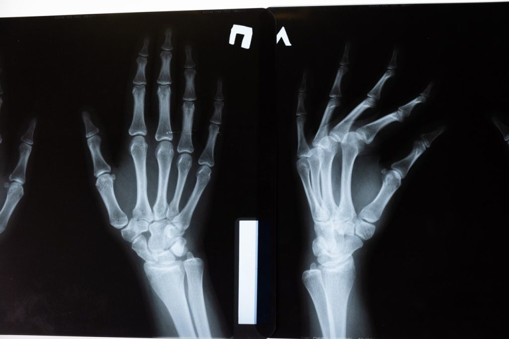
Portable ultrasound devices could provide an alternative to x-ray machines for diagnosing forearm fractures in children, which could alleviate waiting times for families in hospital emergency departments (ED).

Griffith University researchers Professor Robert Ware and Senior Lecturer Peter Snelling compared functional outcomes in children given an ultrasound and those who received an x-ray on a suspected distal forearm fracture. Dr Snelling said the ultrasounds were performed by nurses, physiotherapists and emergency physicians at four south-east Queensland hospitals.

“They treated 270 children, aged between five and 15 years, during the randomised trial, which included a check-up 28 days later and another check-in at eight weeks,” Dr Snelling said. “The findings show the majority of children had similar recoveries and returned to full physical function.”

Less than one-third of children who were given an ultrasound needed a follow-up x-ray and care at an orthopaedic clinic. Those who didn’t have a buckle fracture or fractured arm were discharged from hospital without the need for further imaging.

Professor Ware said children who had an ultrasound initially had fewer x-rays, and shorter stays in the ED. “Families were also more satisfied with the treatment they received,” he said. “The results are promising and have wider implications beyond in hospital diagnosis and follow up care.

“By using a bedside ultrasound, this frees up the x-ray machine for patients who really need it and can potentially be a cost-cutting measure for hospitals as they reduce the number of x-rays without comprising the safety of patients.

“It also would be extremely beneficial in rural or remote areas eliminating the need for children and their families to travel to a larger hospital for an x-ray.”

Source: Griffith University

Amino Acid Supplementation after Fracture Surgery Reduces Complications

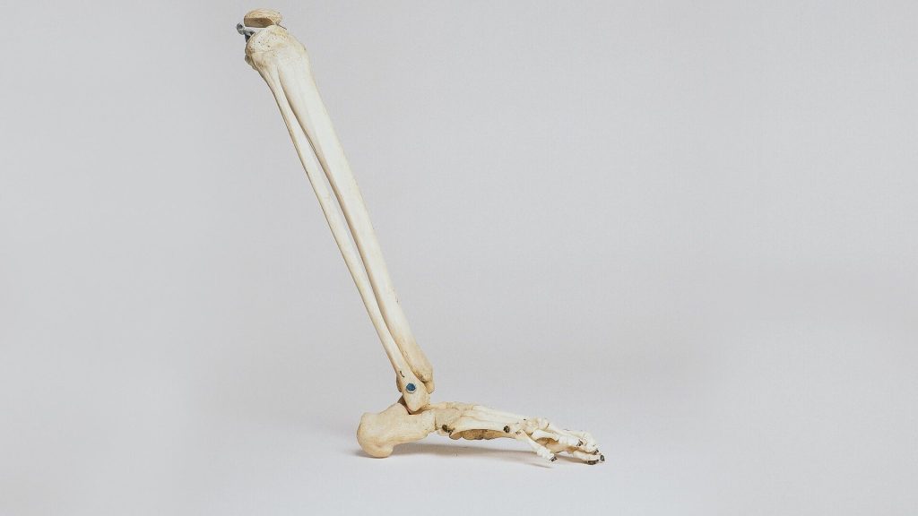
Lower leg bones
Photo by Nino Liverani on Unsplash

By adding conditionally essential amino acids (CEAA) to the diets of patients recovering from fracture fixation surgery, complications are reduced and skeletal muscle wasting is prevented, concludes a study published in The Journal of Bone & Joint Surgery.

The study supports the use of widely available CEAA supplements to promote recovery and preserve function in patients undergoing surgery for repair of major fractures. “Our results suggest that this inexpensive, low-risk intervention has considerable potential to improve outcomes after fracture fixation,” according to the report by Michael Willey, MD, and colleagues of University of Iowa Hospital and Clinics, Iowa City.

The study included 400 patients undergoing operative fixation of fractures in the limbs and/or pelvis at the researchers’ trauma centre. In equal numbers and stratified by fracture severity, patients were randomises to either standard postoperative nutrition or standard nutrition plus CEAA supplementation.

CEAAs are termed “conditionally essential” because the body doesn’t usually require them. However, during times of illness or stress, the need for these conditional amino acids increases dramatically. Previous studies have reported that CEAA supplementation can improve wound-healing and other outcomes in patients with a variety of conditions, including postoperative recovery. In the new trial, patients assigned to the CEAA group received a standard supplement that included arginine, leucine, and glutamine.

At follow-up, the overall complication rate was significantly lower for patients who received CEAA supplementation (30.5%) compared with those who did not receive CEAA (43.8%). The CEAA group also had a lower rate of nonunion (5.1 vs 13.2%, respectively). Some other types of complications, including surgical-site infections, were similar between groups.

Patients who undergo operative fracture fixation are at risk of skeletal muscle wasting, which often results in weight loss as a result of reduced muscle mass. In the new study, patients receiving CEAA supplements had little or no change in fat-free body mass. In contrast, patients receiving standard nutrition had a 1kg reduction in fat-free mass at 6 weeks postoperatively, which took until 12 weeks to return to normal.

An unexpected finding was a sharply reduced mortality rate in the CEAA group (0.5% compared to 4.1 % for the control). Although the authors could not explain the lower risk of death in patients receiving CEAA, they suggest it might result from “unidentified confounding factors.”

Despite advances in surgical techniques, trauma patients undergoing operative fixation of extremity and pelvic fractures remain at risk of complications and prolonged loss of function. “Malnutrition is a potentially modifiable risk factor for mortality, fracture nonunion, wound complications, and increased length of stay,” the authors wrote.

CEAA supplementation therefore appears to be a simple, risk-free, and inexpensive means of promoting good nutrition after fracture fixation surgery. Controlling for other factors, the relative risk of complications is about 40% lower in patients receiving CEAA, with no reduction in fat-free mass during the early weeks of recovery. The researchers concluded: “This study will serve as the foundation for multicentre [randomised controlled trials] that are designed to assess the impact of CEAA nutrition supplementation in reducing complications and loss of functional muscle mass in high-risk populations.”

Source: EurekAlert!

Regenerating Bone with Messenger RNA

Photo by Cottonbro on Pexels

Researchers have developed new way to get bone to regenerate with messenger RNA, which promises to be cheaper and less expensive while having fewer side effects than the current treatment.

Although fractures normally heal, bone will not regenerate under several circumstances. When bone does not regenerate, major clinical problems could result, including amputation.

One treatment is recombinant human bone morphogenetic protein-2, or BMP-2. However, it is expensive and only moderately effective. It also produces side effects, which can be severe.

Researchers at Mayo Clinic, along with colleagues in the Netherlands and Germany, may have a viable, less risky alternative: messenger RNA. 

A study conducted on rats and published in Science Advancesshows that messenger RNA can be used at low doses to regenerate bone – and without side effects. The resulting new bone quality and biomechanical properties are also superior to that of BMP-2. Additionally, messenger RNA is a good choice for bone regeneration because it may not need repeat administrations.

Human bone develops in one of two ways: direct formation of bone cells from mesenchymal progenitor cells, or through endochondral ossification, in which cartilage forms first and then converts to bone. The BMP-2 therapy uses the former method, and the messenger RNA approach uses the latter. In general, the researchers say their work proves that this method “can heal large, critical-sized, segmental osseous defects of long bones in a superior fashion to its recombinant protein counterpart.”

Further studies are required in larger animals than rats before any translation can be considered for clinical trials.

Source: Mayo Clinic

New Biomaterial Produced from Frog Skin and Fish Scales

Photo by Robert Zunikoff on Unsplash

Researchers at Nanyang Technological University, Singapore (NTU Singapore) have developed a new biomaterial made entirely from discarded bullfrog skin and fish scales that could help in bone repair.

The porous biomaterial, which contains the same compounds that are predominant in bones, acts as a scaffold for osteoblasts, or bone-forming cells, to adhere to and multiply, leading to new bone formation. Bone-forming cells successfully latched onto the biomaterial and started growing, and it was found to have a low inflammatory risk.

This kind of scaffold could help regenerate bone tissue lost to disease or injury, such as jaw defects from trauma or cancer surgery. It could also assist bone growth around surgical implants such as dental implants.

The current standard practice of using a patient’s own tissues means extra surgery is needed for bone extraction. The biomaterial used, frog skin and fish scales, are a significant waste stream produced by Singapore’s aquaculture industry and using them helps repurpose this waste.

‘Waste-to-resource’

“We took the ‘waste-to-resource’ approach in our study and turned discards into a high-value material with biomedical applications, closing the waste loop in the process,” said Dalton Tay, Assistant Professor, Nanyang Technological University. “Our lab studies showed that the biomaterial we have engineered could be a promising option that helps with bone repair. The potential for this biomaterial is very broad, ranging from repairing bone defects due to injury or ageing, to dental applications for aesthetics. Our research builds on NTU’s body of work in the area of sustainability and is in line with Singapore’s circular economy approach towards a zero-waste nation.”

To make the biomaterial, the team first extracted Type 1 tropocollagen (many molecules of which form collagen fibres) from the discarded skins of the American bullfrog and hydroxyapatite (a calcium-phosphate compound) from the scales of snakehead fish, commonly known as the Toman fish.

Collagen and hydroxyapatite (HA) are two predominant components found in bones, thus conferring on the biomaterial a structure, composition, and ability to promote cell attachment similar to bone, as well as toughness.

The scientists removed all impurities from the bullfrog skin, then blended it to form a thick collagenous paste that is diluted with water, from which collagen was extracted. “Using this approach, we were able to obtain the highest ever reported yield of collagen of approximately 70 per cent from frog skin, thus making this approach commercially viable,” said Asst Prof Tay, who is also from the NTU School of Biological Sciences (SBS).

HA was harvested from discarded fish scales through calcination – a purification process that requires high heat – to remove the organic matter, and then air-dried.

The biomaterial was synthesised by adding HA powder to the extracted collagen, then cast into a mould to make a 3D porous scaffold — a two-week process which the team believes can be shortened.

Testing the biomaterial

To assess the biological performance of the porous biomaterial scaffold for bone repair, the scientists seeded bone-forming cells onto the scaffold.

The cells proliferated, and after a week, the cells were uniformly distributed across the scaffold – an indicator that the scaffold could promote proper cellular activities and eventually lead to tissue formation. The scientists also found that the presence of HA in the biomaterial significantly enhanced bone formation.

The biomaterial was also tested for its tendency to cause an inflammatory response, which is common after a biomaterial is implanted in the body.

Using real-time polymerase chain reaction, the scientists found that the expression of pro-inflammatory genes in human immune cells exposed to the biomaterial stayed “relatively modest” compared to a control exposed to endotoxins, a compound known to stimulate immune response, said Asst Prof Tay.

For instance, the expression of the gene IL6 in the biomaterial group was negligible and at least 50 times lower than that of the endotoxins-exposed immune cells. This suggests that the risk of the NTU-developed biomaterial to trigger an excessive acute inflammatory response is low.

The team is now further evaluating the long-term safety and efficacy of the biomaterial as dental products. Further research would involve studying how the body responds to this biomaterial in the long term, as well its use in other applications such as skin wounds, along with further development of the waste-to-resource pipeline.

A preprint copy of the article is available as a PDF for download.

Source: Nanyang Technical University