Tag: ferroptosis

A New Way to Kill Cancer Cells via Ferroptosis

Human colon cancer cells. Credit: National Cancer Institute

In a first, a team in Germany has produced a substance capable of sending cancer cells into ferroptosis, a form of cell death discovered only in recent years. This could pave the way for the development of new drugs.

Conventional cancer drugs work by triggering apoptosis, that is programmed cell death, in tumour cells. However, tumour cells have the ability to develop strategies to escape apoptosis, rendering the drugs ineffective. In the journal Angewandte Chemie, a research team from Ruhr University Bochum, Germany, describes a new mechanism of action that kills cancer cells through ferroptosis. Ferroptosis is another form of programmed cell death that wasn’t discovered until the 2010s. The Bochum group synthesised a metal complex, demonstrated its effectiveness in cell cultures and on microtumours and identified the chemical processes underlying the mechanism of action.

Two types of programmed cell death

In programmed cell death, certain signaling molecules initiate a kind of suicide program to cause cells to die in a controlled manner. This is an essential step to eliminate damaged cells or to control the number of cells in certain tissues, for example. Apoptosis has long been known as a mechanism for programmed cell death. Ferroptosis is another mechanism that has recently been discovered which, in contrast to other cell death mechanisms, is characterised by the accumulation of lipid peroxides. This process is typically catalysed by iron which is where the name ferroptosis derives from.

“Searching for an alternative to the mechanism of action of conventional chemotherapeutic agents, we specifically looked for a substance capable of triggering ferroptosis,” explains Johannes Karges. His group synthesized a cobalt-containing metal complex that accumulates in the mitochondria of cells and generates reactive oxygen species, more precisely hydroxide radicals. These radicals attack polyunsaturated fatty acids, resulting in the formation of large quantities of lipid peroxides, which in turn trigger ferroptosis. The team was thus the first to produce a cobalt complex designed to specifically trigger ferroptosis.

Effectiveness demonstrated on artificial microtumours

The researchers from Bochum used a variety of cancer cell lines to show that the cobalt complex induces ferroptosis in tumour cells. On top of that, the substance slowed down the growth of artificially produced microtumours .

“We are confident that the development of metal complexes that trigger ferroptosis is a promising new approach for cancer treatment,” as Johannes Karges sums up the research, adding: “However, there’s still a long way to go before our studies result in a drug.” The metal complex must first prove effective in animal studies and clinical trials. What’s more, the substance doesn’t currently selectively target tumour cells, but would also attack healthy cells. This means that researchers must first find a way to package the cobalt complex in such a way that it damages nothing but tumour cells.

Source: Ruhr-University Bochum

Possible Cause of COVID Arrhythmias Discovered

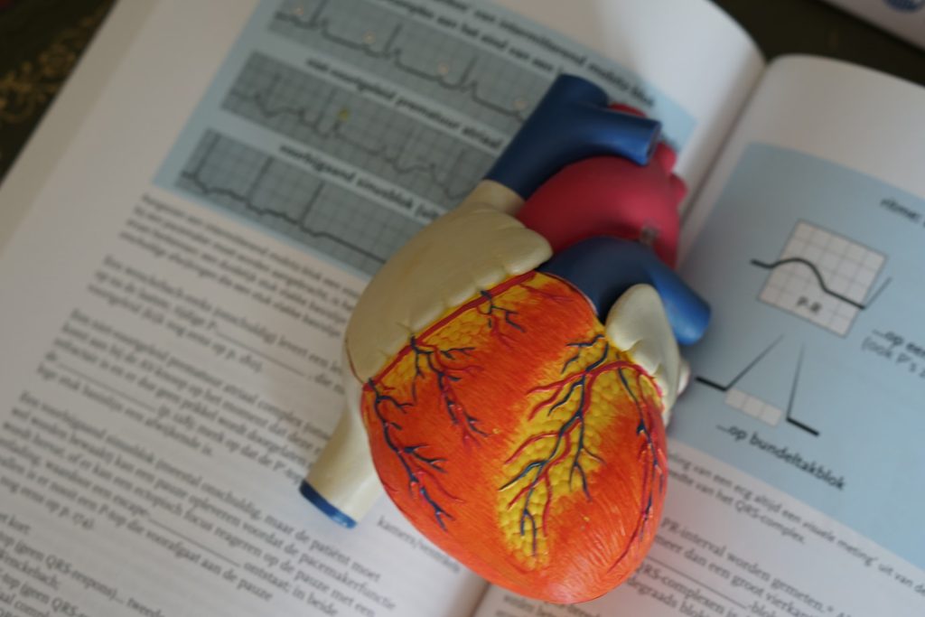
Anatomical model of a human heart
Photo by Robina Weermeijer on Unsplash

The SARS-CoV-2 virus can infect cardiac pacemaker cells, causing the cells to undergo self-destruct by ferroptosis according to a preclinical study reported in Circulation Research. This may explain the heart arrhythmias that are commonly observed in COVID patients.

In the study, the researchers used an animal model as well as human stem cell-derived pacemaker cells to show that SARS-CoV-2 can readily infect pacemaker cells and trigger a process called ferroptosis, where cells self-destruct, releasing damaging reactive oxygen molecules.

“This is a surprising and apparently unique vulnerability of these cells — we looked at a variety of other human cell types that can be infected by SARS-CoV-2, including even heart muscle cells, but found signs of ferroptosis only in the pacemaker cells,” said study co-senior author Professor Shuibing Chen.

Arrhythmias, including tachycardia and bradycardia, has been observed in some COVID patients, and multiple studies link these arrhythmias to worse COVID outcomes. But how the coronavirus caused these remained unclear.

In the new study, the researchers examined golden hamsters (one of the only lab animals that reliably develops COVID-like signs from SARS-CoV-2 infection) and found evidence that following nasal exposure, the virus can infect the sinoatrial node, which is the natural cardiac pacemaker.

The researchers then induced human embryonic stem cells to mature into cells closely resembling sinoatrial node cells. They showed that these induced human pacemaker cells can be infected by SARS-CoV-2 as they express ACE2 receptors. Large increases in inflammatory immune gene activity were also seen in the infected cells.

The team’s most surprising finding, however, was that the pacemaker cells, in response to the stress of infection, showed clear signs of a cellular self-destruct process called ferroptosis, which involves accumulation of iron and the runaway production of reactive oxygen molecules. The scientists were able to reverse these signs in the cells using compounds that are known to bind iron and inhibit ferroptosis.

“This finding suggests that some of the cardiac arrhythmias detected in COVID patients could be caused by ferroptosis damage to the sinoatrial node,” said co-senior author Dr Robert Schwartz

While COVID patients could in principle be treated with ferroptosis inhibitors specifically to protect sinoatrial node cells, antiviral drugs that block the effects of SARS-CoV-2 infection in all cell types would be preferable, the researchers said.

The researchers plan to continue to use their cell and animal models to investigate sinoatrial node damage in COVID and other settings.

“There are other human sinoatrial arrhythmia syndromes we could model with our platform,” said co-senior author Dr. Todd Evans. “And, although physicians currently can use an artificial electronic pacemaker to replace the function of a damaged sinoatrial node, there’s the potential here to use sinoatrial cells such as we’ve developed as an alternative, cell-based pacemaker therapy.”

Source: Weill Cornell Medicine