Tag: female sex hormones

The Risks of Various Menopausal Hormone Treatments Vary

Photo by cottonbro studio:

Researchers have analysed the effects of seven different hormone treatments for menopausal symptoms, and the risk of blood clots, stroke and heart attack. The risks differ depending on the active substance and how the medicine is taken, according to the study findings which appear in the BMJ. In this world’s largest and most comprehensive study of currently prescribed hormonal substances, researchers analysed the risks for one million women aged 50–58.

“There is concern among women that menopausal hormone therapy increases the risk of cardiovascular disease. This concern is based on older studies conducted more than 20 years ago that only looked at one type of treatment. Since then, many new preparations have been introduced and our study shows that the previous conclusions do not apply to all types of treatments,” says Therese Johansson, postdoctoral researcher and lead author of the study, which was part of her thesis at Uppsala University.

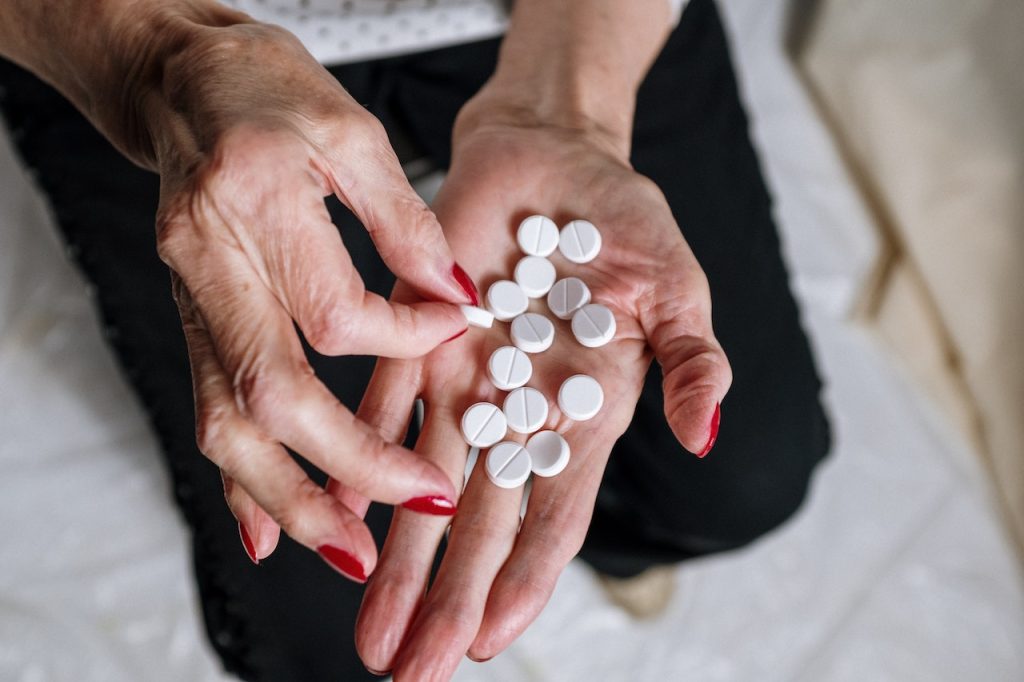
To counteract the health effects of menopause, such as hot flashes and osteoporosis, women may be prescribed hormone replacement therapy which consists of hormones or hormone-like substances.

Treatment available since the 1970s

In Sweden alone, hundreds of thousands of women currently use hormone replacement therapy and this type of treatment has been available since the 1970s. At that time, there was only one type of hormone replacement therapy and when a major study in the 1990s showed that it increased the risk of cardiovascular disease, its use rapidly declined. Since then, new preparations have entered the market, and following this, the use of hormone replacement therapy in connection with menopause has increased significantly in recent years.

In the new study, the researchers looked at seven different types of currently used hormone replacement treatments, administered via tablets, hormone patches or hormone-releasing IUDs. The study is based on all prescriptions for hormone replacement therapy in Sweden from 2007 to 2020 and covers nearly one million women aged 50 to 58. The women were monitored for two years after starting hormone replacement therapy. The risk of blood clots and cardiovascular disease was compared between women who had and had not collected a prescription medicine for hormone replacement therapy.

Different therapies, different risks

The results show clearly that the risks of hormone replacement therapy vary depending on the type of treatment.

For example, the synthetic hormone tibolone, which mimics the effects of the body’s natural hormones, was linked to an increased risk of both heart attack and stroke, but not to an increased risk of blood clots. The risk of heart attack or stroke due to tibolone is estimated at one in a thousand women.

Combined preparations containing both oestrogen and progesterone instead increase the risk of blood clots, including deep vein thrombosis and pulmonary embolism. The researchers estimate that the risk of deep vein thrombosis resulting from this combined preparation is about 7000 women per year.

“It is important that both doctors and women are aware of the risks of menopausal hormone therapy and, in particular, that the existing drugs carry different risks of blood clots and cardiovascular disease. Tibolone in particular was associated with an increased risk of stroke and heart attack. Tibolone is used in Europe but is not approved in countries such as the United States. We hope that our study will lead to the drug being withdrawn from use here as well,” says Åsa Johansson, research group leader at Uppsala University and SciLifeLab, and the study’s senior author.

During the period of the study, 2007–2020, a roughly 50% increase hormone patch use was observed, and these preparations were not linked to the same higher risk. The increased use of safer alternatives, such as patches, is an important step forward in reducing the risk of cardiovascular disease among menopausal women.

Identify individual increased risk

“The next step in our research will be to develop strategies to identify which women are at increased risk of certain diseases in connection with using hormonal drugs. In this way, we can guide patients to the most appropriate medicine for each individual and drastically reduce the number of side effects,” Åsa Johansson says.

Source: Uppsala University

How Oestrogen can Trigger Nerve Impulses in Milliseconds

Photo by Julian Jagtenberg on Pexels

Oestrogen, the major female ovarian hormone, can trigger nerve impulses within milliseconds to regulate a variety of physiological processes. At Baylor College of Medicine, Louisiana State University and collaborating institutions, researchers discovered that oestrogen’s fast actions are mediated by the coupling of the oestrogen receptor-alpha (ER-alpha) with an ion channel protein called Clic1.

Clic1 controls the fast flux of electrically charged chloride ions through the cell membrane, which neurons use for receiving, conducting and transmitting signals. The researchers propose that interacting with the ER-alpha-Clic1 complex enables oestrogen to trigger fast neuronal responses through Clic1 ion currents. The study appeared in Science Advances.

“Oestrogen can act in the brain to regulate a variety of physiological processes, including female fertility, sexual behaviours, mood, reward, stress response, cognition, cardiovascular activities and body weight balance. Many of these functions are mediated by oestrogen binding to one of its receptors, ER-alpha,” said co-corresponding author Dr Yong Xu, professor of pediatrics – nutrition and associate director for basic sciences at the USDA/ARS Children’s Nutrition Research Center at Baylor. 

Fast and slow

It is well known that, upon stimulation by oestrogen, ER-alpha enters the cell nucleus where it mediates the transcription of genes. This classical mode of action as a nuclear receptor takes minutes to hours.

“Oestrogen also can change the firing activity of neurons in a manner of milliseconds, but it was not clear how this happens,” Xu said. “In this case, it did not make sense to us that the minutes-long nuclear receptor function of ER-alpha was involved in such a rapid action. We explored the possibility that ion channels, proteins in the cell membrane that regulate the fast flux of ions, mediated oestrogen’s quick actions.”

In the current study, working with cell lines and animal models, the team searched for cell membrane proteins that interact with ER-alpha. They found that protein Clic1, for chloride intracellular channel protein-1, can physically interact with ER-alpha. Clic1has been implicated in the regulation of neuronal excitability, so the researchers considered it a candidate to mediate oestrogen-triggered fast actions.

“We discovered that oestrogen enhances Clic1-mediated ion currents, and eliminating oestrogen reduced such currents,” Xu said. “In addition, Clic1 currents are required for oestrogen to induce rapid responses in neurons. Also, disrupting the Clic1 gene in animal models blunted oestrogen regulation of female body weight balance.”

The findings suggest that other nuclear receptors could also interact with ion channels, a possibility the researchers look forward to studying in the future.

“This study was conducted with female mice. However, Clic1 is also present in males. We are interested in investigating its role in male physiology,” Xu said.

Chloride channels are not as well studied as other ion channels, such as potassium, sodium or calcium channels. “We are among the first to study the role Clic1 plays in female physiology,” Xu said. “We hope that our findings will inspire other groups in the field to expand these promising investigations.”

Source: Baylor College of Medicine

Oestrogens are Implicated in More than Just Breast Cancers

Photo by National Cancer Institute on Unsplash

Oestrogens are known to drive tumour growth in breast cancer cells that carry its receptors, but a new study by Duke Cancer Institute researchers unexpectedly finds that oestrogens play a role in fuelling the growth of breast cancers without the receptors, as well as numerous other cancers.

Writing in the journal Science Advances, the researchers describe how oestrogens not only decrease the ability of the immune system to attack tumours, but also reduce the effectiveness of immunotherapies that are used to treat many cancers, notably triple-negative breast cancers. Triple-negative breast cancers are an aggressive form of disease that are negative for oestrogen, progesterone, and the HER2 receptor proteins.

Informed by retrospective analysis of patient data and experiments in mice, the researchers found that anti-oestrogen drugs reversed the effects of oestrogens, restoring potency to immunotherapies.

“The treatment for triple-negative breast cancer has been greatly improved with the advent of immunotherapy,” said senior author Donald McDonnell, PhD, professor at Duke University School of Medicine.

“Developing ways to increase the anti-cancer activity of immunotherapies is a primary goal of our research,” McDonnell said. “Here we have found a simple way bolster the effectiveness of immunotherapy for this type of breast cancer and the benefit was even seen in other cancers, including melanoma and colon cancers.”

McDonnell and colleagues, including lead author Sandeep Artham, a postdoctoral associate in the McDonnell lab, focused on a type of white blood cell called eosinophils, which are typically activated during allergic reactions and inflammatory diseases.

Eosinophils have recently been identified as important in tumours, and a phenomenon called tumour associated tissue eosinophilia, or TATE, is associated with better outcomes among patients with multiple types of cancer, including colon, oesophageal, gastric, oral, melanoma and liver cancers.

In their studies, the Duke team described how oestrogens decrease the number of eosinophils and TATE in mice. The hormone contributes to increased tumour growth in oestrogen receptor-negative breast cancer tumours and in melanoma tumours, which do not rely on oestrogen receptors for tumour growth.

Conversely, anti-oestrogen therapies inhibited oestrogen receptor signalling and enhanced the efficacy of immunotherapies, slowing tumour growth.

“These findings highlight the importance of oestrogen-receptor signalling as a regulator of eosinophil biology and TATE and highlight the potential near-term clinical application of anti-oestrogen drugs to increase the benefits of immunotherapies in multiple tumour types,” McDonnell said.

He said clinical trials are being planned using an investigational anti-oestrogen drug called lasofoxifene among patients with triple-negative breast cancers.

Source: Duke University Medical Center

Menstrual Cycle Phases Linked to Increased Injury Risk for Female Athletes

Photo by Ashley Williams

Football players in England’s top-tier WSL were six times more likely to experience a muscle injury in the days leading up to their period compared to when they were on their period, according to a new study published in Medicine & Science in Sports & Exercise.

This the first prospective longitudinal study monitoring menstrual cycles alongside injuries in female footballers. The findings suggest there could be increased injury risk windows at particular times in the cycle.

Despite being a relatively small sample size, the data demonstrates the need to consider the menstrual cycle in elite sports, to reduce injury risk and to support the wellbeing of athletes.

Menstrual cycle symptoms are common and around two thirds of elite athletes feel that these can have negative impacts on their performance. There has been little previous research tracking injuries alongside the menstrual cycle in female sport, despite much speculation and anecdotal evidence suggesting that there may be some key times for increased injury risk. Given the increased professionalism, interest, growth, and investment in women’s sport, the authors say further research in this area is needed.

In this study, researchers at UCL and the University of Bath recorded time-loss injuries and menstrual cycle data for elite female football players across three seasons. All of the players were based at one Women’s Super League (WSL) club, the top tier of women’s football in England. During the study they tracked 593 cycles across 13 390 days, in which time 26 players experienced 74 injuries.

The authors divided each cycle into four main phases in their study. Each phase comes with assumed hormonal changes that have the potential to influence different aspects of a woman’s health and wellbeing.

Ally Barlow, first author of the study from the University of Bath and a physiotherapist at the WSL club, said: “We have been tracking player’s menstrual cycles for a number of seasons to observe trends in terms of symptoms and cycle characteristics. We were interested to learn more about the potential association between injury risk across the menstrual cycle. This study set out to collect specific scientific data so that we could learn more about the menstrual cycle and player’s injury risk.”

Analysis of the data found that players were six times more likely in the pre-menstrual phase (oestrogen and progesterone decrease to bring about the onset of menstruation) and five times more likely in the early-mid luteal phase (after ovulation when both oestrogen and progesterone are assumed to increase and remain high) to experience a muscle injury, compared to when they were in the menstrual phase.

Dr Georgie Bruinvels, senior author of the study from UCL Surgery & Interventional Science and the Institute of Sport, Exercise & Health (ISEH), said: “While these results must be viewed with caution, this data highlights a need to investigate this area further. Given the growth of women’s sport it’s an exciting time to be working in female physiology, but there are a number of known challenges when conducting research with female athletes, in part explaining why there is such a significant sex data gap.

“Conducting large-scale research is complex but must be prioritised to best support female athletes, and we hope studies like this will pave the way for this. Every woman has their own unique physiology, so it’s crucial to support and empower them in the right ways. If future research demonstrates that there are risk windows for certain injury types, we should be proactive in mitigating these risks to enable female athletes to exercise and compete on any given day.”

The authors emphasise that further data collected in a standardised manner is needed before the sports science community can start to look for biological explanations for this increased injury risk.

Dr Jo Blodgett, an author of the study from UCL Surgery & Interventional Science and the Institute of Sport, Exercise & Health (ISEH), said: “Though our sample size for this research was relatively small, we observed clear links between cycle phase and injury prevalence, and the size of the association – six times higher in the premenstrual phase and five times higher in the early-mid luteal phase for muscular injuries – was quite large.

“To better understand the variability in injury risk across the cycle we need more players and teams to continually track injury incidence, menstrual cycle and symptoms in a standardised manner. At the elite level, injuries to your squad can mean the difference between winning and losing, the difference between being crowned champions and runners-up. But perhaps more importantly, it means pain and suffering for players that could perhaps be avoided with better player-centred support.”

Source: University College London

The Resilience of Females’ Kidneys is Down to Hormones

Photo by Robina Weermeijer on Unsplash

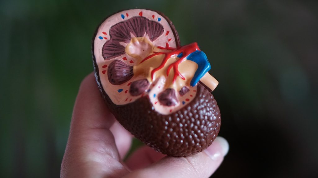
Females’ kidneys are known to be more resilient to disease and injury, so what about them can be applied to treat males’ kidneys? A new USC Stem Cell-led study published in Developmental Cell describes not only how sex hormones drive differences in male and female mouse kidneys, but also how lowering testosterone can “feminise” this organ and improve its resilience.

“By exploring how differences emerge in male and female kidneys during development, we can better understand how to address sex-related health disparities for patients with kidney diseases,” said Professor Andy McMahon, the study’s corresponding author, and the director of the Eli and Edythe Broad Center for Regenerative Medicine and Stem Cell Research at the Keck School of Medicine of USC.

First authors Lingyun “Ivy” Xiong and Jing Liu from the McMahon Lab and their collaborators identified more than 1000 genes with different levels of activity in male and female mouse kidneys, in a study supported by the National Institutes of Health. The differences were most evident in the section of the kidney’s filtering unit known as the proximal tubule, responsible for reabsorbing most of the nutrients such as glucose and amino acids back into the blood stream. Most of these sex differences in gene activity emerged as the mice entered puberty and became even more pronounced as they reached sexual maturity.

Because female kidneys tend to fare better in the face of disease or injury, the researchers were interested how the gene activity of kidneys becomes “feminised” or “masculinised” – and testosterone appeared to be the biggest culprit.

To feminize the kidneys of male mice, two strategies worked equally well: castrating males before puberty and thus lowering their natural testosterone levels, or removing the cellular sensors known as androgen receptors that respond to male sex hormones.

Intriguingly, three months of calorie restriction – which is an indirect way to lower testosterone – produced a similar effect. Accordingly, calorie restriction has already been shown to mitigate certain types of kidney injuries in mice.

To re-masculinize the kidneys of the castrated males, the researchers only needed to inject testosterone. Similarly, testosterone injection masculinised the kidneys of females who had their ovaries removed before puberty.

The scientists performed some similar experiments with mouse livers. Although this organ also displays sex-related differences, the hormones and underlying factors driving these differences are very different than those at play in the kidney. This suggests that these sex-related organ differences emerged independently during evolution.

To test whether the same genes are involved in sex-related kidney differences in humans, the scientists analysed a limited number of male and female donor kidneys and biopsies. When it came to genes that differed in their activity between the sexes, there was a modest overlap of the human genes with the mouse genes.

“There is much more work to be done in studying sex-related differences in normal human kidneys,” said McMahon. “Given the divergent outcomes for male and female patients with kidney disease and injury, this line of inquiry is important for making progress toward eventually closing the gap on these sex-related health disparities.”

Source: Keck School of Medicine of USC

    Oestriol Shown to Reverse Cortex Damage from MS in Mouse Model

    Source: Pixabay CC0

    Treating a mouse model of multiple sclerosis with the pregnancy hormone oestriol reversed the breakdown of myelin in the brain’s cortex, a key region affected in multiple sclerosis, according to a new UCLA Health study.

    In multiple sclerosis, inflammation spurs the immune system to strip away the protective myelin coating around nerve fibres in the brain’s cortex, hampering electrical signals sent and received by the brain. Atrophy of the cortex in MS patients is associated with permanent worsening of disability, such as cognitive decline, visual impairment, weakness and sensory loss.

    No currently available treatments for MS can repair damage to myelin. Instead, these treatments target inflammation to reduce symptom flare-ups and new nerve tissue scarring. Previous UCLA-led research found that oestriol, a type of oestrogen hormone produced in pregnancy, reduced brain atrophy and improved cognitive function in MS patients.

    In the new study, researchers treated a mouse model of MS with oestriol and found that it prevented brain atrophy and induced remyelination in the cortex, indicating that the treatment can repair damage caused by MS, rather than just slow the destruction of myelin.

    This is the first study to identify a treatment that could repair myelin in the cortex, undoing some of the damage caused by MS.

    Source: University of California – Los Angeles Health Sciences

    Intermittent Fasting does not Impact Female Sex Hormones

    Bathroom scale
    Photo by I Yunmai on Unsplash

    Intermittent fasting has been shown to be an effective way to lose weight, but critics have worried that the practice may have a negative impact on women’s reproductive hormones. Now, researchers bring new evidence to the table in a study published in Obesity.

    The researchers, led by Krista Varady, University of Illinois Chicago professor of nutrition, followed a group of pre- and post-menopausal obese women for a period of eight weeks on the ‘warrior diet’ method of intermittent fasting.

    The warrior diet prescribes a time-restricted feeding window of four hours per day, during which dieters can eat without counting calories before resuming a water fast until the next day.

    They measured the differences in hormone levels, obtained by analysing blood sample data, in groups of dieters who stuck to four- and six-hour feeding windows against a control group that followed no diet restrictions.

    Varady and her team found that levels of sex-binding globulin hormone, a protein that carries reproductive hormones throughout the body, was unchanged in the dieters after eight weeks. The same held true for both testosterone and androstenedione, a steroid hormone that the body uses to produce both testosterone and oestrogen.

    However, dehydroepiandrosterone or DHEA, a hormone that fertility clinics prescribe to improve ovarian function and egg quality, was significantly lower in both pre-menopausal and post-menopausal women at the end of the trial, dropping by about 14%.

    While the drop in DHEA levels was the most significant finding of the study, in both pre- and post-menopausal women, DHEA levels remained within the normal range by the end of the eight-week period.

    “This suggests that in pre-menopausal women, the minor drop in DHEA levels has to be weighed against the proven fertility benefits of lower body mass,” Varady said. “The drop in DHEA levels in post-menopausal women could be concerning because menopause already causes a dramatic drop in estrogen, and DHEA is a primary component of estrogen. However, a survey of the participants reported no negative side effects associated with low estrogen post-menopause, such as sexual dysfunction or skin changes.”

    As an added benefit, since high DHEA has been linked to breast cancer risk, Varady said a moderate drop in levels might be helpful in reducing that risk for both pre- and post-menopausal women.

    The study measured levels of oestradiol, oestrone and progesterone as well, but only in post-menopausal women, due to the changing levels of these hormones throughout pre-menopausal women’s menstrual cycles. Among post-menopausal women, there was no change in these hormones at the end of eight weeks.

    Women in both the four-hour and six-hour dieting groups experienced weight loss of 3% to 4% of their baseline weight throughout the course of the study, compared with the control group, which had almost no weight loss. The dieters also saw a drop in insulin resistance and in biomarkers of oxidative stress.

    Perimenopausal women, who are typically in their 40s, were excluded from the study.

    Still, Varady said, “I think this is a great first step. We’ve observed thousands of pre- and post-menopausal women through different alternate-day fasting and time-restricted eating strategies. All it’s doing is making people eat less. By shortening that eating window, you’re just naturally cutting calories. Much of the negative information on intermittent fasting reported has come from studies on mice or rats. We need more studies to look at the effects of intermittent fasting on humans.”

    Source: University of Illinois Chicago