Tag: e. coli

Steroid Drugs Used for HRT could be Repurposed to Combat E. coli and MRSA

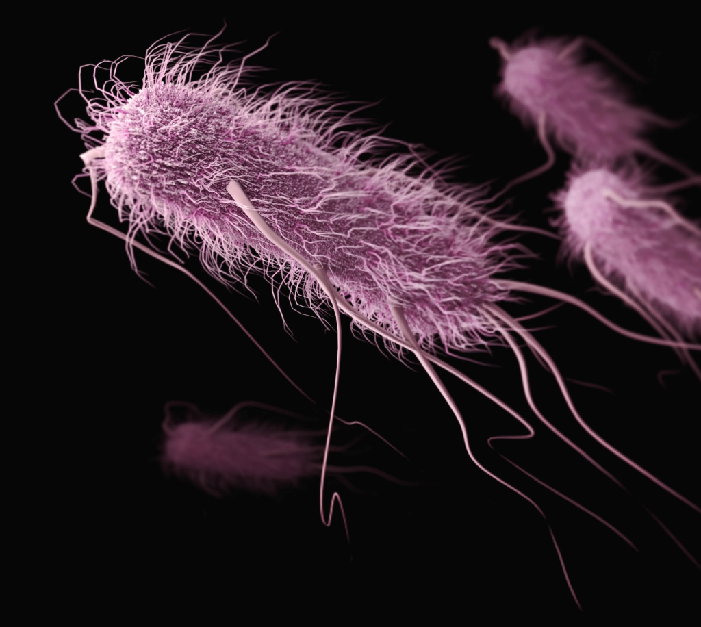
Methicillin resistant Staphylococcus aureus (MRSA) – Credit: CDC

Researchers from the University of Kent’s School of Biosciences have combined computational and microbiology laboratory approaches to identify existing drugs that can be repurposed to combat antibiotic-resistant bacterial infections, instead of developing new ones.

This research, which has been published in the Journal of Infectious Diseases, revealed that a class of steroid drugs currently used in hormone replacement therapy (HRT) can also stop the growth of antibiotic-resistant E. coli and effectively kill MRSA.

These drugs are particularly good at binding to a protein complex, cytochrome bd, which is important for the growth and survival of a range of disease-causing bacterial species. The researchers made an in silico screening for drugs that could inhibit bd activity, and identified quinestrol, ethinyl estradiol and mestranol, then evaluated their effectiveness in vitro.

The steroid drugs ethinyl estradiol and quinestrol inhibited E. coli bd-I activity. The IC50 of quinestrol for inhibiting oxygen consumption in E. coli bd-I-only membranes as 0.2µg/mL, although residual activity remained at around 20% at higher concentrations Quinestrol exhibited potent bactericidal effects against S. aureus but not E. coli.

It is expected that steroids may provide an alternative to conventional antibiotics that are becoming increasingly ineffective.

Dr Mark Shepherd, Reader in Microbial Biochemistry at Kent and the corresponding author on the paper, said: “These exciting developments will help to advance research into new antimicrobials, and we are enthusiastic to use our powerful experimental approach to discover drugs that can target other bacterial proteins and combat a wide range of antibiotic-resistant infections.”

Source: University of Kent

Antibiotic-resistant Bacteria can Persist in the Body for Years

Photo by CDC on Unsplash

People with pre-existing conditions in particular can carry resistant germs and suffer from repeated infections for years, according to a study in the scientific journal Nature Communications.

Some bacteria have developed the ability to break down beta-lactam antibiotics like penicillins and cephalosporins, making them ineffective.

Once a patient’s body has been colonised by these resistant bacteria, they can persist for a long time, reports Professor Sarah Tschudin Sutter’s research group at the Department of Clinical Research of the University of Basel and University Hospital Basel.

The researchers looked at a much longer period of time than previous studies and focused on older people with pre-existing conditions, analysing multiple samples taken from over 70 individuals over a period of ten years.

Their key question: whether and how resistant Klebsiella pneumoniae and Escherichia coli bacteria in the body change over this long period and how they differ in various parts of the body.

Recurring illness

DNA analysis indicates that the bacteria initially adapt quite quickly to the conditions in the colonized parts of the body, but undergo few genetic changes thereafter.

The resistant bacteria could still be detected in the patients up to nine years later.

“These patients not only repeatedly become ill themselves, they also act as a source of infection for other people — a reservoir for these pathogens,” says Dr Lisandra Aguilar Bultet, the study’s lead author.

“This is crucial information for choosing a treatment,” explains Professor Tschudin Sutter.

If someone has previously been infected with resistant bacteria and later requires another course of treatment because of a new infection, there is a risk that standard antibiotics will again fail to work.

Transmission of resistance

In addition, the researchers found that in some patients, bacterial strains of the same species, as well as of different species (specifically, Klebsiella pneumoniae and Escherichia coli), share identical genetic mechanisms of resistance through what are known as mobile genetic elements (such as plasmids). The most likely explanation is that they have transmitted these elements to each other.

Hospitals use special protective measures if a patient has been infected with resistant bacteria in the past.

In everyday life, however, it is difficult to reduce the risk of pathogen transmission.

These findings about the bacterial genetic diversity expected to develop in individual patients over time are a valuable basis for future studies to analyse factors found in both bacteria and patients that correlate with duration of colonisation and progression from colonisation to infection.

Source: University of Basel

Multidrug-resistant E. Coli Can Take Over the Gut

Photo by CDC on Unsplash

A study published in PLOS Biology reveals that different strains of Escherichia coli can outcompete one another to take over the gut. The researchers found that a particular strain, known as MDR ST131, can readily colonise new hosts, even if those hosts are already have commensal E.coli in their healthy gut.

The international team, led by experts at the University of Birmingham, used a mouse model to help understand why strains of E.coli that live in a healthy gut are rapidly overtaken of when challenged with a multi-drug resistant strain.

Lead author Professor Alan McNally, from the Institute of Microbiology and Infection at the University of Birmingham, commented: “Antibiotic resistance has been hailed as one of the biggest health problems of our time by the World Health Organisation. There are further problems looming unless we get a better understanding of what is happening so that further drug resistance can be halted in its tracks.

“Scientists have long questioned what makes certain types of E. coli successful multi-drug resistant pathogens. It seems that extra-intestinal pathogenic E. coli, which cause urinary tract and bloodstream infections, are particularly successful when it comes to developing resistance and are therefore especially tricky to treat. Our study provides evidence that certain types of E. coli are more prone to develop antibiotic resistance than others.”

According to previous research, multi-drug resistance alone is not sufficient to drive strains to complete dominance. This most recent study demonstrates that regardless of multi-drug resistant status, certain types of E.coli will outcompete others to live in the human gut.

The work was completed in parts. First, both multi-drug resistant and non-resistant gut-dwelling E. coli were found to easily colonise a mammalian gut. In a second part of the study, the multi-drug resistant strain was found to efficiently displace an already established gut-dwelling E. coli from the mouse intestinal tract. The study provided further details to demonstrate that multidrug resistant lineages of extraintestinal E. coli have particular genetic differences that appear to give them a competitive advantage.

Successful strains of E.coli need to be able to spread between individuals or from the environment into individual hosts. The new study demonstrates that a particular strain, known as MDR ST131, can readily colonise new hosts, even if those hosts are already have E. coli in their healthy gut.

Source: EurekAlert!

Ciprofloxacin-resistant E. coli Incidence Grows Despite Slashed Prescriptions

Photo by CDC on Unsplash

A US study found that, despite prescriptions for the antibiotic ciprofloxacin dropping by two-thirds between 2015 and 2021, the rates of ciprofloxacin-resistant E. coli bacteria circulating in the community did not decline.

In fact, a study of women over age 50 who had not taken any antibiotics for at least a year discovered that the incidence of gut-colonising ciprofloxacin-resistant E. coli actually increased. About 1 in 5 women in the study were affected.

Scientists at the University of Washington School of Medicine, Kaiser Permanente Washington Health Research Institute and Seattle Children’s Hospital conducted the study. Their findings appear in Communications Medicine.

Their results are consistent with theoretical models indicating that, once a drug-resistant form of E.coli emerges, it will continue to spread by taking up long-term residence in individuals’ gut microbiomes. E. coli is among an alarming number of disease-causing bacteria that have become resistant to several types of antibiotics. Resistance means that the antibiotics can’t kill the bacteria.

Pathogenic E. coli from the gut occasionally enters the urinary tract opening and causes infections. The female pelvic anatomy makes women more vulnerable to these mobile bacteria. Postmenopausal women are especially susceptible to severe, drug-resistant infection. Some drug-resistant E. coli infections are associated with considerable risk of hospitalization and death from sepsis.

Urinary tract infections from antibiotic-resistant E. coli can be frustrating to treat, even with third-generation cephalosporins, the newer types of antibiotics that are being prescribed more frequently for some populations of patients. Resistance to cephalosporins among ciprofloxacin-resistant E. coli also rose between 2015 and 2021.

Ciprofloxacin and similar drugs in its class were once the most prescribed antibiotic for urinary tract infections. In 2015, recommendations from the Centers for Disease Control and Prevention, Food and Drug Administration and Infectious Disease Society of America discouraged broad use of this class of drugs for uncomplicated urinary tract infections, partly due to rising resistance.

“However, it appears to be questionable whether a reduction in antibiotic use can be effective in reducing the rates of resistance in E. coli infections,” the research paper’s authors noted.

“Evidence from studies such as this one may be changing lots of paradigms on how to fight the rise in antibiotic resistance,” said physician scientist Dr. Evgeni V. Sokurenko, professor of microbiology at the University of Washington School of Medicine, who headed this latest research.

In the study, the scientists examined participants’ positive samples to determine which antibiotic-resistant strains of E. coli were present.

They found that the rate of a particularly virulent strain, ST1193, rose during the study period. Together with E. coli strain ST131-H30, these strains are the major causes of a global pandemic of multi-drug-resistant urinary tract infections among all women.

If ST1193 makes its home in more people’s guts, the situation could lead to more urinary tract infections with this more virulent strain, regardless of the curbing of fluoroquinolones prescriptions.

Another strain with a troubling increase in the participant samples was ST69, known to more frequently cause urinary tract infections in children.

tize discovering better ways to control drug-resistant E. coli’s ability to colonize the gut before it causes these infections, the authors wrote. They mentioned potential strategies of deploying probiotic bacteria and anti-bacterial viruses (bacteriophages).

The researchers added that these approaches might be offered to high-risk patients or deployed against the most clinically relevant strains. More investigation is needed on the epidemiology and ecology of antibiotic-resistant gut E. coli, they said, to help determine how these bacteria skillfully colonize human guts and how to target them most effectively to reduce antibiotic-resistant infections.

Source: University of Washington School of Medicine/UW Medicine

Novel Metal Complex Treatment Kills Antibiotic-resistant Bacteria

Photo by CDC on Unsplash

An innovative treatment paves the way for reducing antimicrobial resistance in the treatment of a deadly infection in chickens, according to a new study in Veterinary Microbiology. The ground-breaking study investigated the effectiveness of a novel metal-derived complex in treating Avian Pathogenic Escherichia coli (APEC), a serious respiratory infection of chickens which has become increasingly more resistant to antibiotics. A growing body of evidence indicates that the APEC could potentially spread to humans.

University of Surrey’s Professor Roberto La Ragione said: “Antimicrobial resistance is one of the biggest threats to human and animal health. Not being able to use antibiotics to treat an infection not only prolongs an illness and associated welfare issues, but also increases the likelihood of it spreading.

“Coronavirus demonstrated how easily a pandemic can happen, and the threat of another is looking more likely as antibiotics to treat simple bacterial infections are no longer working.”

To test the effectiveness of the metal complex, manganese carbonyl, researchers worked with the Greater Wax Moth larvae and APEC. Split into two groups, the first received manganese carbonyl, whilst the second, the controls, received either a phosphate-buffered saline (PBS) or dimethyl sulfoxide (DMSO). After four days, the survival rate for the larvae which received manganese carbonyl was between 56–75%, whereas in the control group, the survival rate was between 25–45% (PBS) and 19-45 per cent (DMSO), demonstrating the protective effect of the complex.

The test was repeated in chickens infected with APEC, who again received either manganese carbonyl or PBS. Bacterial shedding identified in the faeces of the chickens was significantly lower 24 hours post-treatment in those who received manganese carbonyl compared to the PBS control group, indicating bacterial killing induced by the compound. This is supported by caecal samples taken three days post-treatment which again found significantly fewer bacteria in those that received manganese carbonyl. Examination of tissue samples from the livers of the birds indicated no toxic effects from the metal compound, which was observed in the larvae.

Dr Jonathan Betts, a Research Fellow at the University of Surrey School of Veterinary Medicine, said:

“The development of alternatives to antibiotics is vital to safeguard our future health. Metal complexes such as manganese carbonyl could do this, as we have shown not only are they effective, but they are much cheaper to produce than traditional antibiotics.

“Discovering the effectiveness of manganese carbonyl in treating APEC is a monumental step forward in tackling antimicrobial resistance as it shows we don’t necessarily need more antibiotics; we just need to think more innovatively in developing treatments.”

The international research team also included the University of Surrey, the Animal and Plant Health Agency, the University of Connecticut, the University of Sheffield and Institut für Anorganische Chemie, Julius-Maximilians-Universität Würzburg.

This study was made possible by a BBSRC grant to Professor La Ragione and Professor Poole.

Source: University of Surrey

Pathogenic Bacteria Use A Sugar from Intestinal Mucus to Dig Themselves in

Source: CC0

A new study shows the sugar sialic acid, which makes up part of the protective intestinal mucus layer, fuels disease-causing bacteria in the gut. The findings, published in PNAS, suggest a potential treatment target for intestinal bacterial infections and a range of chronic diseases linked to gut bacteria, including inflammatory bowel disease (IBD), celiac disease, irritable bowel syndrome and short bowel syndrome.

The research by researchers at the University of British Columbia (UBC) and BC Children’s Hospital, used a mouse model of gut infections.

“Bacteria need to find a place in our intestines to take hold, establish and expand, and then they need to overcome all the different defences that normally protect our gut,” says Dr Bruce Vallance, a professor in the department of paediatrics at UBC and investigator at BC Children’s Hospital. “In the future, we can potentially target this sugar, or how pathogens sense it, to prevent clinically important disease.”

Inflammatory diseases such as IBD are on the rise in children, who are more susceptible to gut infections because of their immature immune systems. Dr Vallance and his team sought to understand what enables these bacterial pathogens to survive and expand inside our intestines.

For the study, the researchers examined Citrobacter rodentium, an intestinal bacterial pathogen of mice that’s used to model infections with human E. coli. The team discovered that the bacteria have genes involved in sialic acid consumption, and when these genes are removed, the bacteria’s growth is impaired.

Further investigation revealed that upon consuming the sugars, the bacteria produced two special virulence proteins that help the bacteria cross the colonic mucus layer and stick to the underlying epithelial cells. The findings reveal how the bacteria can change over time and actually worsen disease.

“You start off with IBD, your microbes change, they start digging their way into the cells lining your gut, causing more inflammation, and that may be one reason why IBD becomes chronic,” says Dr Vallance. “Specific nutrients such as sialic acid or other sugars might be the Achilles heels for them in terms of things you could target to remove dangerous bacteria from the intestine.”

Dr Vallance and his team are now examining the role other sugars in the gut may play in feeding pathogenic bacteria. They’re also looking for probiotics that could outcompete the dangerous bacteria, stealing the sugars away from them.

They also plan to explore potential interactions between resident and pathogenic bacteria. Pathogenic bacteria can’t access the sugars on their own and thus, some of the normally harmless resident bacteria must serve as accomplices.

“Basically, these accomplices cut the sugar off the mucus, and then either they hand it to the dangerous bacteria or the dangerous bacteria have come up with a way of stealing it from them,” he explains.

A better understanding of these interactions could provide new ways to block pathogenic bacteria, something Dr. Vallance says is urgently needed.

“In the past, our ancestors were constantly assaulted by dangerous bacteria,” says Dr. Vallance. “With the advent of more and more antibiotic resistance in bacteria, these bacterial infections are going to become a growing problem again. Without new antibiotics, we need to come up with novel ways to fight these bacteria, like starving them.”

Source: University of British Columbia

A New Way to Vaccinate Against Diarrhoeal Diseases: ‘Bribe’ the Bacteria

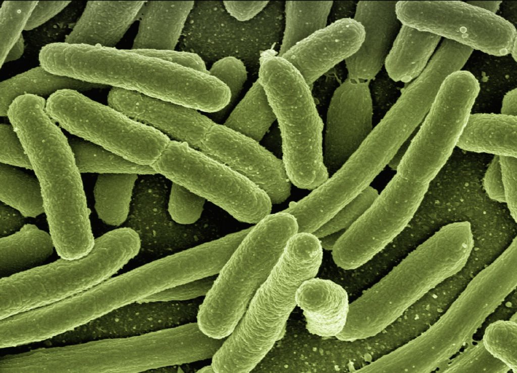
Scanning electron micrograph image of E. Coli bacteria. Credit: NIH

Scientists have found that pairing specific diets with disease-causing bacteria can create lasting immunity in mice without the costs of developing sickness, revealing a new potential vaccination strategy. Their findings, published in Science Advances, may lead to new vaccines that could promote immunity for those with diarrhoeal diseases and possibly other infections.

The body takes one of two defence strategies against bacterial infections: kill the intruders or impair the intruders but keep them around. If the body chooses to impair the bacteria, then the disease can occur without the diarrhoea, but the infection can still be transmitted – also known as asymptomatic carriage.

“We discovered that immunisation against diarrhoeal infections is possible if we allow the bacteria to retain some of its disease-causing behaviour,” says senior author Professor Janelle Ayres at Salk Institute. “This insight could lead to the development of vaccines that could reduce symptoms and mortality, as well as protect against future infections.”

In 2018, Ayres’ lab looked at how dietary interventions can create an asymptomatic infection, which Ayres calls a cooperative, asymptomatic relationship between bacteria and host. They discovered that an iron-rich diet enabled mice to survive a normally lethal bacterial infection without ever developing signs of sickness or disease. The high-iron diet increased unabsorbed glucose in the mice’s intestines, which the bacteria could feast on. The excess glucose served as a ‘bribe’ for the bacteria, keeping them full and incentivised to not attack the host.

This process produced long-term asymptomatic infection with the bacteria, leading the researchers to believe that the adaptive immune system (which ‘remembers’ infections) may be involved.

“Being able to generate lasting immunity against bacteria like C. rodentium or E. coli has not been possible using established vaccination strategies. We wanted to figure out what mechanism was sustaining this lasting immunity, so we could use that mechanism to create an impactful solution to these diarrheal diseases,” says first author Grischa Chen, a former postdoctoral researcher in Ayres’ lab.

The researchers moved to figure out how the body suppresses infection symptoms, whether infection without symptoms can create long-term immunity, and whether that immunity is reproducible as a vaccination strategy.

The team compared mice with iron-rich and normal diets after C. rodentium infection to find whether the diet impacted symptomless infection. Immediately after infection, mice fed an iron-rich diet had no symptoms whereas mice fed a normal diet did have symptoms. All mice were then put on a normal diet to see whether the asymptomatic infection would last.

Mice without functional adaptive immune systems, regardless of whether they had ever been on an iron-rich diet, could not continue maintaining a cooperative relationship with the bacteria. Although the iron-rich diet suppressed symptoms immediately after infection, the adaptive immune system was required for lasting cooperation. Importantly, the mice with functional adaptive immune systems had the disease without any symptoms, with lasting immunity, as demonstrated by survival upon reinfection after a month.

Ayres and team concluded that an iron-rich diet alone can prevent bacteria from creating deadly symptoms in mice during active infection. But a functional adaptive immune system is required for immunity against future infection in the absence of dietary supplementation.

Some bacterial strains, if mutated enough, don’t cause symptoms. To test whether such bacteria could produce lasting immunity, the team repeated their iron-diet versus normal-diet experiment in mice, but this time using bacteria that could cause disease and bacteria that could not cause disease. They found that only mice that received disease-causing, unmutated bacteria were able to support immunity upon reinfection.

The scientists note that this is only a preliminary study and people shouldn’t consume large amounts of iron after reading it. They also hope their insights will provide a basis for future research in humans and the creation of a vaccination regiment that protects and prevents against diarrhoeal illness.

Source: Salk Institute

Food Poisoning Bacteria Species Swap Pathogenic Genes

Escherichia coli. Image by CDC on Unsplash

Researchers have identified how pathogenic genes in some Providencia spp., which have gained attention as causes of food poisoning as well as enterohaemorrhagic Escherichia coli. O157 and Salmonella, are transferred within bacterial cells. Their findings are expected to provide new insights into the identification of infection routes of Providencia spp. and the establishment of preventive methods for food poisoning.

Recently, Providencia spp. which have been detected in patients with gastroenteritis, and similar to enterohemorrhagic Escherichia coli. O157 and Salmonella spp., have been attracting attention as causative agents of food poisoning. For children with low immunity, food poisoning can be lethal as it causes severe symptoms such as diarrhoea and dehydration, so clarifying the source of infection and pathogenic factors of Providencia spp., and establishing preventive methods are urgent issues worldwide.

A joint research group led by Professor Shinji Yamasaki, Dr Sharda Prasad Awasthi, a Specially Appointed Lecturer, and graduate student Jayedul Hassan from the Graduate School of Veterinary Science, Osaka Metropolitan University, determined how the pathogenic genes in some Providencia spp. such as Providencia alcalifaciens and Providencia rustigianii are transferred within bacterial cells of genus Providencia. The group has also elucidated that the pathogenic genes of Providencia rustigianii are also transferred to other bacterial cells belonging to Enterobacteriaceae.

Professor Yamasaki concluded, “This achievement is expected to provide new insights into the identification of infection routes of Providencia spp. and the establishment of preventive methods for food poisoning.”

Source: Osaka Metropolitan University

Patients Themselves may be the Source of New Strains of HAIs

E. Coli bacteria. Image by CDC

Despite stringent infection-control efforts around the world, hospital-acquired infections (HAIs)keep on popping up from new strains of bacteria. In Science Translational Medicine, researchers report evidence pointing to an unexpected source of such bacteria: the hospitalised patients themselves.

From experiments with mice, researchers at Washington University School of Medicine in St. Louis discovered that urinary tract infections (UTIs) can arise after sterile tubes, called catheters, are inserted into the urinary tract, even when no bacteria are detectable in the bladder beforehand. Such tubes are commonly used in hospitals to empty the bladders of people undergoing surgery. In the mice, inserting the tubes activated dormant Acinetobacter baumannii bacteria hidden in bladder cells, triggering them to emerge, multiply and cause UTIs, the researchers said.

The findings suggest that screening patients for hidden reservoirs of dangerous bacteria could supplement infection-control efforts and help prevent deadly HAIs.

“You could sterilise the whole hospital, and you would still have new strains of A. baumannii popping up,” said co-senior author Mario Feldman, PhD, a professor of molecular microbiology. “Cleaning is just not enough, and nobody really knows why. This study shows that patients may be unwittingly carrying the bacteria into the hospital themselves, and that has implications for infection control. If someone has a planned surgery and is going to be catheterised, we could try to determine whether the patient is carrying the bacteria and cure that person of it before the surgery. Ideally, that would reduce the chances of developing one of these life-threatening infections.”

The notoriously multidrug-resistant A. baumannii is a major threat to patients, causing many cases of UTIs in people with urinary catheters, pneumonia in people on ventilators, and bloodstream infections in people with central-line catheters into their veins.

The researchers set out to investigate why so many A. baumannii UTIs develop after people receive catheters.

Most UTIs among otherwise healthy people are caused by the bacterium Escherichia coli. Research has shown that E. coli can hide out in bladder cells for months after a UTI seems to have been cured, and then re-emerge to cause another infection.

The researchers investigated whether A. baumannii can hide inside cells like E. coli can. They studied mice with UTIs caused by A. baumannii. They used mice with weakened immune systems because, like people, healthy mice can fight off A. baumannii.

Once the infections had resolved and no bacteria were detected in the mice’s urine for two months, the researchers inserted catheters into the mice’s urinary tracts with a sterile technique. Within 24 hours, about half of the mice developed UTIs caused by the same strain of A. baumannii as the initial infection.

“The bacteria must have been there all along, hiding inside bladder cells until the catheter was introduced,” said co-senior author Scott J. Hultgren, PhD, a professor and expert on UTIs. “Catheterisation induces inflammation, and inflammation causes the reservoir to activate, and the infection blooms.”

Since A. baumannii rarely causes symptoms in otherwise healthy people, many people who carry the bacteria may never know they’re infected, the researchers said. According to the researchers’ literature search, 2% of healthy people carry A. baumannii in their urine.

“I wouldn’t put much weight on the precise percentage, but I think we can say with certainty that some percentage of the population is walking around with A. baumannii,” Feldman said. “As long as they’re basically healthy, it doesn’t cause any problems, but once they’re hospitalised, it’s a different matter. This changes how we think about infection control. We can start considering how to check if patients already have Acinetobacter before they receive certain types of treatment; how we can get rid of it; and if other bacteria that cause deadly outbreaks in hospitals, such as Klebsiella, hide in the body in the same way. That’s what we’re working on figuring out now.”

Source: Washington University School of Medicine

Artificial Sweeteners Can Turn Gut Bacteria Bad

Source: Breakingpic on Pexels

Scientists have found that common artificial sweeteners can turn previously healthy gut bacteria pathogenic, invading the gut wall and potentially leading to serious health issues.

This study is the first to show the pathogenic effects of some of the most widely used artificial sweeteners (saccharin, sucralose, and aspartame) on two types of gut bacteria, Escherichia coli and Enterococcus faecalisE. faecalis is capable of crossing the intestinal wall to enter the bloodstream and congregate in the lymph nodes, liver, and spleen, causing a number of infections including septicaemia. To top it off, this commensal bacteria has emerged as a multi-drug resistant pathogen.

Previous studies have shown that artificial sweeteners can affect the composition of gut bacteria, but this new molecular research, led by academics from Anglia Ruskin University (ARU), has shown that sweeteners can also induce pathogenic features in certain bacteria. It found that these pathogenic bacteria can latch onto, invade and kill epithelial Caco-2 cells lining the intestinal wall.

This new study discovered that at a concentration equivalent to two cans of diet soft drink, all three artificial sweeteners significantly increased the adhesion of both E. coli and E. faecalis to intestinal Caco-2 cells, and differentially increased biofilm formation. Bacteria growing in biofilms are less sensitive to antimicrobial resistance treatment and are more likely to secrete toxins and express disease-causing virulence factors.

Additionally, all three sweeteners caused the pathogenic gut bacteria to invade Caco-2 cells found in the wall of the intestine, save for saccharin, which had no significant effect on E. coli invasion.

Senior author Dr Havovi Chichger, Senior Lecturer in Biomedical Science at ARU, said: “There is a lot of concern about the consumption of artificial sweeteners, with some studies showing that sweeteners can affect the layer of bacteria which support the gut, known as the gut microbiota.

“Our study is the first to show that some of the sweeteners most commonly found in food and drink—saccharin, sucralose and aspartame—can make normal and ‘healthy’ gut bacteria become pathogenic. These pathogenic changes include greater formation of biofilms and increased adhesion and invasion of bacteria into human gut cells.

“These changes could lead to our own gut bacteria invading and causing damage to our intestine, which can be linked to infection, sepsis and multiple-organ failure.

“We know that overconsumption of sugar is a major factor in the development of conditions such as obesity and diabetes. Therefore, it is important that we increase our knowledge of sweeteners versus sugars in the diet to better understand the impact on our health.”
Source: EurekAlert!

Journal reference: Shil, A & Chichger, H (2021) Artificial Sweeteners Negatively Regulate Pathogenic Characteristics of Two Model Gut Bacteria, E. coli and E. faecalis. International Journal of Molecular Sciences. doi.org/10.3390/ijms22105228.