Tag: Down syndrome

Groundbreaking Study Discovers Differences in Oxygen Physiology in Down Syndrome
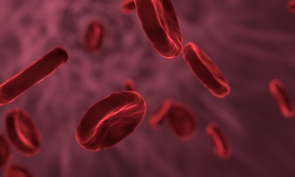
Source: Pixabay CC0

A groundbreaking new study published in Cell Reports by researchers from the University of Colorado Anschutz Medical Campus reports important differences in oxygen physiology and red blood cell function in individuals with Down syndrome. The study is part of the ongoing Human Trisome Project, a large and detailed cohort study of the population with Down syndrome, including deep annotation of clinical data, the largest biobank for the study of Down syndrome to date, and multi-omics datasets.

The Crnic Institute team first analysed hundreds of blood samples to identify physiological differences between research participants with Down syndrome versus controls from the general population. They observed that triplication of chromosome 21, or trisomy 21, the chromosomal abnormality that causes Down syndrome, leads to a physiological state reminiscent of hypoxia. They identified major changes in gene expression indicative of low oxygen availability, including induction of many hypoxia-inducible genes and proteins, as well as increased levels of factors involved in the synthesis of haeme, the molecule that transports oxygen inside red blood cells.

“These results reveal that hypoxia and hypoxic signalling should be front and centre when we talk about the health of people with Down syndrome,” says Dr Joaquín Espinosa, executive director of the Crnic Institute, professor of pharmacology, principal investigator of the Human Trisome Project, and one of the senior authors of the paper. “Given the critical role of oxygen physiology in health and disease, we need to understand the causes and consequences of hypoxia in Down syndrome, which could lead to effective interventions to improve oxygen availability in this deserving population.”

“The results are remarkable, it is safe to say that the blood of people with Down syndrome looks like that of someone who was quickly transported to a high altitude or who was injected with erythropoietin (EPO), the master regulator of erythropoiesis, the process of new red blood cell formation,” explains Dr Micah Donovan, lead author of the paper. “Although it has been known for many years that people with Down syndrome have fewer and bigger red blood cells, this is the first demonstration that they overproduce EPO and that they are undergoing stress erythropoiesis, a phenomenon whereby the liver and the spleen need to start producing red blood cells to supplement those arising from the bone marrow.”

The team discovered that these phenomena are also observed in a mouse model of Down syndrome, thus reinforcing the idea that these important physiological changes arise from triplication of genetic material and overexpression of specific genes.

“The fact that hypoxic signaling and stress erythropoiesis are conserved in the mouse model paves the way for mechanistic investigations that could identify the genes involved and reveal therapeutic interventions to improve oxygen physiology in Down syndrome,” explains Dr. Kelly Sullivan, associate professor of pediatrics, director of the Experimental Models Program at the Crnic Institute and co-author in the study.

The study team also investigated whether the elevated hypoxic signaling and associated stress erythropoiesis was tied to the heightened inflammatory state characteristic of Down syndrome. Although individuals with the stronger hypoxic signatures show more pronounced dysregulation of the immune system and elevated markers of inflammation, their results indicate that lowering inflammation does not suffice to reverse the hypoxic state.

“We will need a lot more data to understand what is causing the hypoxic state and its impacts on the health of people with Down syndrome,” says Dr Matthew Galbraith, assistant research professor of pharmacology, director of the Data Sciences Program at the Crnic Institute, and one of the senior authors of the paper. “The hypoxic state could be caused by obstructive sleep apnoea (which is common in Down syndrome), cardiopulmonary malfunction, or even perhaps defects in red blood cell function. We are very excited about several ongoing clinical trials funded by the NIH INCLUDE Project for obstructive sleep apnea in Down syndrome, which we believe will be very informative.”

The Crnic Institute study team is already planning several follow up studies, with the explicit goal of illuminating strategies to improve oxygen physiology in the population with Down syndrome.

Down Syndrome Research Should Expand Focus to the Whole Cell

Human chorosomes. Source: NIH

Researchers propose in The American Journal of Human Genetics a new way of looking at Down syndrome, suggesting that when an extra chromosome is present, the impact on the cell depends less on which chromosome is duplicated and more on the presence of extra DNA.

“Understanding the complexity and general nature of disease phenotypes allows us to see a bigger picture and not get stuck focusing on a single gene, due to its presence on the extra chromosome,” says lead author Maria Krivega, developmental biologist at Heidelberg University.

Every cell starts out with extra chromosomes during early embryogenesis; however, this DNA gets sorted into pairs after about a week of growth. When this process goes awry, it often leads to death of the embryo, with only a few being able to survive with the extra DNA, like in the case of Down syndrome.

By taking a step back and looking at the entire cell, researchers were able to create a new understanding of these syndromes. Krivega and her collaborators took a critical look at recent evidence suggesting that Down syndrome phenotypes arise not only because of increased dosage of genes on chromosome 21 but also because of global effects of chromosome gain.

The researchers sifted through published datasets of proteins and RNA of individuals with Down syndrome and compared these to laboratory made cells with trisomies of chromosomes 3, 5, 12, and 21. What they found from this comparison was that it didn’t matter which chromosome was in excess, the cells all had decreased ability to replicate, survive, and maintain their DNA.

“We were interested to find out why cells with imbalanced chromosomal content – in other words, aneuploid – are capable of surviving,” says Krivega. “It was particularly exciting to me to learn if viable aneuploid embryonic cells have similarities with aneuploid cancer cells or cell lines, derived in the laboratory.”

Additionally, they found that the adaptive T cell immune system was underdeveloped in all cells, while the innate immune system seemed to be overactive. The authors suggest that this is a consequence of general chromosome gain. This research can be expanded into autoimmune diseases, such as Alzheimer disease or acute leukemias in trisomy chr. 8 or 21, that also exist without any connection to aneuploidy.

“We hope that our work elucidating a complex trisomy phenotype should help to improve such kids’ development,” says Krivega.

Source: Cell Press

Alzheimer’s Prions also Appear in Down Syndrome

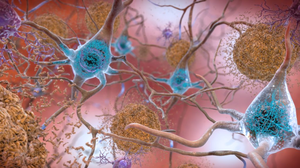
Plaques and neurons. Source: NIAH

The brains of people with Down syndrome develop the same neurodegenerative tangles and plaques associated with Alzheimer’s disease and they frequently demonstrate signs of the neurodegenerative disorder in their 40s or 50s. A new study in the journal PNAS shows that these tangles and plaques are driven by the same amyloid beta (Aß) and tau prions that they showed are behind Alzheimer’s disease.

Prions begin as normal proteins that become misshapen and self-propagate. They spread through tissue like an infection by forcing normal proteins to adopt the same misfolded shape. In both Alzheimer’s and Down syndrome, as Aß and tau prions accumulate in the brain, they cause neurological dysfunction that often manifests as dementia.

Tau tangles and Aß plaques are evident in most people with Down syndrome by age 40, according to the National Institute on Aging, with at least 50% of this population developing Alzheimer’s as they age.

The new study highlights how a better understanding of Down syndrome can lead to new insights about Alzheimer’s, as well.

“Here you have two diseases – Down syndrome and Alzheimer’s disease – that have entirely different causes, and yet we see the same disease biology. It’s really surprising,” said Stanley Prusiner, MD, the study’s senior author, who was awarded the Nobel Prize in 1997 for his discovery of prions.

Down syndrome is the most common neurodegenerative disease among younger people in the United States, while Alzheimer’s is the most common among adults.

Down syndrome occurs because of an extra copy of chromosome 21. Among the many genes on that chromosome is one called APP, which codes for one of the major components of amyloid beta. With an extra copy of the gene, people with Down syndrome produce excess APP, which may explain why they develop amyloid plaques early in life.

A clearer picture in young brain

It’s been known for some time that Aß plaques and tau tangles are present in both Down syndrome and Alzheimer’s. Having shown earlier that these neurodegenerative features are provoked by prions in Alzheimer’s, the researchers wanted to know whether the same aberrant proteins were present in the brains of people with Down syndrome.

While these plaques and tangles in the brains of people with Alzheimer’s disease have been well-studied, it can be challenging to discern which changes in the brain are from old age and which are from prion activity, said Prusiner.

“Because we see the same plaques-and-tangles pathology at a much younger age in people with Down syndrome, studying their brains allows us to get a better picture of the early process of disease formation, before the brain has become complicated by all the changes that go on during aging,” he said. “And ideally, you want therapies that address these early stages.”

Employing a variation on the novel assay they used in the Alzheimer’s study, the team looked at donated tissue samples from deceased people with Down syndrome, which they obtained from biobanks around the world. Of the 28 samples from donors aged 19 to 65 years old, the researchers were able to isolate measurable amounts of both Aß and tau prions in almost all of them.

New insights could yield preventative measures

The results confirm not only that prions are involved in the neurodegeneration seen in Down syndrome, but that Aß drives the formation of tau tangles as well as amyloid plaques, a relationship that has been suspected but not proven.

“The field has long tried to understand what the intersection is between these two pathologies,” said lead author Carlo Condello, PhD, also a member of the UCSF Institute for Neurodegenerative Diseases. “The Down syndrome case corroborates the idea; now you have this extra chromosome that’s driving the Aß, and there’s no tau gene on the chromosome. So, it’s truly by increasing the expression of Aß that you kick off production of the tau.”

These and other insights gained from studying the brains of people with Down syndrome will lead to a much better picture of how prions begin to form in the first place, said Condello.

Whether the Down syndrome brain tissue will prove to be the ultimate model for developing treatments for Alzheimer’s remains to be seen, the researchers said. While the two disorders share many similarities in their prion pathobiology, there are some differences that may be limiting.

Still, the researchers said, studying the plaques and tangles in Down syndrome is a promising route to identifying the specific prions that arise at the very earliest stages of the disease process. That insight could open new vistas on not only treating but perhaps even fending off Alzheimer’s disease.

“If we can understand how this neurodegeneration begins, we are one big step closer to being able to intervene at a meaningful point and actually prevent these large brain lesions from forming,” Condello said.

Source: University of California – San Francisco