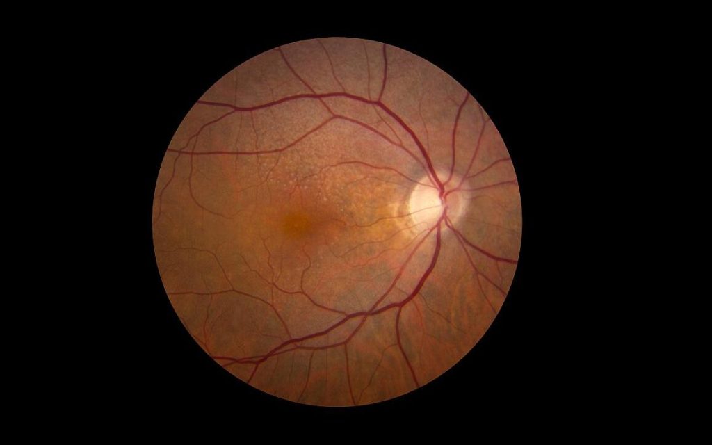
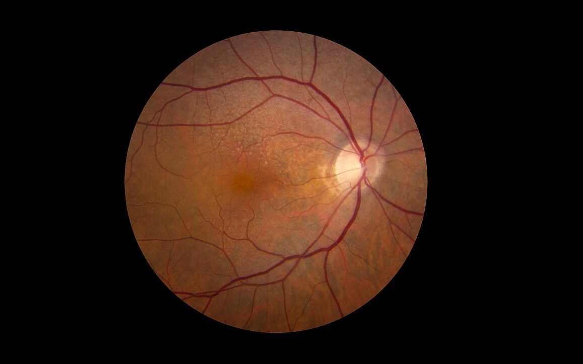
Tag: diabetic retinopathy

Don’t Overlook Latent Autoimmune Diabetes in Adults, Researchers Caution
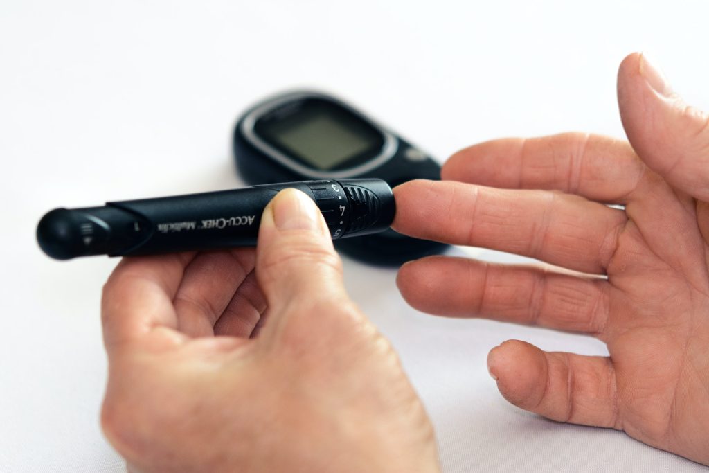
Photo by Photomix Company on Pexels

To reduce the risk of complications, it is important to measure antibodies those with adult onset diabetes, while also considering the levels of these antibodies.

In a study published in the journal Diabetes Care, researchers demonstrate that individuals with Latent Autoimmune Diabetes in Adults (LADA) have an equally high risk of developing cardiovascular disease as people with type 2 diabetes, but a higher risk of developing retinopathy and poorer glucose control. Many also lack adequate treatment.

LADA is a common but relatively unknown form of diabetes. Similar to type 1 diabetes, it is an autoimmune disease characterised by antibodies against insulin-producing cells. It develops in adulthood, and the autoimmune process progresses more slowly than in type 1 diabetes. LADA also shares features with type 2 diabetes, which means those affected risk getting the wrong diagnosis if antibodies are not measured. Incorrect diagnosis can result in inadequate treatment. Previous studies suggest that between five and ten percent of all individuals initially diagnosed with type 2 diabetes actually have LADA. Researchers at Karolinska Institutet, and the Universities of Lund and Helsinki set out to examine the risk of complications in LADA.

Our results emphasise the importance of diagnosing LADA correctly and careful monitoring of glucose control in these individuals, so that treatment can be intensified if needed, thereby reducing the risk of complications.

Yuxia Wei, PhD-student and Sofia Carlsson, senior lecturer, Institute of Environmental Medicine, Karolinska Institutet

According to the study LADA was characterised by fewer metabolic risk factors than type 2 diabetes, such as high blood pressure and high blood lipids. However, a lower proportion of individuals with LADA achieved good glucose control. The lack of glucose control was most evident in LADA patients with high levels of the antibody GADA (glutamic acid decarboxylase antibody). A significant portion of individuals with LADA lacked any glucose-lowering treatment.

The results of the new study are based on the ESTRID study, where researchers followed over 4000 individuals with diabetes, of whom 550 had LADA, for up to 12 years after diagnosis. According to the researchers, it is the most comprehensive study to date regarding the risk of complications in LADA.

Source: Karolinska Institutet

Experimental Drug may Prevent Diabetic Vision Loss

Researchers at Wilmer Eye Institute, Johns Hopkins Medicine say they have evidence that an experimental drug may prevent or slow vision loss in people with diabetes. The results are from a study published in the Journal of Clinical Investigation, that used mouse models as well as human retinal organoids and eye cell lines.

The team focused on models of two common diabetic eye conditions: proliferative diabetic retinopathy and diabetic macular enema, both of which affect the retina, the light-sensing tissue at the back of the eye that also transmits vision signals to the brain. In proliferative diabetic retinopathy, new blood vessels overgrow on the retina’s surface, causing bleeding or retinal detachments and profound vision loss. In diabetic macular enema, blood vessels in the eye leak fluid, leading to swelling of the central retina, damaging the retinal cells responsible for central vision.

Results of the study show that a compound called 32-134D, previously shown to slow liver tumour growth in mice, prevented diabetic retinal vascular disease by decreasing levels of a protein called HIF, or hypoxia-inducible factor. Doses of 32-134D also appeared to be safer than another treatment that also targets HIF and is under investigation to treat diabetic eye disease.

Current treatment for both proliferative diabetic retinopathy and diabetic macular enema includes eye injections with anti-vascular endothelial growth factor (anti-VEGF) therapies. Anti-VEGF therapies can halt the growth and leakiness of blood vessels in the retina in patients with diabetes. However, these treatments aren’t effective for many patients, and may cause side effects with prolonged use, such as increased internal eye pressure or eye tissue damage.

Study author Akrit Sodhi, MD, PhD, says that in general, the idea of inhibiting HIF, a fundamental protein in the body, has raised concerns about toxicity to many tissues and organs. But when his team screened a library of HIF inhibitor drugs and conducted extensive testing, “We came to find that the drug examined in this study, 32-134D, was remarkably well tolerated in the eyes and effectively reduced HIF levels in diseased eyes,” says Sodhi.

HIF, a type of protein known as a transcription factor, has the ability to switch certain genes, including vascular endothelial growth factor (VEGF), on or off throughout the body. In the eye, elevated levels of HIF cause genes like VEGF to increase blood vessel production and leakiness in the retina, contributing to vision loss.

To test 32-134D, researchers dosed multiple types of human retinal cell lines associated with the expression of proteins that promote blood vessel production and leakiness. When they measured genes regulated by HIF in cells treated with 32-134D, they found that their expression had returned to near-normal levels, which is enough to halt new blood vessel creation and maintain blood vessels’ structural integrity.

Researchers also tested 32-134D in two different adult mouse models of diabetic eye disease. In both models, injections were administered into the eye. Five days post-injection, the researchers observed diminished levels of HIF, and also saw that the drug effectively inhibited the creation of new blood vessels or blocked vessel leakage, therefore slowing progression of the animals’ eye disease. Sodhi and his team said they also were surprised to find that 32-134D lasted in the retina at active levels for about 12 days following a single injection without causing retinal cell death or tissue wasting.

“This paper highlights how inhibiting HIF with 32-134D is not just a potentially effective therapeutic approach, but a safe one, too,” says Sodhi. “People facing diabetic eye disease and vision loss include our family members, friends, co-workers – this is a disease that impacts a large group of people. Having safer therapies is critical for this growing population of patients.”

Sodhi says that further studies in animal models are needed before moving to clinical trials.

Source: Johns Hopkins Medicine

Periods of Hypoglycaemia Worsen Progression of Diabetic Retinopathy

Credit: National Eye Institute

People with diabetes who experience periods of hypoglycaemia, a common event in those new to blood sugar management, are more likely to have worsening diabetic eye disease. Now, researchers say they have linked such low blood sugar levels with a molecular pathway that is activated in hypoxic cells in the eye.

The research, involving human and mouse eye cells and intact retinas grown in a low glucose environment in the laboratory, as well as mice with low glucose levels, was published in Cell Reports.

“Temporary episodes of low glucose happen once or twice a day in people with insulin-dependent diabetes and often among people newly diagnosed with the condition,” says Akrit Sodhi, MD, PhD, Johns Hopkins Medicine professor. Low glucose levels can also occur during sleep in people with non-insulin dependent diabetes. “Our results show that these periodic low glucose levels cause an increase in certain retinal cell proteins, resulting in an overgrowth of blood vessels and worsening diabetic eye disease,” adds Sodhi.

Up to a third of diabetic patients will develop diabetic retinopathy, which is characterised by the overgrowth of abnormal blood vessels in the retina.

Sodhi says the current study suggests that people with diabetic retinopathy may be particularly vulnerable to periods of low glucose, and keeping glucose levels stable should be an important part of glucose control.

For the study, the researchers analysed protein levels in human and mouse retinal cells and intact retinas grown in an environment of low glucose in the laboratory, as well as in mice that had occasional low blood sugar.

In human and mouse retinal cells, low glucose levels triggered a cascade of molecular changes that can lead to blood vessel overgrowth. First, the researchers saw that low glucose caused a decrease in retinal cells’ ability to break down glucose for energy.

When the researchers focused on Müller glial cells, which are supportive cells for neurons in the retina and rely primarily on glucose for energy production, they found that the cells increased the expression of the GLUT1 gene, which makes a protein that transports glucose into cells.

The researchers found that, in response to low glucose, the cells increased levels of a transcription factor, hypoxia-inducible factor (HIF)-1α. This turned on the cellular machinery, including GLUT1, needed to improve their ability to utilise available glucose, preserving the limited oxygen available for energy production by retinal neurons.

However, in hypoxic environments, as occurs in the retinas of patients with diabetic eye disease, this normal, physiologic response to low glucose triggered a flood of HIF-1α protein into the nucleus.

This resulted in an increase in the production of proteins such as VEGF and ANGPTL4, which cause the growth of abnormal, leaky blood vessels – the key culprit of vision loss in people with diabetic eye disease.

The researchers plan to study whether low glucose levels in people with diabetes may impact similar molecular pathways in other organs, such as the kidney and brain.

Sodhi says the HIF-1α pathway may serve as an effective target for developing new treatments for diabetic eye disease.

Source: Johns Hopkins Medicine

30-year Diabetes Study Determines HbA1c Threshold for Complications

Diabetes - person measures blood glucose
Photo by Photomix Company from Pexels

The levels of long-term blood sugar, HbA1c, can be used to accurately determine the risk of a person with type 1 diabetes developing eye- and kidney complications. A Swedish study, published in Diabetes Care, followed individuals after the onset type 1 diabetes for 30 years and showed that this level should under 53mmol/mol (7%).

People with diabetes may experience damage to the small blood vessels in various organs. The reasons for this are unclear, but it has been known since the 1990s that good control of blood sugar levels reduces the risk of complications. It has, however, not been clear what level of long-term sugar, HbA1c, people with type 1 diabetes should have in order to avoid serious damage to blood vessels in the eyes and kidneys.

“Our study determines accurately the levels of long-term sugar that can avoid complications. This knowledge can increase a person’s motivation to keep their blood sugar level under control,” said study leader Hans Arnqvist, professor emeritus at Linköping University.

Researchers in the current study, known as VISS (Vascular Diabetic Complications in Southeast Sweden), have followed all children and adults under 35 who developed type 1 diabetes during the period 1983–1987 in Southeast Sweden. All 447 newly diagnosed persons in the region during this period were included in the study. The researchers have followed the patients’ HbA1c values, which reflect their average blood sugar levels during a longer period. They have also monitored the development of eye- and kidney damage in these patients for a period of between 32 and 36 years after diagnosis.

In type 1 diabetes, the small blood vessels in the eye are particularly susceptible to damage. Nearly all patients experience small haemorrhages in the eye that do not affect their vision. In some cases, proliferative retinopathy develops, forming new blood vessels which can lead to blindness. The macula of the retina can also be damaged, leading to blurred vision.

While the kidneys are not as sensitive to high blood sugar levels as the eye, the important small blood vessels here can also be damaged, leading to albuminuria. The damage to the kidneys eventually leads to impaired kidney function and, in serious cases, kidney failure.

In healthy individuals, the blood sugar level is very closely controlled, with a maximum HbA1c level of 42mmol/mol (6.0%).

“The results of our study show that people with type 1 diabetes for at least 32 years should keep their mean long-term sugar level below 53mmol/mol (7.0%), if they are to completely avoid serious damage. The risk of eye- and kidney complications increases as the level increases. Our conclusions relate to avoiding complications arising from blood vessel damage. But if a patient has problems with low blood sugar, hypoglycaemia, it’s not possible to control the blood sugar level so strictly,” said Prof Arnqvist.

The target level for HbA1c that is suggested by the results of the VISS study agrees with the individual targets recommended by the American Diabetes Association. In Sweden, target levels are given for groups, rather than individuals.

The previous follow-up by the research group was conducted 20 years after the onset of disease. Now after 30 years, the results show that damage has arisen at lower blood sugar levels than was the case after 20 years.

More patients have experienced damage, despite having blood sugar levels that are not higher than those they have previously had. In other words, it seems that the threshold for developing complications falls gradually with time. This means that the study does not allow any conclusions for the recommended blood sugar levels of people with type 1 diabetes longer than 30 years after diagnosis.

Source: Linköping University

Fenofibrate Confers Modest Risk Reduction for Diabetic Retinal Disease

Retina showing reticular pseudodrusen. Credit: National Eye Institute

Taking the cholesterol-lowering drug fenofibrate had a modest but statistically significant association with reduced risk of vision-threatening diabetic retinopathy (VTDR), according to results of a large study published in JAMA Ophthalmology. In the study, fenofibrate use was associated with an 8% lower risk of progression compared to non-use.

Fenofibrate use had a greater effect on the risk of proliferative diabetic retinopathy, with a 24% decrease in progression (PDR) but did not significantly affect the risk of developing diabetic macular oedema (DME).

The researchers noted that these findings are in line with evidence showing fenofibrate may protect against diabetes-associated breakdown of the blood-retinal barrier, although ophthalmologists rarely use the drug to treat diabetic eye disease.

“Our positive association for progression to PDR coincides with results of previous clinical trials and adds new information with regards to the impact on DME,” the researchers stated.

Protection against progression to PDR “was found without regards to underlying NPDR [nonproliferative diabetic retinopathy] severity level, which is not well coded within the claims database,” the researchers continued. “Understanding this limitation and how the inclusion of NPDR severity levels that may not benefit from fenofibrates would bias our findings to the null means that the positive association seen in our study is actually an underestimate of the true association.”

While fenofibrate’s mechanism of action in diabetic retinopathy is not well understood, “interest in the use of this oral agent has become substantial,” noted Robert N. Frank, MD. author of an accompanying editorial.

“From the point of view of a clinician with a long-time interest in diabetic retinopathy, its causal mechanisms, and its evolving treatments, the possibility that an oral medication originally used for a different disease may be beneficial for the management of diabetic eye disease is exciting,” Dr Frank wrote.

“The evidence that fenofibrate can slow the progression of diabetic retinopathy is growing, but it has not yet become a widely accepted treatment,” he added. “It will be interesting to see how this large population analysis and the results from the ongoing DRCR Retina Network  randomised clinical trial will affect clinical practice in the years to come.”

Two clinical trials that evaluated fenofibrate’s effect on diabetic eye disease, the FIELD study and the ACCORD-Eye trial yielded conflicted findings regarding DME, PDR and progression of diabetic retinopathy. Both trials suggested that only patients with mild nonproliferative eye disease were likely to benefit.

To help inform decision-making on fenofibrate in eye disease, researchers drew on a large health insurer database for 150 252 adults who had NPDR-associated lab values from January 2002 through June 2019. The primary outcomes were a new diagnosis of VTDR (composite of PDR or DME) or DME and PDR individually.

The analysis showed 5835 (3.9%) used fenofibrate. During follow-up, 27 325 patients progressed to VTDR, including 4 086 to PDR and 22 750 to DME. While men accounted for a larger proportion of fenofibrate users (61.1% vs 51.0% of nonusers), patients had similar baseline characteristics.

Study limitations included lack of clinical applicability, not accounting for duration of fenofibrate use, and data being drawn from a single database.

Source: MedPage Today

Stem Cells Research Hints at Ways to Prevent Diabetic Retinopathy

Old man with magnifying glass
Image by Mar Lezhava on Unsplash

Researchers are investigating novel stem cell approaches that could lead to treatments for early retinal vascular dysfunction in diabetic patients, which could help prevent diabetic retinopathy.

Diabetic complications cause major metabolic disturbances that damage the cardiovascular, visual, peripheral nerve and renal systems through harming small and large microvessels that feed these tissues. New treatments are needed to treat the growing number of people who develop such retinal vascular dysfunction.

Research strategies include identifying and using new methods to differentiate or mature human induced pluripotent stem cells (hiPSCs) into the specific mesoderm subset of cells that display vascular reparative properties.

“Vascular diseases afflict hundreds of millions of people in the world,” said Chang-Hyun Gil, MS, PhD, a postdoctoral fellow in the Department of Surgery and co-first author of the study. “In this study, we focused on the retinal vessel in type 2 diabetes. Our results demonstrate the safe, efficient and robust derivation of hiPSC-derived specific mesoderm subset for use as a novel therapy to rescue ischemic tissues and repair blood vessels in individuals with vascular diseases. The results provide a foundation for an early phase clinical trial.”

In the study, published in Science Advances, investigators genetically reprogrammed diabetic and non-diabetic peripheral blood cells into hiPSCs and matured the cells into special blood vessel reparative cells. Upon injection into animal models with type 2 diabetic murine (T2D) retinal dysfunction, results showed significant improvement in visual acuity and electroretinograms with restoration of vascular perfusion. They hypothesised that hiPSC-derived vascular reparative cells could work as endothelial precursors that will display in vivo vessel reparative properties in these diabetic subjects.

“Unlike the use of embryonic stem cells (ESCs), genetically engineered hiPSCs do not carry the ethical challenges ESCs possess that limit their possible usage, and hiPSCs are being increasingly recognised as a viable alternative in study design and application as a cell therapy for human disorders,” Dr Gil said.

Researchers converted hiPSC into a specific mesoderm subset that was enriched to generate endothelial cells with vessel reparative properties similar to endothelial colony forming cells (ECFC).

Dr Gil said certain mesoderm subsets were better able to differentiate into ECFC and form functional blood vessels in vivo. and that mesoderm populations corrected vasodegeneration of injured retinal vessels. Tests showed enhanced function of neural retina and improved vision.

Source: Indiana University