Tag: cytokines

Turning off Pro-inflammatory Cytokine IL-11 Extends Healthy Lifespan in Mice

Credit: MRC LMS, Duke-NUS Medical School

Scientists have discovered that ‘turning off’ the cytokine IL-11 can significantly increase the healthy lifespan of mice by almost 25%.

The scientists, at the Medical Research Council Laboratory of Medical Science (MRC LMS), Imperial College London and Duke-NUS Medical School in Singapore, tested the effects of IL-11 by creating mice with the gene for IL-11 (interleukin 11) deleted. This extended the lives of the mice by over 20% on average. The cytokine has for years been misidentified as an anti-inflammatory and anti-fibrotic.

They also treated 75-week-old mice, equivalent to the age of about 55 years in humans, with an injection of an anti-IL-11 antibody, a drug that stops the effects of the IL-11 in the body.

Median lifespan extended

The results, published in Nature, were dramatic, with mice given the anti-IL-11 drug from 75 weeks of age until death having their median lifespan extended by 22.4% in males and 25% in females. The mice lived for an average of 155 weeks, compared with 120 weeks in untreated mice.

The treatment largely reduced deaths from cancer in the animals, as well as reducing the many diseases caused by fibrosis, chronic inflammation and poor metabolism, which are hallmarks of ageing. There were very few side effects observed.

Fewer cancers and free from the usual signs of ageing and frailty

Professor Stuart Cook, who was co-corresponding author, from MRC LMS, Imperial College London and Duke-NUS Medical School in Singapore, said:

These findings are very exciting. The treated mice had fewer cancers, and were free from the usual signs of ageing and frailty, but we also saw reduced muscle wasting and improvement in muscle strength. In other words, the old mice receiving anti-IL11 were healthier.

Previously proposed life-extending drugs and treatments have either had poor side-effect profiles, or don’t work in both sexes, or could extend life, but not healthy life, however this does not appear to be the case for IL-11.

While these findings are only in mice, it raises the tantalising possibility that the drugs could have a similar effect in elderly humans. Anti-IL-11 treatments are currently in human clinical trials for other conditions, potentially providing exciting opportunities to study its effects in ageing humans in the future.

The researchers have been investigating IL-11 for many years and in 2018 they were the first to show that IL-11 is a pro-fibrotic and pro-inflammatory protein, overturning years of incorrect characterisation as anti-fibrotic and anti-inflammatory.

Levels of IL-11 increases with age

Assistant Professor Anissa Widjaja, who was co-corresponding author, from Duke-NUS Medical School, Singapore, said:

This project started back in 2017 when a collaborator of ours sent us some tissue samples for another project. Out of curiosity, I ran some experiments to check for IL-11 levels. From the readings, we could clearly see that the levels of IL-11 increased with age and that’s when we got really excited!

We found these rising levels contribute to negative effects in the body, such as inflammation and preventing organs from healing and regenerating after injury. Although our work was done in mice, we hope that these findings will be highly relevant to human health, given that we have seen similar effects in studies of human cells and tissues.

This research is an important step toward better understanding ageing and we have demonstrated, in mice, a therapy that could potentially extend healthy ageing, by reducing frailty and the physiological manifestations of ageing.

Previously, scientists have posited that IL-11 is an evolutionary hangover in humans, as while it is vital for limb regeneration in some animal species, it is thought to be largely redundant in humans.

IL-11 linked to chronic inflammation and frailty

However, after about the age of 55 in humans, more IL-11 is produced and past research has linked this to chronic inflammation, fibrosis in organs, disorders of metabolism, muscle wasting (sarcopaenia), frailty, and cardiac fibrosis. These conditions are many of the signs we associate with ageing.

When two or more such conditions occur in an individual, it is known as multimorbidity, which encompasses a range of conditions including lung disease, cardiovascular disease, diabetes, vision and hearing decline and a host of other conditions.

Professor Cook said:

The IL-11 gene activity increases in all tissues in the mouse with age. When it gets turned on it causes multimorbidity, which is diseases of ageing and loss of function across the whole body, ranging from eyesight to hearing, from muscle to hair, and from the pump function of the heart to the kidneys.

Multimorbidity among biggest global healthcare challenges

Multimorbidity and frailty are acknowledged to be among the biggest global healthcare challenges of the 21st century, according to many leading health bodies, including the NHS and the World Health Organization.

Currently, no treatment for multimorbidity is available, other than to try to treat the separate multiple underlying causes individually.

The scientists caution that the results in this study were in mice and the safety and effectiveness of these treatments in humans need further establishing in clinical trials before people consider using anti-IL-11 drugs for this purpose.

Source: UK Research & Innovation

A Cellular Switch to Shut off Cytokine Storms

Signalling from inflammatory cytokines activates several protein kinases in a chain – at the end of this process MKK6 (yellow) ‘switches on’ p38α (green) by binding it and adding phosphates. The combination of computer simulations, SAXS, and cryo-EM has shown how the two proteins interact and bind to each other to transmit the signal. Credits: Ella Marushchenko, Isabel Romero Calvo/EMBL

Cytokine storms, where inflammatory cascades during an infection that can spiral out of control and lead to severe disease and even death, were recently highlighted during the COVID-19 pandemic. In a new paper published in Science, researchers report their discovery of a cellular ‘switch’ which may lead to new drugs to prevent deadly cytokine storms.

EMBL Grenoble and University of Geneva researchers provide essential insights on a protein called p38α which is an important cellular ‘switch’ triggering the inflammatory response. They have obtained the first structure of p38α being activated by another regulatory protein kinase, MKK6, opening up new directions to develop drugs to stop cytokine storms.

The final switch: a drug target

Matthew Bowler, a researcher at EMBL Grenoble, has been studying kinases for more than a decade. This group of enzymes plays an important role in regulating complex processes in the cell by acting as a ‘switch’ to transmit signals and activate gene expression. They do so by phosphorylation, in which phosphate is added to other molecules to modulate their function.

Bowler’s work particularly focuses on belonging to the Mitogen Activated Protein (MAP) family of kinases, key players involved in the inflammatory response. Inflammation is switched on via a series of kinases, which activate each other in a cascade of reactions, the final kinase in the series being responsible for activating gene transcription required for inflammation. This process releases cytokines, pro-inflammatory signalling molecules, which, in case of overactivation, can lead to cytokine storms.

This kinase chain reaction is well regulated and is similar to a logic circuit: the inflammation response requires specific buttons to be switched on, ultimately activating p38α – the convergence point for all the signals and the last switch of the inflammatory process.

Because the kinase chain reaction can come from different ‘branches’ of the logic circuit, this last switch is a particularly relevant drug target. The inflammatory response is regulated by p38α and is highly activated during a cytokine storm. Inactivating it could prevent inflammation from occurring, instead of trying to treat it while it is already underway.

Protein kinases, including p38α, have therefore been heavily studied. The first protein kinase structure was solved 30 years ago and many more structures have followed, with over 7000 structures now available in the Protein Data Bank.

However, important parts of the puzzles are still missing. “Structural biologists have obtained detailed information on the structure and functions of protein kinases, but mostly in isolation. So we don’t really know how these enzymes are activated along the chain reaction,” explained Bowler.

Without this essential information about how the activation is triggered, drugs have mostly targeted the kinases’ nucleotide-binding site – a common and well-known pocket present in all kinases, where the phosphate transfer occurs. The lack of drug specificity due to a common binding site across kinases means that a drug designed to stop one kinase from signalling could also stop others. This presents a problematic side effect, considering the essential role of kinases as key regulators in cellular processes.

“There are many molecules that have been designed to target p38α, especially its nucleotide-binding site, but none have yet made it past clinical trials due to this lack of specificity,” added Bowler.

Cracking the activation mechanism

Since 2009, Bowler and a former PhD student in his lab, Erika Pellegrini, have therefore been investigating the interactions between p38α and MKK6, the kinase which activates it. But studying the interaction between kinases proves to be extremely complex. “These enzymes are very dynamic molecules; they transmit important signals and need to act quickly. In the case of p38α, it has to go into the nucleus and activate lots of other different proteins,” said Bowler.

They were hampered by the fact that the interactions of the MKK6-p38α complex cannot be determined by macromolecular crystallography, a structural biology technique often employed to investigate proteins but that is particularly challenging to apply in the case of such dynamic proteins.

Recent developments in cryo-electron microscopy (cryo-EM), particularly during the last decade, raised new hopes. In 2016, Bowler and new PhD student and first author of the paper, Pauline Juyoux, decided to switch to this technique – even though the protein complex was at the time considered too small for cryo-EM analysis. They were supported by Pellegrini, who had acquired expertise in this technique.

Using cryo-EM and complementary techniques, such as X-ray crystallography and small-angle X-ray scattering (SAXS), the team managed to obtain the 3D structure of the complex and identify a previously unknown docking site where the two enzymes interact – crucial information for understanding how p38? is activated. “This could be an interesting target for inhibitors that block this specific interaction, and consequently the signal triggering the inflammatory response,” explained Juyoux.

A collaboration with the Gervasio Lab from the University of Geneva, which uses molecular dynamics simulations, provided further insight into how the two kinases interact. “They showed that the model we had generated was compatible with the enzymatic activity and that the phosphorylation site was at the right distance from the active site,” explained Juyoux. “They also classified the different types of conformations of the complex to show how they assemble.”

Crucially, by comparing these simulations with the SAXS data they were able to model how the two proteins interact prior to catalysis. “The beauty of combining the state-of-the-art simulations with SAXS and cryo-EM data through advanced statistical approaches is that we can ‘see’ the dance of the two kinases approaching one another, while knowing that what we see in the computer is fully supported by all the experimental data available,” explained Francesco Gervasio. “The simulations required several months of supercomputing time generously allocated by the Swiss National Supercomputing Centre,” he continued, “But it was well worth it, given the relevance of the final results.”

These results provide an alternative drug target site to explore and also open the door to studying similar processes in two other families of MAP kinases: ERK kinases, which are involved in cancer, and JNK kinases, which are also involved in inflammation, especially in Alzheimer’s disease.

“Kinases are very similar to one another in terms of sequence and structure, but we don’t know how and why they respond or send a specific signal,” said Juyoux, whose current research project as a postdoctoral fellow at Institut de Biologie Structurale in Grenoble focuses on JNK kinases. “Comparing these different families of kinases could help explain the specificity of interactions and lead the way to new therapeutic approaches.”

Source: European Molecular Biology Laboratory

Cytokine Storms Were Not the Real COVID Killer After All?

Secondary bacterial pneumonia was extremely common in patients with COVID-19, affecting almost half the patients who required support from mechanical ventilation. In a study published in the Journal of Clinical Investigation, researchers applied machine learning to medical record data and found that secondary bacterial pneumonia that does not resolve was a key driver of death in COVID patients.

Bacterial infections may even exceed death rates from the viral infection itself, according to the findings. The study’s researchers at Northwestern University Feinberg School of Medicine also found evidence that COVID does not cause a “cytokine storm,” so often believed to cause death.

“Our study highlights the importance of preventing, looking for and aggressively treating secondary bacterial pneumonia in critically ill patients with severe pneumonia, including those with COVID-19,” said senior author Benjamin Singer, MD, professor at Northwestern.

The investigators found nearly half of COVID patients develop a secondary ventilator-associated bacterial pneumonia.

“Those who were cured of their secondary pneumonia were likely to live, while those whose pneumonia did not resolve were more likely to die,” Singer said. “Our data suggested that the mortality related to the virus itself is relatively low, but other things that happen during the ICU stay, like secondary bacterial pneumonia, offset that.”

The study findings also negate the cytokine storm theory, said Singer, also a professor of Biochemistry and Molecular Genetics.

“The term ‘cytokine storm’ means an overwhelming inflammation that drives organ failure in your lungs, your kidneys, your brain and other organs,” Singer said. “If that were true, if cytokine storm were underlying the long length of stay we see in patients with COVID-19, we would expect to see frequent transitions to states that are characterised by multi-organ failure. That’s not what we saw.”

The study analysed 585 patients in the intensive care unit (ICU) at Northwestern Memorial Hospital with severe pneumonia and respiratory failure, 190 of whom had COVID. The scientists developed a new machine learning approach called CarpeDiem, which groups similar ICU patient-days into clinical states based on electronic health record data. This novel approach, which is based on the concept of daily rounds by the ICU team, allowed them to ask how complications like bacterial pneumonia impacted the course of the illness.

These patients or their surrogates consented to enrol in the Successful Clinical Response to Pneumonia Therapy (SCRIPT) study, an observational trial to identify new biomarkers and therapies for patients with severe pneumonia. As part of SCRIPT, an expert panel of ICU physicians used state-of-the-art analysis of lung samples collected as part of clinical care to diagnose and adjudicate the outcomes of secondary pneumonia events.

“The application of machine learning and artificial intelligence to clinical data can be used to develop better ways to treat diseases like COVID and to assist ICU physicians managing these patients,” said study co-first author Catherine Gao, MD.

“The importance of bacterial superinfection of the lung as a contributor to death in patients with COVID-19 has been underappreciated, because most centres have not looked for it or only look at outcomes in terms of presence or absence of bacterial superinfection, not whether treatment is successful or not,” said study co-author Richard Wunderink, MD.

The next step in the research will be to use molecular data from the study samples and integrate it with machine learning approaches to understand why some patients go on to be cured of pneumonia and some don’t. Investigators also want to expand the technique to larger datasets and use the model to make predictions that can be brought back to the bedside to improve the care of critically ill patients.

Source: Northwestern University

Rare T Cell Could Guide Precision Treatment of Allergies

In a new Nature Immunology study, researchers sheds light on how a rare type of helper T cell, called Th9, can drive allergic disease, suggesting new precision medicine approaches to treating allergies in patients with high levels of Th9.

“Th9 cells are kind of like the black sheep of helper T cells,” said senior author Daniella Schwartz, MD, assistant professor of rheumatology at Pitt’s School of Medicine. “They need a perfect storm of occurrences to pop up, and they aren’t long-lived, which makes them hard to study. The other weird thing about Th9 cells is that they remain functional without seeing their antigen.”

T cells switch on when they encounter viruses, bacteria or other pathogens, causing them to ramp up production of inflammatory proteins called cytokines, which control a suite of immune responses via the JAK-STAT signalling pathway. The main “on” switch for T cells is when the T cell receptor recognises an antigen, a specific identifying feature of a threat. Beyond this specific form of activation, there’s also another type of switch known as bystander activation, which doesn’t involve the T cell receptor.

“Bystander activation usually requires other types of dangerous signals that indicate a threat,” said Schwartz. “What’s really unusual about Th9 cells is that they can be turned on even without these dangerous signals.”

To learn more about how Th9 cells are activated in allergic responses, the team measured the cytokine IL9, produced by Th9 cells, in T cells from patients with atopic dermatitis, and healthy volunteers. They found that Th9 cells from the allergy patients responded to bystander activation, but not those from healthy volunteers.

“This told us that there’s some sort of checkpoint that prevents non-specific activation of Th9 cells in healthy people,” explained Schwartz. “In allergy patients, we hypothesised that the checkpoint breaks down, so you’re getting production of the cytokine even without restimulating the cells with antigen.”

In most helper T cells, when antigen binds to T cell receptor, this highly specific recognition process causes DNA in the T cell’s nucleus to unwind like thread on a spool, opening up regions of DNA that encode the production of cytokines that unleash a suite of immune responses. When the threat is eliminated, there’s no more antigen to stimulate T cell receptors and the cells turn off. But the DNA structure remains open so that the cell is poised for a possible future encounter.

Schwartz and her team found that Th9 cells have a different type of regulation. These cells are activated by transcription factors called STAT5 and STAT6, which bind to the open region DNA around IL9 to activate the gene. Unusually, the DNA closes over time, shutting down production of IL9.

In healthy people, this opening and closing mechanism acts like a checkpoint to manage immune responses being on all the time. But when this checkpoint breaks down in allergy, the DNA remains open, keeping the IL9 gene switched on and driving allergic inflammation.

In a mouse model of allergic asthma driven by Th9, blocking JAK-STAT signaling with a drug called tofacitinib, which is approved for treating rheumatoid arthritis, atopic dermatitis and other inflammatory disorders, improved disease symptoms

Analysing data from allergic asthma patients, the researchers found that those with higher levels of Th9 cells had greater activation of STAT5 and STAT6-related genes. This finding supports the idea that Th9 could act as a biomarker to predict patients who are likely to respond to JAK inhibitors, pointing to new approaches for allergy precision medicine.

Source: University of Pittsburgh

A Molecular Mechanism for Hydrocephalus may Enable a Non-surgical Treatment

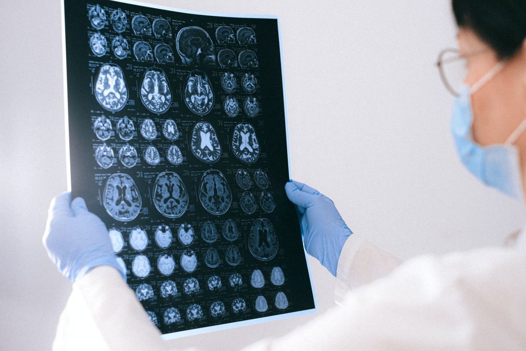
MRI images of the brain
Photo by Anna Shvets on Pexels

Researchers at Massachusetts General Hospital have discovered a novel molecular mechanism behind the most common forms of acquired hydrocephalus – which could lead to the first non-surgical treatments for the life-threatening disease. Research in animal models uncovered a pathway through which infection or bleeding in the brain triggers inflammation, causing increased production of cerebrospinal fluid (CSF) by the choroid plexus and lead to swelling of the brain ventricles.

“Finding a nonsurgical treatment for hydrocephalus, given the fact neurosurgery is fraught with tremendous morbidity and complications, has been the holy grail for our field,” says Kristopher Kahle, MD, PhD, a paediatric neurosurgeon at MGH and senior author of the study in the journal Cell. “We’ve identified through a genome-wide analytical approach the mechanism that underlies the swelling of the ventricles which occurs after a brain bleed or brain infection in acquired hydrocephalus. We’re hopeful these findings will pave the way for approval of an anti-inflammatory drug to treat hydrocephalus, which could be a game-changer for populations in the US and around the world that don’t have access to surgery.”

Occurring in about 0.2% of births, acquired hydrocephalus is the most common cause of brain surgery in children, though it can affect people at any age. In underdeveloped regions where bacterial infection is the most prevalent form, hydrocephalus is often deadly for children due to the lack of surgical intervention. Brain surgery, where a shunt is implanted to drain fluid from the brain, is the only known treatment. But about half of all shunts in paediatric patients fail within two years of placement, according to the Hydrocephalus Association, requiring repeat neurosurgical operations and a lifetime of brain surgeries.

Pivotal to the process is the choroid plexus, the brain structure that routinely pumps cerebrospinal fluid into the four ventricles of the brain to keep the organ buoyant and injury-free within the skull. An infection or brain bleed, however, can create a dangerous neuroinflammatory response where the choroid plexus floods the ventricles with cerebral spinal fluid and immune cells from the periphery of the brain in a cytokine storm, swelling the brain ventricles.

“Scientists in the past thought that entirely different mechanisms were involved in hydrocephalus from infection and from haemorrhage in the brain,” explains co-author Bob Carter, MD, PhD, chair of the Department of Neurosurgery at MGH. “Dr Kahle’s lab found that the same pathway was involved in both types and that it can be targeted with immunomodulators like rapamycin, a drug that’s been approved by the US Food and Drug Administration for transplant patients who need to suppress their immune system to prevent organ rejection.”

MGH researchers are continuing to explore how rapamycin and other drugs which quell the inflammation seen in acquired hydrocephalus could be repurposed. “What has me most excited is that this noninvasive therapy could provide a way to help young patients who don’t have access to neurosurgeons or shunts,” says Kahle. “No longer would a diagnosis of hydrocephalus be fatal for these children.”

Source: Massachusetts General Hospital

Why People with Asthma are Less Vulnerable to COVID

Photo by Sahej Brar on Unsplash

Researchers have revealed biological reasons for how disease progression happens and why a certain population of asthma patients are less susceptible to severe COVID.

This research, published in PNAS, shows the importance of the well-known cytokine interleukin-13 (IL-13) in protecting cells against SARS-CoV-2, something which helps explain why people with allergic asthma fare better than the general population despite having a chronic lung condition. However, the same cannot be said for individuals with other diseases, such as chronic obstructive pulmonary disease (COPD) or emphysema, who are at very high risk of severe COVID.

“We knew there had to be a bio-mechanistic reason why people with allergic asthma seemed more protected from severe disease,” said Assistant Professor Camille Ehre, PhD, senior author of the paper. “Our research team discovered a number of significant cellular changes, particularly due to IL-13, leading us to conclude that IL-13 plays a unique role in defence against SARS-CoV-2 infection in certain patient populations.”

Although cytokines like IL-13 cannot be used as therapies because they trigger inflammation, it is important to understand natural molecular pathways that cells use to protect themselves from pathogen invasion, as these studies have the potential to reveal new therapeutic targets.

Many health factors increase a person’s risk of severe COVID, but during the pandemic, epidemiologists found that people with allergic asthma were less susceptible to severe disease.

“These are patients with asthma caused by allergens, such as mould, pollen, and dander,” said A/Prof Ehre. “To find out why they are less susceptible, we investigated specific cellular mechanisms in primary human airway epithelial cell cultures.”

Genetic analysis human airway cell cultures infected with SARS-CoV-2 revealed that the expression of ACE2 governed which cell types were infected and their viral load.

Electron microscopy (EM) identified an intense exodus of virus from infected ciliated cells, which move mucus along the airway surface. EM also revealed severe cytopathogenesis – changes inside human cells due to viral infection. And these changes culminating in ciliated cells (packed with virions) shedding away from the airway surface.

“This shedding is what provides a large viral reservoir for spread and transmission of SARS-CoV-2,” A/Prof Ehre said. “It also seems to increase the potential for infected cells to relocate to deeper lung tissue.”

Further experiments on infected airway cells revealed that a major mucus protein called MUC5AC was depleted inside cells, likely because the proteins were secreted to try to trap invading viruses. But the virus load kept increasing because the cells tasked with producing MUC5AC were overwhelmed in the face of a rampant viral infection.

The researchers knew from epidemiological studies that allergic asthma patients—known to overproduce MUC5AC—were less susceptible to severe COVID. A/Prof Ehre and colleagues also knew the cytokine IL-13 increased MUC5AC secretion in the lungs when asthma patients faced an allergen.

The scientists decided to mimic asthmatic airways by treating human airway cells with IL-13. They then measured viral titres, viral mRNA, the rate of infected cell shedding, and the overall number of infected cells. Each one was significantly decreased. They found this remained true even when mucus was removed from the cultures, suggesting other factors were involved in the protective effects of IL-13 against SARS-CoV-2.

Bulk RNA-sequencing analyses revealed that IL-13 upregulated genes that control glycoprotein synthesis, ion transport, and antiviral processes – all of which are important in airway immune defence. They also showed that IL-13 reduced the expression of the viral receptor, ACE2, as well as reducing the amount of virus inside cells and cell-to-cell viral transmission.

Taken together, these findings indicate that IL-13 significantly affected viral entry into cells, replication inside cells, and spread of virus, thus limiting the virus’s ability to find its way deeper into the airways to trigger severe disease.

“We think this research further shows how important it is to treat SARS-CoV-2 infection as early as possible,” A/Prof Ehre said. “And it shows just how important specific mechanisms involving ACE2 and IL-13 are, as we try our best to protect patients from developing severe infections.”

Source: University of North Carolina Health Care