Tag: contrast agent

Amid Shortage, Suggested Ways to Conserve Contrast Agent

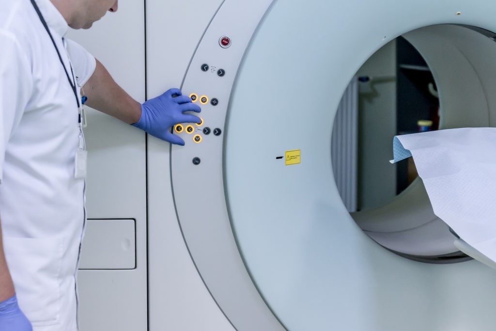
Technician and patient with MRI machine
Source: Mart Production on Pexels

Amid an ongoing worldwide shortage of contrast agent for medical imaging, a new UC San Francisco research letter in JAMA described strategies that can be used to safely reduce contrast agent use in computed tomography (CT) by up to 83%.

The three conservation strategies are weight-based (rather than fixed) dosing, reducing contrast dose while reducing tube voltage on scanners, and replacing contrast-enhanced CT with nonenhanced CT when it will minimally affect diagnostic accuracy.

That third strategy – not using the contrast agent in certain CT scans where there is only a small improvement in accuracy – yielded the most dramatic reduction of contrast agent use: 78%.

“Contrast is essential in any situation where we need to assess the blood vessels – for example, for some trauma patients or those with a suspected acute gastrointestinal bleed – and it is also needed for evaluation of certain cancers, such as in the liver or pancreas,” said senior study author Rebecca Smith-Bindman, MD, professor at UCSF.

“However, most CT scans are done for less specific indications such as abdominal pain in a patient with suspected appendicitis,” Prof Smith-Bindman added. “These can and should be done without contrast during the shortage, because the loss of information in these patients will be acceptable for most patients.”

The global shortage of contrast agent started in April with a COVID-related supply chain disruption of GE Healthcare in Shanghai and is expected to last at least several more weeks. More than 54 million diagnostic imaging exams using contrast agents are done every year in the US, a majority being CT scans, and these conservation methods could continue past the current shortage to reduce the use of contrast agent in general, the authors noted.

Referring clinicians are key to conservation
Researchers modelled the three strategies individually and in combination using a sample of 1.04 million CT exams in the UCSF International CT Dose Registry from January 2015 to March 2021.

On its own, weight-based dosing for abdomen, chest, cardiac, spine and extremity imaging reduced contrast agent use by 10%; reducing the tube voltage in appropriate patients allowed a contrast agent reduction of 25%. These two measures combined with using non-contrast CT when possible led to a total reduction of 83%.

Following all three strategies at once may not be possible for some facilities, but each can help conserve supply, Prof Smith-Bindman said. And it is not just radiologists who need to know about them.

“Given the acute shortage, it’s important that clinicians who order imaging exams coordinate with radiology to cancel scans that aren’t absolutely necessary, postpone exams that can be safely delayed, replace CT with MRI and ultrasound where possible, and order an unenhanced scan where possible. Further, clinicians should communicate with their patients about why this is necessary. It is crucial that contrast be conserved for clinical situations where its use is essential for accurate diagnosis,” said Prof Smith-Bindman.

After the shortage ends, medical facilities should consider continuing some of these practices that conserve contrast agent, she added. For example, reducing the tube voltage not only reduces the contrast agent used but also lowers the radiation dose. Tailoring doses weight allows lower dosing volumes for many patients.

In addition, Prof Smith-Bindman noted that this analysis highlights the large amount of contrast agent that is wasted when single-dose vials are used Hospitals and imaging centres that routinely use single-dose contrast agent vials should consider using larger multi-dose vials, which allows for exact dosing and obviates the need to discard unused portions, she said.

“By carrying some of these practices forward, we can mitigate future supply-chain risk and reduce overall waste,” said Smith-Bindman.

Source: University of California – San Francisco

Hypersensitivity Link Between MRI and X-Ray Contrast Agents

Photo by Mart Production on Pexels

People with a history of hypersensitivity to iodine-based contrast agents for X-ray based scans, are also susceptible to similar reactions from commonly used MRI contrast agents, according to a large, eight-year cohort study. The study, published in the journal Radiology, also found that premedication or switching to a different MRI contrast agent may reduce risk in patients who have had previous contrast agent reactions.

For a long time, gadolinium-based contrast agents (GBCA) have been used to enhance visualisation of organs, tissues and blood vessels on MRI and provide a more accurate depiction of disease. Though GBCA are relatively safe, recent studies have reported several adverse reactions related to their use, including allergic-like hypersensitivity reactions, such as rash and flushing.

These reactions are increasing in incidence with the widespread use of GBCA, prompting an urgent need for research into risk factors, according to the study’s senior author Hye-Ryun Kang, MD, PhD.

Analysing more than 330 000 cases of GBCA exposure in 154 539 patients over an eight-year period, the researchers found 1304 cases of allergic-like hypersensitivity reactions, for a rate of 0.4%. In patients who had a previous GBCE reaction, the average recurrence rate was 15%.

Acute allergic-like hypersensitivity reactions, or those that occur within one hour of contrast administration, accounted for 1178 cases, while a far smaller number of 126 cases were delayed allergic-like hypersensitivity reactions, or those that occur beyond the first hour and mostly within one week after exposure.

The risk of allergic-like hypersensitivity reactions to GBCAs was higher in those with a history of similar reactions to iodinated contrast media. Normally, having a history of iodinated contrast media hypersensitivity was not thought to be a risk factor for hypersensitivity to GBCAs and vice versa, because of their structural and compositional differences.

“The results of our study challenge this idea,” Dr Kang said.

An underlying predisposition to drug allergies in susceptible patients could be the cause, Dr Kang said, as opposed to any cross-reactivity associated with structural similarities between iodinated contrast media and GBCA. In fact, the risk of hypersensitivity reactions to iodinated contrast media was also higher in those who previously experienced a similar reaction to GBCA.

“Thus, physicians should be aware that patients with a history of hypersensitivity to one of iodinated contrast media or GBCA are at greater risk of developing hypersensitivity reactions to the other,” she said.

Analysis of the data showed that premedication, typically with steroids and antihistamines, and changing the GBCA showed preventive effects in patients with a history of acute allergic-like hypersensitivity reactions. Patients who received premedication and before MRI or were switched to a different GBCA showed the lowest rate of recurrence. Only premedication significantly reduced the incidence of reactions in patients with a history of delayed reactions.

“As the most important preventive measure is avoidance of the culprit agent, a precise record of previously used GBCA should be kept for all patients,” Dr. Kang said. “Physicians should discuss appropriate premedication strategies with their patients prior to MRI procedures.”

Dr Kang nevertheless stressed that contrast-enhanced MRI examinations are invaluable in the diagnosis and follow-up of various diseases, and the overall risk remains low.

“As most of these reactions are mild, we believe the benefits of MRI outweigh the potential risks associated with GBCA use,” she said.

Dr Kang recommended that in all patients receiving an MRI with GBCA exposure, a detailed history of previous hypersensitivity allergic reactions be conducted, and when necessary, appropriate prevention measures should be implemented, such as using premedication and switching to different GBCA types.

Future work would be to perform studies with larger populations to identify possible risk factors and effective preventive strategies for delayed hypersensitivity reactions to GBCA.

Source: Radiological Society of North America

Manganese Sharpens MRI Scans of Heart Attacks

Clinician prepares an MRI scanner. Image by Michal Jarmoluk from Pixabay

Manganese, a common trace mineral, could improve MRI scans of hearts after a heart attack and guide therapy, according to a new study.

By far the most widely used contrast agent for MRI is gadolinium, which improves the visibility of different organs and tissue types in MRI scans. However, it is taken up equally by cells regardless of their activity, and spreads out in damaged tissue. Furthermore, there are also extremely rare instances of serious kidney damage from its use. 

Manganese, besides being less toxic, has a useful property in that it competes with calcium uptake. Calcium handling is highly sensitive to altered heart muscle viability and changes rapidly after damage. Manganese ions enter heart muscle cells through calcium channels, and thus give a useful surrogate for heart tissue viability.

The contrast agent was tested first in vitro with heart muscle cells, and then in mice which had a myocardial infarction (heart attack) induced. The manganese contrast agent was administered with a calcium supplement or administered slowly to negate the effects of manganese interfering with the heart’s calcium channel. Findings were evaluated by examining the infarct size and blood supply at three key intervals: one hour, one day and 14 days after a myocardial infarction was induced. Overall, the manganese contrast agent was superior to gadolinium.

These findings could have major implications for heart attack treatment, if confirmed. They could also be greatly useful in preclinical evaluation of treatments for patients with cardiac ischaemia – where blood supply to the heart muscle is reduced, possibly leading to cardiac arrest.

Furthermore, if manganese-enhanced MRI is performed within the first few hours of a heart attack it could be used to determine the optimal treatment regime for individual patients – helping to regulate changes in the cardiac muscle and thereby further improving survival chances. 

“Magnetic resonance imaging (MRI) is increasingly used to diagnose and give information on heart conditions,” said lead researcher Dr Patrizia Camelliti, Senior Lecturer in Cardiovascular Science, University of Surrey. “This research using mice allows us to measure the health status of the heart muscle rapidly after a heart attack and could provide important information for optimizing treatments in patients.”

Source: News-Medical.Net

Journal reference: Jasmin, N.H., et al. (2021) Myocardial Viability Imaging using Manganese‐Enhanced MRI in the First Hours after Myocardial Infarction. Advanced Science. doi.org/10.1002/advs.202003987.