Tag: circadian rhythm

Scientists Discover a Secret to Regulating our Circadian Rhythm

Photo by Mert Kahveci on Unsplash

A team of scientists in Singapore and the US uncovered how a protein that controls our biological clock modifies its own function, offering new ways for treating jet lag and seasonal adjustments

Scientists from Duke-NUS Medical School and the University of California, Santa Cruz, have discovered the secret to regulating our internal clock. They identified that this regulator sits right at the tail end of Casein Kinase 1 delta (CK1δ), a protein which acts as a pace setter for our internal biological clock or the natural 24-hour cycles that control sleep-wake patterns and other daily functions, known as circadian rhythm.

Published in the journal PNAS, their findings could lead to new treatments for circadian rhythm disorders.

CK1δ regulates circadian rhythms by tagging other proteins involved in circadian rhythm to fine-tune the timing of these rhythms. In addition to modifying other proteins, CK1δ itself can be tagged, thereby altering its own ability to regulate the proteins involved in running the body’s internal clock.

Previous research identified two distinct versions of CK1δ, known as isoforms δ1 and δ2, which vary by just 16 building blocks or amino acids right at the end of the protein in a part called the C-terminal tail. Yet these small differences significantly impact CK1δ’s function. While it was known that when these proteins are tagged, their ability to regulate the body clock decreases, no one knew exactly how this happened.

Using advanced spectroscopy and spectrometry techniques to zoom in on the tails, the researchers found that how the proteins are tagged is determined by their distinct tail sequences.

Professor Carrie Partch at the University of California, Santa Cruz and corresponding author of the study explained:

“Our findings pinpoint to three specific sites on CK1δ’s tail where phosphate groups can attach, and these sites are crucial for controlling the protein’s activity. When these spots get tagged with a phosphate group, CK1δ becomes less active, which means it doesn’t influence our circadian rhythms as effectively. Using high-resolution analysis, we were able to pinpoint the exact sites involved—and that’s really exciting.”

Having first studied this protein more than 30 years ago while investigating its role in cell division, Professor David Virshup, the director of the Cancer and Stem Cell Biology Programme at Duke-NUS and co-corresponding author of the study, elaborated:

“With the technology we have available now, we were finally able to get to the bottom of a question that has gone unanswered for more than 25 years. We found that the δ1 tail interacts more extensively with the main part of the protein, leading to greater self-inhibition compared to δ2. This means that δ1 is more tightly regulated by its tail than δ2. When these sites are mutated or removed, δ1 becomes more active, which leads to changes in circadian rhythms. In contrast, δ2 does not have the same regulatory effect from its tail region.”

This discovery highlights how a small part of CK1δ can greatly influence its overall activity. This self-regulation is vital for keeping CK1δ activity balanced, which, in turn, helps regulate our circadian rhythms.

The study also addressed the wider implications of these findings. CK1δ plays a role in several important processes beyond circadian rhythms, including cell division, cancer development, and certain neurodegenerative diseases. By better understanding how CK1δ’s activity is regulated, scientists could open new avenues for treating not just circadian rhythm disorders but also a range of conditions.

The researchers plan to further investigate how real-world factors, such as diet and environmental changes, affect the tagging sites on CK1δ. This could provide insights into how these factors affect circadian rhythms and might lead to practical solutions for managing disruptions.

Source: Duke-NUS Medical School

The Colours of the Sunset Reset Circadian Clocks

Photo by Matteo Vistocco on Unsplash

Those mesmerising blue and orange hues in the sky at the start and end of a sunny day might have an essential role in setting humans’ internal clocks. In new research from the University of Washington in Seattle, a novel LED light that emits alternating wavelengths of orange and blue outpaced two other light devices in advancing melatonin levels in a small group of study participants. 

Published in the Journal of Biological Rhythms, the finding appears to establish a new benchmark in humans’ ability to influence their circadian rhythms, and reflects an effective new approach to counteract seasonal affective disorder (SAD). 

A raft of health and mood problems have been attributed to out-of-sync circadian rhythms. Such asynchrony is encouraged by seasonal changes, a lack of exposure to natural light, graveyard-shift jobs and flights across multiple time zones.  

“Our internal clock tells us how our body’s supposed to act during different times of day, but the clock has to be set, and if our brain is not synced to the time of day, then it’s not going to work right,” said Jay Neitz, a coauthor on the paper and a professor of ophthalmology at the UW School of Medicine. 

Circadian rhythms are trained and reset every day by the 24-hour solar cycles of light and dark, which stimulate circuits in the eyes that communicate to the brain. With that information, the brain produces melatonin, a hormone that helps organisms become sleepy in sync with the solar night. 

People who spend many daily hours in artificial light often have circadian rhythms whose melatonin production lags that of people more exposed to natural light. Many commercial lighting products are designed to offset or counteract these lags. 

Most of these products, Neitz said, emphasise blue wavelength because it is known to affect melanopsin, a photopigment in the eyes that communicates with the brain and is most sensitive to blue. 

By contrast, “the light we developed does not involve the melanopsin photopigment,” Neitz explained. “It has alternating blue and orange wavelengths that stimulate a blue-yellow opponent circuit that operates through the cone photoreceptors in the retina. This circuit is much more sensitive than melanopsin, and it is what our brains use to reset our internal clocks.”

The paper’s lead author was James Kuchenbecker, a research assistant professor of ophthalmology at the UW School of Medicine. He sought to compare different artificial lights’ effects on the production of melatonin.  

He and colleagues devised and conducted a test of three devices:

  • a white light of 500 lux (a brightness appropriate for general office spaces)
  • a short-wavelength blue LED designed to trigger melanopsin
  • the newly developed LED of blue and orange wavelengths, which alternate 19 times a second to generate a soft white glow

The goal was to see what lighting approach was most effective at advancing the phase of melatonin production among six study participants. All participants underwent the following regimen with exposure to each of the three test lights:

The first evening, multiple saliva samples were taken to discern the baseline onset and peak of the participants’ melatonin production. For each subject, the onset of this phase dictated when they were exposed to the test light for two hours in the morning. That evening, saliva samples were again taken to see whether subjects’ melatonin phase had started earlier relative to their individual baselines.

During each test, exposure to other light sources was controlled. The three test spans were spaced such that subjects could return to their normal baseline phases before starting a new device.

In terms of shifting the melatonin-production phase, the alternating blue-orange LED device worked best, with a phase advance of 1 hour, 20 minutes. The blue light produced a phase advance of 40 minutes. The white, 500-lux light elicited an advance of just 2.8 minutes. 

Gesturing toward the light that his team developed, Neitz explained. 

“Even though our light looks like white to the naked eye, we think your brain recognizes the alternating blue and orange wavelengths as the colours in the sky. The circuit that produced the biggest shift in melatonin wants to see orange and blue.” 

Source: UW Medicine

When is the Best time of Day for Chemotherapy?

Photo by Malvestida on Unsplash

Researchers from Charité are developing new methods to use the internal clock inside tumour cells to optimise cancer therapies

One of the factors determining the effectiveness of certain medications depends on various factors, including the time of day when they are administered. This is due to circadian rhythms, which vary across individuals and makes it difficult to tailor medication schedules. Researchers at Charité – Universitätsmedizin Berlin have now developed a method for determining the optimum time of cancer treatment based on certain breast cancer cell lines. They describe their approach in the journal Nature Communications.

As well as bodily functions and metabolic processes, such as sleep and digestion, individual cells also follow a circadian rhythm. This is hugely important to chemotherapy. Previous studies have shown that chemotherapy is most effective when the tumour cells are dividing. But this finding has been hardly used at all in clinical treatment to date.

An interdisciplinary team at Charité headed by Dr. Adrián Enrique Granada from the Charité Comprehensive Cancer Center (CCCC) set out to close this gap. The team began looking for the optimum time to administer medication, based on the individual circadian rhythms of the tumors.

Triple-negative breast cancer as an example

“We cultured cells from patients with triple-negative breast cancer to observe how they respond at different times of day to the medications administered,” explains Carolin Ector, a research associate in Granada’s working group. Triple-negative breast cancer is a highly aggressive form of breast cancer, with few effective treatments available. “We used live imaging, a method of continuously monitoring living cells, and complex data analysis techniques to monitor and evaluate the circadian rhythms, growth cycles, and medication responses of these cancer cells in detail.”

In this way, the researchers identified certain times of the day at which cancer cells are most responsive to medication-based treatments. For example, the chemotherapeutic drug 5-fluorouracil (5-FU) turned out to have peak efficacy against a certain cancer cell line between eight and ten a.m. As the study also shows, the crucial aspects here are certain cellular and genetic factors. The scientists were even able to identify which genes are key to the circadian effects of certain medications. “We call them ‘core clock genes’. They have a significant impact on how responsive cancer cells are to treatments administered at different times of day,” Granada explains.

Profiles show how cancer cell types respond to medications

This approach can be used to create detailed profiles showing how different types of cancer cells respond to different medications at various times. “This can help to identify the most effective combinations of drugs,” Granada says. “Overall, our findings indicate that personalized treatment plans based on individual circadian rhythms could substantially improve the efficacy of cancer treatment”, he concludes. Moreover, undesirable side effects could also be reduced.

For these findings to contribute to clinical practice soon, the results should be validated in studies involving larger groups of patients. “We’re also planning to study the molecular mechanisms behind the circadian influences on medication sensitivity to further optimize treatment times and identify new therapeutic targets,” Granada says.

Source: Charité – Universitätsmedizin Berlin

Timed Therapy with Intense Light can Benefit Cardiovascular Health

Photo by Stormseeker on Unsplash

Managing circadian rhythms through intense light and chronologically timed therapy can help prevent or treat a variety of circulatory system conditions including heart disease, according to a new study published in Circulation Research.

“The impact of circadian rhythms on cardiovascular function and disease development is well established,” said the study’s lead author Tobias Eckle, MD, PhD, professor of anaesthesiology at the University of Colorado School of Medicine.

“However, translational preclinical studies targeting the heart’s circadian biology are just now emerging and are leading to the development of a novel field of medicine termed circadian medicine.”

The senior author is Professor Tami A. Martino, PhD, distinguished chair in molecular and cardiovascular research at the University of Guelph in Ontario, Canada.

The study reviews current circadian medicine research, focusing on the use of intense light therapy following surgery, utilizsng light to treat cardiac injury, exploring how cardiovascular disease can differ between men and women and administering drugs at specific times of day to coincide with the body’s internal clock to speed healing.

It also urges more aggressive use of this therapy in humans, rather than relying on mostly animal models.

“There are literally millions of patients who could benefit from this,” Eckle said.

“The treatments are almost all low-risk. Some involve using light boxes and others use drugs that are already on the market.”

Circadian rhythms significantly influence how the cardiovascular system operates. Timing is everything. Blood pressure and heart rates follow distinct patterns, peaking during the day and ebbing at night. When this is disrupted, it leads to worse cardiovascular disease outcomes including myocardial infarction and heart failure. Light is critical in maintaining the proper balance and functioning of the body. Shift employees who may work night hours then day hours often have worse cardiac outcomes.

Eckle, who has studied circadian rhythm and health for years, said intense light can help heal the body after heart surgery while protecting it from injury during surgery, including reducing the chances of cardiac ischemia.

According to the researchers, when light hits the human eye it is transmitted to the suprachiasmatic nucleus, a structure in the brain’s hypothalamus that regulates most circadian rhythms in the body.

Intense light stabilizes the PER2 gene and increases levels of adenosine, which blocks electrical signals in the heart that cause irregular rhythms, making it cardiac protective.

Eckle has used light therapy with patients after surgery and seen positive results including lower levels of troponin, a key protein whose elevation can signal a heart attack or stroke.

Given the mounting evidence that intense light and timed drug treatments are effective, he said, it is time to move forward with more clinical trials.

“Circadian rhythms play a crucial role in cardiovascular health, influencing the timing of onset and severity of cardiovascular events and contributing to the healing process from disease,” Eckle said. “Studies in humans are clearly required. Regarding intense light therapy, chronotherapy and restricted feeding are low-risk strategies that should be tested sooner than later.”

Source: University of Colorado Anschutz Medical Campus

In Type 2 Diabetics, Toxic Lipids and a Beneficial One Surge at Certain Times

Credit: Cell Reports Medicine (2023).

While sugar is most frequently blamed in the development of type 2 diabetes, a better understanding of the role of fats is also essential. By analysing the blood profiles of dozens of people suffering from diabetes or pre-diabetes, or who have had their pancreas partially removed, researchers at the University of Geneva (UNIGE) and Geneva University Hospitals (HUG) have made two major discoveries.

Firstly, the lipid composition of blood and adipose tissues fluctuates during the day, and is altered in a day-time dependent manner in diabetics, who have higher levels of toxic lipids. In addition, one type of lipid, lysoPI, is capable of boosting insulin secretion when the beta cells that normally produce it fail. These results, published in the journals Cell Reports Medicine and Diabetes, may have important implications for the treatment of diabetic patients.

The role of lipids in the physiological and pathological processes of human metabolism is gradually becoming clearer, particularly in type 2 diabetes, one of the most widespread serious metabolic disorders. Thanks to cutting-edge tools, in particular mass spectrometry, researchers are now able to simultaneously measure the levels of several hundred different types of lipids, each with its own specific characteristics and beneficial or harmful effects on our metabolism.

‘‘Identifying which lipids are most present in type 2 diabetics could provide a basis for a wide range of interventions: early detection, prevention, potential therapeutic targets or personalised recommendations – the possibilities are immense,’’ says Charna Dibner, a professor in the Department of Surgery and a specialist in circadian rhythms in metabolic disorders, . ‘‘This is why we carried out a detailed analysis of the blood profiles of patients recruited in four European countries and confirmed some of our results on a mouse model of the disease.’’

Dibner led the studies along with Pierre Maechler, a professor in the Department of Cell Physiology and Metabolism, at the UNIGE Faculty of Medicine, and members of the Diabetes Faculty Centre.

Chronobiology to better identify diabetes

The team carried out a ‘‘lipidomic’’ analysis of two groups of patients in order to establish the profile, over a 24-hour cycle, of multiple lipids present in the blood and adipose tissues. ‘‘The differences between the lipid profiles of type 2 diabetics and people without diabetes are particularly pronounced in the early morning, when there is an increase in certain toxic lipids,’’ explains Dibner. ‘‘Why? We don’t know yet. But this could be a marker of the severity of diabetes and paves the way for personalised care according to each patient’s specific chronotype.”

And implications go beyond diabetes: if samples are taken at very different times of the day, the results can be distorted and give contradictory results. ‘‘It’s the same thing in the clinic: an examination carried out in the morning or evening, or a treatment taken at different times, can have an impact on diagnosis and even on the effectiveness of treatments.’’

A crutch for beta cells

Charna Dibner and Pierre Maechler extended their lipidomic analyses to include not only people with type 2 diabetes but also a mouse model of pre-diabetes and patients who had lost around half their insulin-producing beta cells after a surgery. ‘‘We discovered that a type of lipid, lysoPIs, increases when there is a sharp decrease in functional β cells, even before the onset of clinical symptoms of diabetes.’’

The scientists then administered lysoPI to diabetic mice and observed an increase in insulin production. ‘‘The same phenomenon occurred in vitro, on pancreatic cells from diabetic patients,’’ adds Pierre Maechler. ‘‘The lysoPIs therefore have the capacity to reinforce insulin secretion by acting as a crutch when the number of beta cells decreases or when these cells malfunction. Yet, certain foods, such as legumes, naturally contain lysoPI precursors.’’

By bringing to light the unsuspected role of lysoPIs, researchers will be able to explore new avenues opened by their discoveries. The development of dietary supplements or even molecules specific to lysoPI receptors could be an interesting strategy for controlling diabetes, as could taking better account of the chronobiological profiles of patients. Diabetes is a complex disease that calls for much more personalised management than is currently the case.

Source: University of Geneva

Night Owls have Nearly Double the Incidence of Atherosclerosis

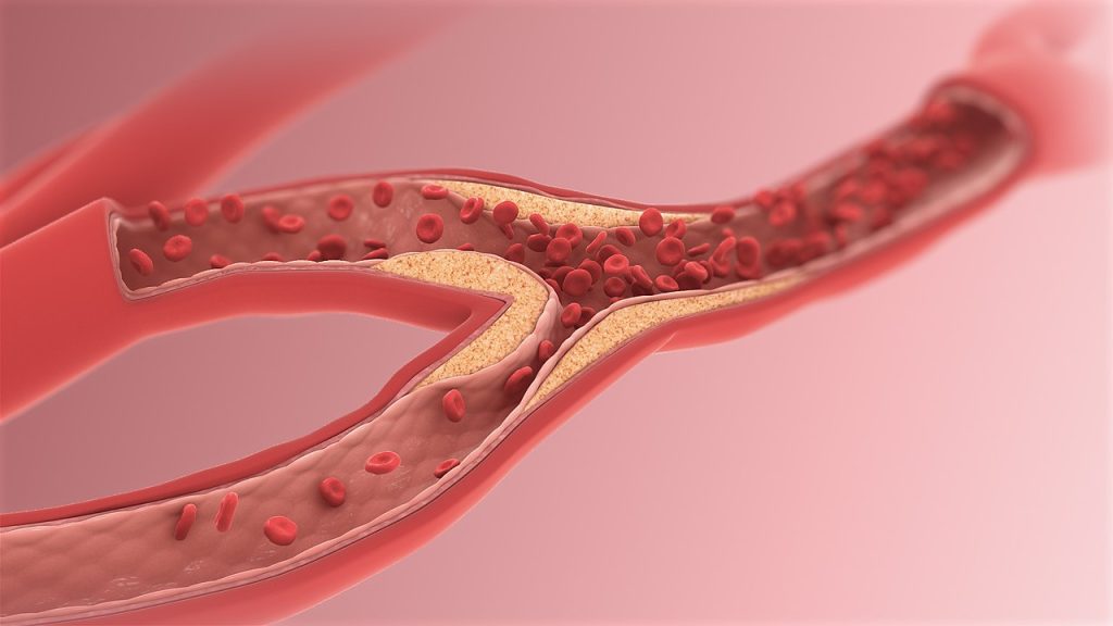
Image by Scientific Animations, CC4.0

Atherosclerosis is almost twice as common in night owls compared to early birds, according to a study from the University of Gothenburg, Sweden. Circadian function appears to be particularly important during the early stages of cardiovascular disease.

Atherosclerosis involves fatty deposits gradually accumulating on the inside of the arteries, making it harder for blood to pass through. The disease is usually not noticed until it leads to blood clots causing angina, heart attack, or stroke.

Previous research has shown that people with late-night habits have an increased risk of cardiovascular disease, but this is the first study to show how circadian rhythms specifically affect calcification of the arteries.

Coronary artery calcification

The study, which has been published in the journal Sleep Medicine, involved 771 men and women aged between 50 and 64, all of whom are part of the larger population study SCAPIS.

The degree of artery calcification in the heart’s coronary arteries was examined using computer tomography.

Participants themselves indicated their so called chronotype on a five-point scale: extreme morning type, moderate morning type, intermediate type, moderate evening type, or extreme evening type.

Of the 771 participants, 144 identified as extreme morning types, and 128 as extreme evening types.

Among the group who were most alert in the morning, 22.2% had pronounced artery calcification — the lowest proportion of all five chronotypes.

The extreme evening type group had the highest prevalence of severe coronary artery calcification, at 40.6%.

The first author of the study is Mio Kobayashi Frisk, a doctoral student at Sahlgrenska Academy, University of Gothenburg:

“Our results indicate that extreme evening chronotype may be linked not only to poorer cardiovascular health in general, but also more specifically to calcification in the coronary arteries calcification and atherosclerosis,” Mio Kobayashi Frisk says.

Preventive treatment

The statistical analysis considered a range of other factors that can affect the risk of atherosclerosis, including blood pressure, blood lipids, weight, physical activity, stress level, sleep, and smoking.

The last author of the study is Ding Zou, a researcher at Sahlgrenska Academy, University of Gothenburg:

“As well as the previously known factors, the individual circadian rhythm also appears to be an important risk factor for atherosclerosis. We interpret our results as indicating that circadian rhythms are more significant early in the disease process. It should therefore particularly be considered in the preventive treatment of cardiovascular diseases,” says Ding Zou.

Self-reported chronotype

Those who had experienced a heart attack were excluded from the study, meaning that the study participants were healthier than the general population.

Another weakness identified by the researchers is that participants themselves provided their chronotype.

Each chronotype can be said to have an average time when half of the night’s sleep has passed.

In a previous study on the same population, though not necessarily the same individuals, this time occurred at 02:55 AM for the extreme morning type group and at 04:25 AM for the extreme evening type group.

With the remaining chronotype groups’ mid-sleep times were somewhere in between these extremes.

Source: University of Gothenburg

Brain Changes from Shift Work Increase Appetite

Photo by Ernest Brillo on Unsplash

Scientists have uncovered why night shift work is associated with changes in appetite in a new University of Bristol-led study. The study shows that circadian disruption can disrupt the brain’s regulation of appetite hormones. The findings, published in Communications Biology, could help the millions of people that work through the night and struggle with weight gain.

Scientists from Bristol and the University of Occupational and Environmental Health in Japan, sought to understand how ‘circadian misalignment’ – a phenomenon commonly associated with ‘jet-lag’ whereby the body’s biological clock is disrupted – affects the hormones responsible for regulating appetite.

Prevalent in night shift workers, in this new study, the international team reveal how circadian misalignment can profoundly alter the brain’s regulation of hormones controlling hunger to the detriment of metabolic health.

The team focused on glucocorticoid hormones in the adrenal gland which regulate many physiological functions including metabolism and appetite. Glucocorticoids are known to directly regulate a group of brain peptides controlling appetitive behaviour, with some increasing appetite (orexigenic) and some decreasing appetite (anorexigenic).

In an experiment using animal models, comprising a control group and a out-of-phase ‘jet-lagged’ group, the team found misalignment between light and dark cues led the out-of-phase group’s orexigenic hypothalamic neuropeptides (NPY) to become dysregulated, driving an increased desire to eat significantly more during the inactive phase of the day.

Strikingly, the team discovered that rats in the control group ate 88.4% of their daily intake during their active phase, and only 11.6% during their inactive phase. In contrast, the ‘jet-lagged’ group consumed 53.8% of their daily calories during their inactive phase (without an increase in activity during this time). This equated to nearly five-times more (460% more) than what the control group consumed during the inactive phase. These results show that it is timing of consumption that has been affected.

This new discovery revealed how completely, and significantly, disordered the neuropeptides become when daily glucocorticoid levels are out of synch with light and dark cues. However, the authors suggest the neuropeptides identified in this study may be promising targets for pharmacological treatments for eating disorders and obesity.

Research Fellow Dr Becky Conway-Campbell, the study’s senior author, said: “For people working throughout the night, a reversed body clock can play havoc with their health.

“For those who are working night shifts long-term, we recommend they try to maintain daylight exposure, cardiovascular exercise and mealtimes at regulated hours. However, internal brain messages to drive increased appetite are difficult to override with ‘discipline’ or ‘routine’ so we are currently designing studies to assess rescue strategies and pharmacological intervention drugs. We hope our findings also provide new insight into how chronic stress and sleep disruption leads to caloric overconsumption.”

Professor Stafford Lightman, co-senior author on the study, added: “The adrenal hormone corticosterone, which is normally secreted in a circadian manner, is a major factor in the daily control of brain peptides that regulate appetite. Furthermore when we disturb the normal relationship of corticosterone with the day to night light cycle it results in abnormal gene regulation and appetite during the period of time that the animals normally sleep.

“Our study shows that when we disturb our normal bodily rhythms this in turn disrupts normal appetite regulation in a way that is at least in part a result of desynchrony between adrenal steroid hormone production and the timing of the light and dark cycle.”

Dr Benjamin Flynn, one of the study’s co-authors who conducted the study while at Bristol but is now based at the University of Bath, added: “This is further evidence of how phase shift ‘jet-lag’ affects feeding behaviours and neuronal gene expression – data important for shift work co-morbidity research.”

Source: University of Bristol

Aripiprazole Improves Sleep in Psychiatric Disorders by Entrainment to Light/Dark Cycles

Photo by Cottonbro on Pexels

Researchers in Japan have shown that the commonly prescribed antipsychotic drug aripiprazole helps reduce sleep disruptions in patients with certain psychiatric disorders by improving their natural entrainment to light and dark cycles. Their findings are published in Frontiers in Neuroscience.

Many patients with psychiatric conditions, such as bipolar disorder and major depressive disorder, frequently experience disruptions in their sleep–wake cycles. Research has shown that the administration of aripiprazole, a commonly prescribed antipsychotic drug, alleviates the symptoms of circadian sleep disorders in these patients. This improvement may be attributed to the effects of aripiprazole on the circadian central clock, specifically the hypothalamic suprachiasmatic nucleus (SCN), which regulates various circadian physiological rhythms, including the sleep–wake cycle, in mammals. However, the precise mechanism through which aripiprazole addresses these sleep disorder symptoms remains elusive.

Researchers from the University of Tsukuba have discovered that aripiprazole can directly affect the mammalian central circadian clock; specifically, it can modulate the photic entrainment in mice. Located in the hypothalamic suprachiasmatic nucleus (SCN), the central circadian clock comprises clock neurons that synchronize with each other, maintaining a roughly 24-hour rhythm. Simultaneously, SCN is receptive to external inputs like light, aligning itself with the environmental light-dark cycle. The researchers have found that aripiprazole disrupts the synchronization among the clock neurons in the SCN, heightening the responsiveness of these neurons to light stimuli in mice. Additionally, aripiprazole influences intracellular signalling within the SCN by targeting the serotonin 1A receptor, a prominent receptor in the SCN.

These findings suggest that the efficacy of aripiprazole in alleviating circadian rhythm sleep disorder symptoms in psychiatric patients might be attributed to the modulation of the circadian clock by the drug. This study expands the potential clinical usage of aripiprazole as a treatment for circadian rhythm sleep disorders.

Source: University of Tsukuba

Study Shows that Intermittent Fasting Might Improve Alzheimer’s Symptoms

Photo by Matteo Vistocco on Unsplash

Circadian disruption is a hallmark of Alzheimer’s disease, affecting nearly 80% of patients with issues such as difficulty sleeping and worsening cognitive function at night. Currently there are no treatments for Alzheimer’s that target this aspect of the disease.

A new study in Cell Metabolism from researchers at University of California San Diego School of Medicine has shown in mice that it is possible to correct the circadian disruptions seen in Alzheimer’s disease with time-restricted feeding, a type of intermittent fasting focused on limiting the daily eating window without limiting the amount of food consumed.

In the study, mice that were fed on a time-restricted schedule showed improvements in memory and reduced accumulation of amyloid proteins in the brain. The authors say the findings will likely result in a human clinical trial.

“For many years, we assumed that the circadian disruptions seen in people with Alzheimer’s are a result of neurodegeneration, but we’re now learning it may be the other way around – circadian disruption may be one of the main drivers of Alzheimer’s pathology,” said senior study author Paula Desplats, PhD, professor at UC San Diego School of Medicine. “This makes circadian disruptions a promising target for new Alzheimer’s treatments, and our findings provide the proof-of-concept for an easy and accessible way to correct these disruptions.”

People with Alzheimer’s experience a variety of disruptions to their circadian rhythms, including changes to their sleep/wake cycle, increased cognitive impairment and confusion in the evenings, and difficulty falling and staying asleep.

“Circadian disruptions in Alzheimer’s are the leading cause of nursing home placement,” said Desplats. “Anything we can do to help patients restore their circadian rhythm will make a huge difference in how we manage Alzheimer’s in the clinic and how caregivers help patients manage the disease at home.”

Boosting the circadian clock is an emerging approach to improving health outcomes, and one way to accomplish this is by controlling the daily cycle of feeding and fasting. The researchers tested this strategy in a mouse model of Alzheimer’s disease, feeding the mice on a time-restricted schedule where they were only allowed to eat within a six-hour window each day. For humans, this would translate to about 14 hours of fasting each day.

Compared to control mice who were provided food at all hours, mice fed on the time-restricted schedule had better memory, were less hyperactive at night, followed a more regular sleep schedule and experienced fewer disruptions during sleep. The test mice also performed better on cognitive assessments than control mice, demonstrating that the time-restricted feeding schedule was able to help mitigate the behavioral symptoms of Alzheimer’s disease.

The researchers also observed improvements in the mice on a molecular level. In mice fed on a restricted schedule, the researchers found that multiple genes associated with Alzheimer’s and neuroinflammation were expressed differently. They also found that the feeding schedule helped reduce the amount of amyloid protein that accumulated in the brain. Amyloid deposits are one of the most well-known features of Alzheimer’s disease.

Because the time-restricted feeding schedule was able to substantially change the course of Alzheimer’s in the mice, the researchers are optimistic that the findings could be easily translatable to the clinic, especially since the new treatment approach relies on a lifestyle change rather than a drug.

“Time-restricted feeding is a strategy that people can easily and immediately integrate into their lives,” said Desplats. “If we can reproduce our results in humans, this approach could be a simple way to dramatically improve the lives of people living with Alzheimer’s and those who care for them.”

Light Therapy may Relieve Alzheimer’s Circadian Disruption

Photo by Matteo Vistocco on Unsplash

New Alzheimer’s research suggests that enhanced light sensitivity may contribute to ‘sundowning’, which is the worsening of symptoms late in the day, thereby spurring sleep disruptions thought to contribute to the disease’s progression.

Published in Frontiers in Aging Neuroscience, these new insights from UVA Health into the disruptions of the biological clock seen in Alzheimer’s could lead to new treatments and symptom management, the researchers say. For example, caregivers often struggle with the erratic sleep patterns caused by Alzheimer’s patients’ altered circadian rhythms. Light therapy, the new research suggests, might be an effective tool to help manage that.

Better understanding Alzheimer’s effects on circadian rhythms could have implications for prevention. Poor sleep quality in adulthood is a risk factor for Alzheimer’s, as brains at rest naturally cleanse themselves of amyloid beta proteins that are thought to form harmful tangles in Alzheimer’s.

“Circadian disruptions have been recognised in Alzheimer’s disease for a long time, but we’ve never had a very good understanding of what causes them,” said researcher Thaddeus Weigel, a graduate student working with Heather Ferris, MD, PhD. “This research points to changes in light sensitivity as a new, interesting possible explanation for some of those circadian symptoms.”

Alzheimer’s hallmark is progressive memory loss, to the point that patients can forget their own loved ones, but there can be many other symptoms, such as restlessness, aggression, poor judgment and endless searching. These symptoms often worsen in the evening and at night.

Ferris and her collaborators used a mouse model of Alzheimer’s to better understand what happens to the biological clock in Alzheimer’s disease. They essentially gave the mice “jet lag” by altering their exposure to light, then examined how it affected their behaviour. The Alzheimer’s mice reacted very differently to control mice.

The Alzheimer’s mice, the scientists found, adapted to a six-hour time change significantly more quickly than the control mice. This, the scientists suspect, is the result of a heightened sensitivity to changes in light. While our biological clocks normally take cues from light, this adjustment happens gradually – thus, jet lag when we travel great distances. Our bodies need time to adapt. But for the Alzheimer’s mice, this change happened abnormally fast.

The researchers initially thought this might be because of neuroinflammation. So they looked at immune cells called microglia that have become promising targets in developing better Alzheimer’s treatments. But the scientists ultimately ruled out this hypothesis, determining that microglia did not make a difference in how quickly mice adapted. (Though targeting microglia might be beneficial for other reasons.)

Notably, the UVA scientists also ruled out another potential culprit: “mutant tau,” an abnormal protein that forms tangles in the Alzheimer’s brain. The presence of these tangles also did not make a difference in how the mice adapted.

The researchers’ results ultimately suggest there is an important role for the retina in the enhanced light sensitivity in Alzheimer’s, and that gives researchers a promising avenue to pursue as they work to develop new ways to treat, manage and prevent the disease.

“These data suggest that controlling the kind of light and the timing of the light could be key to reducing circadian disruptions in Alzheimer’s disease,” Ferris said. “We hope that this research will help us to develop light therapies that people can use to reduce the progression of Alzheimer’s disease.”

Source: University of Virginia Health System