Tag: chronic traumatic encephalopathy

Heads up – School Rugby and Head Injuries Explained

The rugby season is kicking off in schools across South Africa and players, parents, coaches and referees are preparing for exciting, yet physically demanding matches. In many sports, injuries are an unfortunate, common occurrence. Rugby, inherently a contact sport, also carries the inevitable risk of head injuries, ranging from minor concussions to severe Traumatic Brain Injuries (TBIs).

The importance of early detection

The early detection of head injuries is essential for effective treatment and preventing further complications. In many cases, the symptoms of a concussion or TBIs may not be immediately apparent and athletes may continue playing which can lead to further damage.

Accurate diagnosis and management of head injuries require a combination of clinical evaluation and advanced imaging techniques. Dr Hofmeyr Viljoen, radiologist at SCP Radiology talks about the nature of these injuries, the critical role radiology plays in diagnosing and managing them and what preventative measures can be taken.

Understanding head injuries in rugby

Dr Viljoen explains that there are several types of head injuries common in rugby. ‘The most frequent is concussion, a mild traumatic brain injury occurring when the brain is jolted inside the skull from an impact or violent movement. Concussions can be mild or lead to significant short and long-term issues. Occasionally, with more severe injuries we see skull fractures, contusions and haemorrhage surrounding the brain. These require urgent diagnosis and management.’

Recognising the symptoms

He emphasises awareness of concussion symptoms, including headaches, dizziness, nausea, confusion, memory problems, sensitivity to light and difficulty concentrating. ‘Immediate recognition is vital,’ he explains. ‘A player with any of these symptoms must be removed from play immediately to prevent further injury.’

The role of radiology

Radiology plays an essential part in accurately diagnosing the extent of head injuries. According to Dr Viljoen, Computed Tomography (CT) scans are always the first imaging method used in emergency settings. Although patients with concussion typically do not have significant imaging findings, it is crucial to image those patients with severe concussion or atypical symptoms. ‘CT scans rapidly detect serious issues like fractures, brain swelling and bleeding, providing crucial information for urgent treatment decisions,’ he explains.

Magnetic Resonance Imaging (MRI) is used in situations requiring more detailed evaluation, particularly when concussion symptoms persist or worsen. ‘MRI excels in identifying subtle injuries, such as microbleeds and brain swelling, often missed by CT scans,’ says Dr Viljoen. Unlike CT scans, MRI does not use radiation, making it a safer option for repeated assessments over time.

Advanced imaging methods

Emerging imaging techniques, such as Diffusion Tensor Imaging (DTI), show promise for better understanding and management of head injuries, especially the subtle effects of concussions. ‘DTI helps identify damage to the brain’s white matter, potentially guiding return-to-play decisions and treatment strategies,’ notes Dr Viljoen.

Understanding possible complications – Second Impact Syndrome (SIS)

SIS is a rare but extremely serious condition that occurs when a person sustains a second concussion before fully recovering from an initial concussion. This second injury doesn’t have to be severe to trigger SIS – it can even be minor – but it causes rapid and severe brain swelling (cerebral oedema).

The brain’s ability to regulate its blood flow and pressure is compromised following the initial concussion, making it vulnerable to catastrophic swelling after a subsequent impact. Symptoms can escalate quickly, often within minutes, including loss of consciousness, severe headache, dilated pupils, respiratory failure and even death. Young athletes are especially vulnerable to SIS. Due to its rapid progression and severity, SIS is considered a medical emergency requiring immediate intervention.

Preventing SIS involves strictly adhering to concussion management protocols, ensuring full recovery after any head injury and carefully monitoring symptoms before returning to sports or high-risk activities.

Addressing Chronic Traumatic Encephalopathy (CTE)

Dr Viljoen says CTE is a long-term degenerative brain condition linked to repeated head impacts. ‘CTE is challenging because currently, it can only be definitively diagnosed after death.  However, ongoing research aims to develop methods to detect CTE in living patients, potentially using advanced imaging techniques like Positron Emission Tomography (PET).’ Most research is focused on advancing non-invasive methods to see what is happening inside the brain of a living person and to track it over time.

Common causes of head injuries in rugby

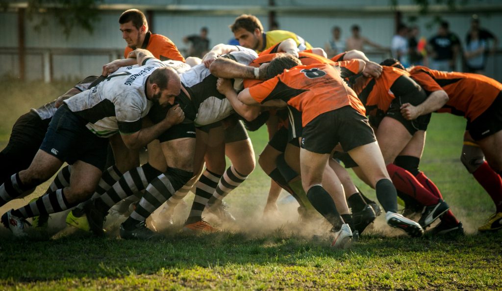
  • These primarily arise from the high-impact nature of the sport, with tackling identified as a significant risk factor. Tackling, particularly when performed incorrectly or at a dangerous height, frequently leads to head trauma. Young players are especially vulnerable as their tackling techniques may not yet be fully developed, increasing the likelihood of injury. Teaching safe and correct tackling methods early is a way to mitigate these risks
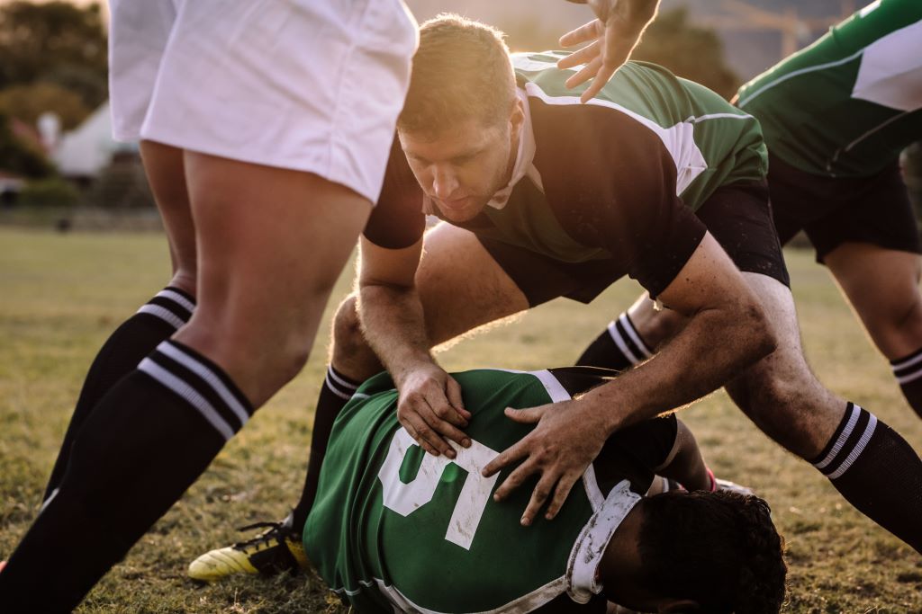
  • Rugby’s dynamic gameplay often results in players being brought down forcefully or falling awkwardly. Even with protective gear, the impact of the head striking the playing surface can lead to concussions or more severe trauma
  • Due to the speed and intensity of the game, unintended impacts between players are inevitable. These include clashes of heads or impacts from knees and elbows, which can result in injuries ranging from mild concussions to more severe brain injuries. Preventative strategies and safer playing practices can reduce these risks

Prevention remains critical

Dr Viljoen emphasises the importance of proper training: ‘Educating young players on safe tackling techniques and enforcing protective protocols significantly reduces injury risks. Protective gear like headguards can minimise superficial injuries, though it does not prevent concussions.’

He also stresses the importance of concussion protocols. ‘Coaches at schools and clubs must rigorously apply concussion management strategies, ensuring players are adequately assessed and cleared by medical professionals before returning to the field.’ Under-reporting in schoolboy ruby often occurs because the player either wants to stay in the game and/or doesn’t recognise the symptoms of concussion.

Dr Viljoen concludes, ‘Rugby is a fantastic sport for building teamwork and resilience but player safety must always come first. Through awareness, timely medical intervention and proper preventative strategies, we can significantly reduce the risk and severity of head injuries, allowing young athletes to safely enjoy the game they love.’

Soccer Headers may be More Damaging to Brain than Thought

Photo by Kenny Webster on Unsplash

Soccer heading may cause more damage to the brain than previously thought, according to a study presented at the annual meeting of the Radiological Society of North America (RSNA).

Heading is a widely used technique in soccer where the players control the direction of the ball by hitting it with their head. In recent years, research has been done that suggests a link between repeated head impacts and neurodegenerative diseases, such as chronic traumatic encephalopathy (CTE).

“The potential effects of repeated head impacts in sport are much more extensive than previously known and affect locations similar to where we’ve seen CTE pathology,” said study senior author Michael L. Lipton, MD, PhD, professor of radiology at Columbia University Irving Medical Center. “This raises concern for delayed adverse effects of head impacts.”

While prior studies have identified injuries to the brain’s white matter in soccer players, Dr Lipton and colleagues used a new approach in diffusion MRI to analyse microstructure close to the surface of the brain.

To identify how repeated head impacts affect the brain, the researchers compared brain MRIs of 352 male and female amateur soccer players, ranging in age from 18 to 53, to brain MRIs of 77 non-collision sport athletes, such as runners.

Soccer players who headed the ball at high levels showed abnormality of the brain’s white matter adjacent to sulci, which are deep grooves in the brain’s surface. Abnormalities in this region of the brain are known to occur in very severe traumatic brain injuries.

The abnormalities were most prominent in the frontal lobe of the brain, an area most susceptible to damage from trauma and frequently impacted during soccer heading. More repetitive head impacts were also associated with poorer verbal learning.

“Our analysis showed that the white matter abnormalities represent a mechanism by which heading leads to worse cognitive performance,” Dr Lipton said.

Most of the participants of the study had never sustained a concussion or been diagnosed with a traumatic brain injury. This suggests that repeated head impacts that don’t result in serious injury may still adversely affect the brain.

“The study identifies structural brain abnormalities from repeated head impacts among healthy athletes,” Dr Lipton said. “The abnormalities occur in the locations most characteristic of CTE, are associated with worse ability to learn a cognitive task and could affect function in the future.”

The results of this study are also relevant to head injuries from other contact sports. The researchers stress the importance of knowing the risks of repeated head impacts and their potential to harm brain health over time.

“Characterising the potential risks of repetitive head impacts can facilitate safer sport engagement to maximise benefits while minimising potential harms,” Dr Lipton said. “The next phase of the study is ongoing and examines the brain mechanisms underlying the MRI effects and potential protective factors.”

Source: Radiological Society of North America

Amnesia from Head Injury Reversed in Early Mouse Study

Photo by Olga Guryanova on Unsplash

A mouse-based study to investigate memory loss in people who experience repeated head impacts, such as athletes, suggests the condition could potentially be reversed. The research in mice finds that amnesia and poor memory following head injury is due to inadequate reactivation of neurons involved in forming memories.

The study, conducted by researchers at Georgetown University Medical Center in collaboration with Trinity College Dublin, Ireland, is reported in the Journal of Neuroscience.

Importantly for diagnostic and treatment purposes, the researchers found that the memory loss attributed to head injury was not a permanent pathological event driven by a neurodegenerative disease.

Indeed, the researchers could reverse the amnesia to allow the mice to recall the lost memory, potentially allowing cognitive impairment caused by head impact to be clinically reversed.

The Georgetown investigators had previously found that the brain adapts to repeated head impacts by changing the way the synapses in the brain operate, which can cause trouble in memory storage and retrieval.

In their new study, investigators were able to trigger mice to remember memories that had been forgotten due to head impacts.

“Our research gives us hope that we can design treatments to return the head-impact brain to its normal condition and recover cognitive function in humans that have poor memory caused by repeated head impacts,” says the study’s senior investigator, Mark Burns, PhD, a professor and Vice-Chair in Georgetown’s Department of Neuroscience and director of the Laboratory for Brain Injury and Dementia.

In the new study, the scientists gave two groups of mice a new memory by training them in a test they had never seen before. One group was exposed to a high frequency of mild head impacts for one week (similar to contact sport exposure in people) and one group were controls that didn’t receive the impacts. The impacted mice were unable to recall the new memory a week later.

“Most research in this area has been in human brains with chronic traumatic encephalopathy (CTE), which is a degenerative brain disease found in people with a history of repetitive head impact,” said Burns.

“By contrast, our goal was to understand how the brain changes in response to the low-level head impacts that many young football players regularly experience.”

Researchers have found that, on average, college football players receive 21 head impacts per week with defensive ends receiving 41 head impacts per week.

The number of head impacts to mice in this study were designed to mimic a week of exposure for a college football player, and each single head impact by itself was extraordinarily mild.

Using genetically modified mice allowed the researchers to see the neurons involved in learning new memories, and they found that these memory neurons (the “memory engram”) were equally present in both the control mice and the experimental mice.

To understand the physiology underlying these memory changes, study first author Daniel P. Chapman, PhD, said, “We are good at associating memories with places, and that’s because being in a place, or seeing a photo of a place, causes a reactivation of our memory engrams. This is why we examined the engram neurons to look for the specific signature of an activated neuron. When the mice see the room where they first learned the memory, the control mice are able to activate their memory engram, but the head impact mice were not. This is what was causing the amnesia.”

The researchers were able to reverse the amnesia to allow the mice to remember the lost memory using lasers to activate the engram cells.

“We used an invasive technique to reverse memory loss in our mice, and unfortunately this is not translatable to humans,” Burns adds.

“We are currently studying a number of non-invasive techniques to try to communicate to the brain that it is no longer in danger, and to open a window of plasticity that can reset the brain to its former state.”

Source: Georgetown University Medical Center

  1. Daniel P. Chapman, Sarah D. Power, Stefano Vicini, Tomás J. Ryan, Mark P. Burns. Amnesia after repeated head impact is caused by impaired synaptic plasticity in the memory engramThe Journal of Neuroscience, 2024; e1560232024 DOI: 10.1523/JNEUROSCI.1560-23.2024