Tag: chemotherapy

Could a Simple Dietary Change Increase Platelet Counts?

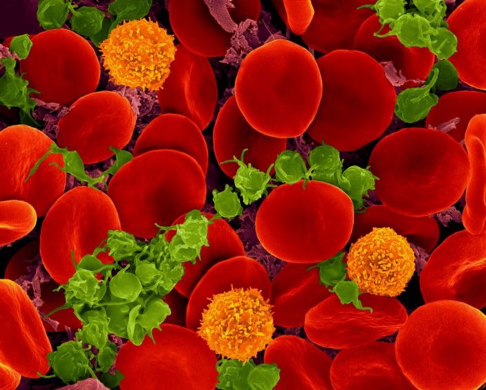
Scanning electron micrograph of red blood cells, T cells (orange) and platelets (green). Source: Wikimedia CC0

Aside from transfusions, there currently is no way to boost people’s platelet counts, which can drop for reasons such as chemotherapy, leaving them at risk for uncontrolled bleeding. But new research published in Nature Cardiovascular Research suggests that there could be a simple alternative: a dietary change in type of fat intake could raise platelet counts in people with low levels.

A study led by Kellie Machlus, PhD, and Maria Barrachina, PhD at Boston Children’s Hospital found that they could raise platelet counts in mice by feeding them polyunsaturated fatty acids (PUFAs) like those found in the Mediterranean diet. In contrast, mice fed a diet high in saturated fatty acids had decreased platelet counts.

“We were honestly surprised at how profound the effects were,” says Machlus, whose lab focuses on studying platelets and their precursor cells, megakaryocytes, and ways to get the body to increase platelet production.

But equally interesting is the apparent reason for the dietary effect.

“What brought me to the idea of diet is that megakaryocytes make these long extensions from their membrane when they form platelets,” Machlus says. “We thought the membrane must have an unusual composition to make it so fluid.”

A fluid megakaryocyte membrane

No one had studied megakaryocyte membranes before, perhaps because megakaryocytes are in the bone marrow and hard to access. Machlus, Barrachina, and their colleagues decided to comprehensively assess the membranes’ fat content with lipidomics.

“We found that PUFAs are enriched in megakaryocytes, especially right before they begin making platelets,” says Machlus. “We think they provide the fluidity necessary for the membrane to move and reshape.”

In culture, the megakaryocytes with higher amounts of PUFAs in their membrane made more platelets. When the cells were instead supplied with saturated fats as their lipid source, platelet production declined. The same thing happened when the team added compounds to inhibit uptake of PUFAs from the blood.

The researchers also identified one of the receptors on megakaryocytes that’s responsible for taking up PUFAs from blood: CD36. When they deleted the gene for CD36 in their mouse model, the animals had low platelet counts.

Serendipitously, the researchers were able to connect the dots to humans. Through a colleague in the U.K., they identified a family in which several members had a mutation in the CD36 gene. Those affected had low platelet counts and, in the mother’s case, bleeding episodes.

An olive oil intervention?

Intrigued by their findings, Barrachina hopes to extend the study by collaborating with a team in her native Spain. The team is studying dietary interventions for cardiovascular disease, including the Mediterranean diet.

“We want to look at platelets from these patients,” she says. She thinks that platelets with more saturated fatty acids in their membranes might be in a more activated state that could lead them to aggregate and form blood clots.

While Machlus thinks it may be worth encouraging patients with thrombocytopenia to consume more olive oil to increase PUFA levels, she recognises that a drug treatment may be more practical.

“Our next steps are to find out the enzymes that create PUFAs,” she says. “Maybe we can target them to make more platelets.”

Source: Boston Children’s Hospital

Game-changing Therapy Targets Colon Cancer that has Spread to the Liver

Source: CC0

Physicians at Cedars-Sinai Cancer are now using a unique therapy, called hepatic artery infusion (HAI) pump chemotherapy, that offers hope to colorectal cancer patients whose disease has spread and who now have inoperable liver tumours. The system, which was developed over two decades ago, is only now being adopted more widely, also spares the rest of the body from much of the chemotherapy drugs’ toxicity.

“Many of these patients are not candidates for curative surgery and we now have a meaningful option for treating them,” said Cristina Ferrone, MD, chair of the Department of Surgery at Cedars-Sinai and a specialist in the care of patients with complex hepato-pancreato-biliary disorders. “This therapy has been shown to extend both life and quality of life.”

Colorectal cancer is the fourth-leading cause of cancer-related death in the US. In as many as 25% of patients diagnosed with the disease, the cancer spreads to the liver, where it can be difficult to treat. However, more than half of patients receiving hepatic artery infusion pump therapy go on to receive curative surgery, studies have shown.

Cedars-Sinai Cancer and associate professor of Surgery at Cedars-Sinai, sat down with the Cedars-Sinai Newsroom to explain this lifesaving therapy.

How do the pumps work?

We surgically place the pump underneath the skin, outside of the abdominal cavity, and it is attached to tubing that enters the abdominal cavity and goes into the gastroduodenal artery. That artery feeds into the hepatic artery, which supplies blood to the liver. During surgery, we block blood flow from the gastroduodenal artery from going into portions of the small intestine so that the therapy flows only to the liver.

The pump has a soft centre, allowing its internal reservoir to be filled through the skin via a syringe. After surgery, the patient comes in every two weeks and we refill the pump, which then allows the chemotherapy drug to flow directly into the liver via the arterial supply.

Which patients are likely to benefit from hepatic artery infusion pump therapy?

This therapy is designed for patients, based on the distribution of the metastatic disease (where are the tumours and how many), for whom curative surgery is not an option at the time of diagnosis. The best we had been able to offer these patients was lifelong chemotherapy that had potential systemic toxicities, and that never quite reduced their tumour size to the point that we could surgically remove it. This therapy offers an additional option for liver-directed therapy that can potentially make patients candidates for surgery by specifically targeting the liver disease.

What are the advantages of the hepatic artery infusion pump over traditional chemotherapy delivery?

A majority of these tumours derive their blood supply from the hepatic arterial system, and delivering chemotherapy to the tumours through the hepatic arterial system allows us to give higher doses of specific chemotherapeutic agents without exposing the patient to their systemic toxicities. Data shows that up to 60% of appropriately selected patients receiving hepatic artery infusion pump chemotherapy were then able to receive curative surgery. Patients can often continue receiving systemic chemotherapy in combination with hepatic artery infusion pump chemotherapy.

Are hepatic artery infusion pumps used to treat other types of liver cancer?

Some patients with cholangiocarcinoma are currently treated with HAI pumps, but this is not yet standard of care. Colon cancer is the second most common cancer, and colon cancer that has metastasized to the liver affects a significant number of patients. And we have seen good outcomes with those patients. Other types of cancers that metastasise to the liver are significantly more challenging to treat, and thus far, we don’t think this therapy will benefit those patients.

Is this a new therapy?

Hepatic artery infusion pumps have actually been around for about 25 to 30 years, but until quite recently only a few medical centres were using them. But more and more centres are realising that this therapy can truly benefit patients, and it is becoming more widely available.

Source: Cedars-Sinai Medical Center

Study Reveals Why Cancer Drugs Cause Cardiotoxicity

While being effective at treating cancer, some cancer treatments can cause cardiotoxicity which can lead to heart failure – a phenomenon unexplained until now. An international study, published in the journal Science Advances, has identified proteins present in the blood that are linked to an increased risk of developing cardiac disease, including heart failure, and which are also affected by drugs used in cancer treatment.

The findings can explain how cancer drugs cause their damaging effects on the heart and could help to identify those at increased risk. In the long run, the researchers believe this will help to improve cancer treatments, with new drugs potentially being developed that can shrink tumours without affecting the identified proteins.

In addition, the study reveals new potential drug targets for treating heart diseases including heart failure. These may work by inhibiting proteins linked to higher disease risk, or activating proteins linked to lower risk.

The researchers first performed a genome-wide association study, searching through the DNA of nearly 37,000 people without heart disease enrolled in the UK Biobank study. This identified genetic variants linked to changes to the structure and function of the pumping chambers of the heart – the ventricles.

The researchers then pinpointed 33 proteins using Mendelian randomisation, coded for by these genetic variants, that are present in the blood and associated with the risk of developing several heart diseases. These included different types of heart failure, and atrial fibrillation (a common abnormal heart rhythm which increases the risk of stroke). Crucially, many of these proteins are the targets of drugs currently used to treat cancer.

Lead author Dr Floriaan Schmidt said: “The proteins identified in our study will help to accelerate future drug development, offering scientists a blueprint for new treatments for both cancer and heart diseases. This can help them to be more confident of the effects of the drugs that they design – whether that’s shrinking tumours without causing damage elsewhere or improving the heart’s pumping action.”

Professor Sir Nilesh Samani, Medical Director at the British Heart Foundation, said: “While there have been advances in treating cancer, one of the consequences has been a risk of heart damage from these drugs.

“This research points the way towards developing safer and more refined drugs so that, one day, worries about developing heart problems after cancer treatment might be a thing of the past.”

Source: University College London

For Women, Lymphoma Chemotherapy more Effective in Afternoon

Photo by Malvestida on Unsplash

While chemotherapy is highly effective at killing cancer cells, it also kills healthy cells, something which medical research is trying minimise. Recently, ‘chronochemotherapy’ has garnered growing attention in the research community. As the name suggests, the aim is timing the delivery of the drug when the body is least vulnerable to the harmful effects of the drug, while the cancer cells are at their most vulnerable.

Chronochemotherapy exploits the fact that human physiological processes, including cell proliferation and differentiation, are regulated by the circadian clock. However, it is not yet widely exploited in real-world clinical settings because there at present is no systematic method to find the optimal chemotherapy delivery time.

This problem was tackled by an interdisciplinary team of researchers from the Institute for Basic Science (IBS), South Korea. They were led by the principal investigators, mathematician Kim Jae Kyoung at IBS and oncologist Koh Youngil at Seoul National University Hospital. The researchers studied a group of patients suffering from diffuse large B-cell lymphoma (DLBCL), which accounts for about 30 to 40% of non-Hodgkin’s lymphoma. Their findings are published in the journal JCI Insights.

The research team noticed that DLBCL patients at Seoul National University Hospital received chemotherapy at two different schedules, with some patients receiving morning treatment (8:30), while others taking the drugs in the afternoon (14:30). All patients received R-CHOP, which is a combination of targeted therapy and chemotherapy, 4 to 6 times in the morning or afternoon at intervals of about 3 weeks.

Figure 1. Chemotherapy in the afternoon can improve treatment outcomes
The daily fluctuation of proliferative activity of bone marrow is larger in females than in males, and it becomes higher in the morning (left). Thus, chemotherapy in the morning strongly inhibits proliferative activity in female lymphoma patients, resulting in a higher incidence of adverse events such as neutropenia and infections. This forces the clinicians to reduce the dose intensity (center). Consequently, female patients undergoing the morning treatment show a lower survival probability than those undergoing the afternoon treatment (right). Specifically, only ~13% of female patients treated in the afternoon had a worse outcome and ~2% of them died while ~37% of female patients treated in the morning had a worse outcome and ~25% of them died. Male patients did not show any difference in treatment outcomes depending on the chemotherapy delivery time.

They analysed 210 patients to investigate differences between morning and afternoon treatment. They found that female patients who received afternoon treatment had 12.5 times reduced mortality rate (25% to 2%), while the cancer recurrence after 60 months was decreased by 2.8 times (37% to 13%). In addition, chemotherapy side effects such as neutropenia were more common in female patients who received morning treatment.

Surprisingly, there was no difference in treatment efficiency depending on the treatment schedule in the case of male patients.

To understand the cause of gender differences, the research team analysed ~14 000 blood samples from the Seoul National University Hospital Health Examination Center. It was found that in females, white blood cell count tends to decrease in the morning and increase in the afternoon. This indicates that the bone marrow proliferation rate is higher in the morning than in the afternoon because there is a ~12hr delay between the bone marrow proliferation and blood cell production.

This means that if a female patient receives chemotherapy in the morning when bone marrow is actively producing blood cells, the possibility of adverse side effects becomes greater. These results are consistent with the findings from recent randomised clinical trials that showed female colorectal cancer patients treated with irinotecan in the morning suffered from higher drug toxicities.

One confounding variable was the drug dose. Since the morning female patients suffered from greater adverse side effects, oftentimes the dose had to be reduced in these patients. On average, the drug dose was reduced by ~10% compared to the dose intensity given to female patients receiving the afternoon treatment.

Unlike female patients, it was found that male patients did not show a significant difference in white blood cell count and bone marrow cell proliferation activity throughout the day, which is the reason why the timing of the treatment had no impact.

Professor Koh Young-il said, “We plan to verify the conclusions of this study again with a large-scale follow-up study that completely controls confounding variables, and to confirm whether chemotherapy has similar effects in other cancers.”

Ci Kim Jae Kyung said, “Because the time of the internal circadian clock can vary greatly depending on the individual’s sleep-wake patterns, we are currently developing a technology to estimate the time of the circadian clock from the patient’s sleep pattern. We hope that it can be used to develop an individualised anti-cancer chronotherapy.”

Source: Institute for Basic Science

Turning a Traditional Chinese Medicinal Plant into a Cancer Fighter

Photo by Bundo Kim on Unsplash

The evolutionary secrets that enable the traditional Chinese medicinal herb known as barbed skullcap to produce cancer fighting compounds have been unlocked by a collaboration of UK and Chinese researchers, who published their research in the journal Molecular Plant.

The researchers used DNA sequencing technology to assemble the genomic sequence of skullcap (Scutellaria barbata) known in China as banzhilian. This gave researchers the genetic information, a microevolutionary history, required to identify how the plant produces the compound scutebarbatine A, which acts against a range of cancer cells.

Professor Cathie Martin, Group Leader at the John Innes Centre, and one of the authors of the study said, “We have found that the primary metabolite has activity against cancer cells but not non-cancer cells which is especially important for an anti-cancer metabolite. Now we are looking to develop synthetic methods for producing more of the lead compound.”

In Traditional Chinese Medicine (TCM), to isolate medicinal chemistry from the plant, the herb is boiled in water for two hours and extract is dried to produce a powder and taken as a decoction (concentrated liquid). Now, with the knowledge of the genes that make up the biochemical pathway behind the anti-cancer activity of the herb, researchers are close to being able to synthesise larger quantities of compounds more rapidly and sustainably by using a host such as yeast.

The research is led by CEPAMS, a partnership between the John Innes Centre and the Chinese Academy of Science and supported by The Royal Society.

“This is a fantastic collaboration about developing interesting drug leads from natural resources and shows the practical value of focusing on the microevolution of a species” said Professor Martin.

The Skullcap genus has been used for centuries in TCM for treatment of different medical conditions. Clinical work has shown that preparations based on Scutellaria barbata during chemotherapy can reduce the risk of metastatic tumours.

CEPAMS Group Leader based at Shanghai Dr Evangelos Tatsis said, “Natural products have long been the lead compounds for the discovery of new drugs. By following the trail of the traditional Chinese plants, we can develop new anti-cancer medicines and this research marks a crucial step in that direction.”

Plant-based traditional medicines have long been used to provide leads for the new drug discovery, leading to drugs such as vinblastine and taxol which are now used clinically as anticancer drugs.

TCM is one of the best catalogued systems with empirical information about the therapeutic properties of herbal remedies.

Anti-cancer drugs obtained from traditional Chinese medicine have higher efficacy than chemical synthetic drugs and with less toxic side effects. The genomes of medicinal skullcaps reveal the polyphyletic origins of clerodane diterpene biosynthesis in the family Laminiaceae, is published in Molecular Plant

Source: John Innes Centre

Extended Chemotherapy Slashes Childhood Leukaemia Recurrence

Photo by National Cancer Institute on Unsplash

Giving three years of chemotherapy to children with acute lymphoblastic leukaemia (ALL) instead of two years lowers the risk of their disease coming back after treatment by three times. The survival rate of all children with ALL, the most common form of childhood cancer, together has further increased to 94%. Less intensive therapy proved safe for three groups of children, resulting in a better quality of life. These findings on a large Dutch study into ALL were reported at the annual conference of the American Society of Hematology (ASH).

Many children with ALL have good outcomes. After two years of chemotherapy treatment, nine out of ten children are cured. But some children have a more aggressive disease, such as having the Ikaros mutation in their leukaemia cells, have a greater risk of recurrence after treatment. In order to improve the chances of survival and quality of life of all children with leukaemia, the treatment protocol has been continuously adapted over the years, based on the latest scientific insights.

Prof Rob Pieters, medical director and paediatric oncologist at the Princess Máxima Center for paediatric oncology in the Netherlands, presented the outcomes of the ALL-11 treatment protocol. The Dutch researchers tested the benefit of an adapted treatment in specific groups of children with leukaemia, including children with the Ikaros mutation. More than 800 children in the Netherlands were treated with this protocol between April 2012 and July 2020.

Threefold lower risk of recurrence

Children with Ikaros leukaemia received an extra year of chemotherapy in the ‘maintenance phase’ on top of the first two years of treatment. This change lowered the risk of their cancer coming back by threefold: this happened in only 9% of them, compared to 26% of the children in the previous treatment protocol.

87% of children with Ikaros leukaemia survived their disease for five years without their cancer coming back, an improvement on the 72% in the previous protocol. Because of the extra year of chemotherapy, this group of children had a slightly higher risk of infection, but these were treatable. The extended therapy did not lead to any additional side effects.

Analysis of data from all children with ALL, regardless of subtype, showed that the five-year survival rate has improved stepwise over the past 30 years from 80% to 94% under the ALL-11 protocol.

Safe reduction of treatment

In the ALL-11 protocol, doctors and researchers also looked at the benefit of a less intensive treatment plan for three groups of children. This included children with a leukaemia mutation linked to a very high chance of recovery, and children with Down syndrome who experience more severe side effects. These children received treatment without or with a lower dose of anthracyclines, a type of leukaemia drug that increases the risk of heart damage and infections. The reduced treatment proved successful: children had the same or even a better chance of survival, while their quality of life improved due to a lower risk of infections and damage to the heart.

Global interest

Globally, there is much interest in the Dutch research as it has been unclear how to improve therapy for children with Ikaros leukaemia. The results have now been presented for the first time at the largest blood cancer conference, and could lead to changes in treatment protocols for these children worldwide.

In the Netherlands, there are about 15 children with ALL each year for whom existing treatments stop working. Since 2019, they have been eligible for treatment with CAR T-cell therapy, a promising form of immunotherapy that now leads to a cure in 40% of these children.

Making a difference

Prof Monique den Boer, medical biologist and group leader at the Princess Máxima Center, played an important role in the adapted therapy for children with the Ikaros gene change. She says: ‘The Ikaros mutation was first discovered about 15 years ago in children with leukaemia who had a poor prognosis, partly thanks to the emergence of new DNA technologies. We saw that the cancer came back in many of these children shortly after the end of the two-year treatment plan. I am very proud that our lab findings have now found their way into the clinic and can make such a big difference for children with leukaemia.”

More cure with fewer side effects

Prof Pieters concludes: The five-year survival rate for children with acute lymphoblastic leukaemia has increased enormously since the 1960s, from zero to 94%, but the last steps are the most difficult. We are now one step closer to curing all children with ALL. We have also largely been able to remove a drug that poses a risk of heart damage from the treatment of children with a less aggressive form of the disease. The latest results for children with leukaemia therefore fit in perfectly with our mission: curing more children with cancer, with fewer side effects.”

Source: Princess Máxima Center

Neurofeedback Could Help Alleviate ‘Chemo Brain’

Using neurofeedback to restore normal functioning in the brains of cancer patients could potentially alleviate the mental fogginess that many report after treatment, according to a new pilot study published in the Journal of Complementary and Integrative Medicine.

The UCLA study is one of the first to indicate that neurofeedback, or electroencephalogram (EEG) biofeedback, could help address cognitive deficits of cancer patients experiencing ‘chemo brain’, a constellation of symptoms that could include problems with memory, concentration and organisation, as well as other symptoms like trouble sleeping and emotional difficulties. In previous studies, neurofeedback, in which brain waves are trained to operate in optimal frequency patterns, has been shown to help improve cognitive function in patients with cognitive impairments like attention-deficit/hyperactivity disorder, stroke and seizures, as well as helped regulate brain activity in patients with substance use and post-traumatic stress disorders.

“The history of neurofeedback shows that it’s helpful for a whole range of disorders and symptoms. This study was an opportunity for seeing whether neurofeedback is something that could be helpful with chemo brain,” said study leader Stephen Sideroff, a UCLA professor who has used neurofeedback training with patients for over 20 years.

The study by Sideroff and UCLA colleagues David Wellisch and Valerie Yarema included nine female breast cancer patients between the ages of 21 and 65 who had completed chemotherapy at least one year earlier and complained of debilitating symptoms of chemo brain, which brought significant disruptions to their work and personal lives. A clinical nurse practitioner conducted a brief mental status interview with each patient to confirm that they had persistent difficulties with concentration, memory, organization and confusion. The patients selected for the study did not have a current breast cancer diagnosis, a present or recent diagnosis of a major depressive disorder or other mental illness, or used cognitive-altering medications that might confound study results.

Before the neurofeedback training sessions began, the study participants received neurocognitive and psychological tests, as well as a quantitative EEG to measure brain wave frequencies that could be compared to normative data. The pre-training quantitative EEGs shows that each study participant had abnormal brain waive activity compared to healthy adult brains.

The study participants received a series of 18 neurofeedback sessions, scheduled for 30 minutes each over a six-week period. During these sessions, sensors were placed on the scalp and earlobe to monitor brain wave frequencies. Patients were shown a monitor displaying these frequencies in bar graphs, and they were told their goal was to increase or decrease the amplitude of specific frequency ranges to turn each bar green. They received audio and visual feedback when they successfully shifted these amplitudes.

Quantitative EEGs taken after completion of the 18 neurofeedback sessions found that brain wave frequencies had significantly normalised in seven of the nine study participants, and significantly improved in the other two.

Neurocognitive tests also showed substantial improvements in the study participants’ information processing, executive set shifting and sustained visual attention. There was improvement for all in everyday functioning and overall psychological.

Study limitations include a small sample size and lack of a control group, and some were unable to complete the study in the allotted period.

“Our results are more impressive given we were not able to have subjects stick to the schedule,” Prof Sideroff said.

Prof Sideroff said the study results were strong enough to support further research into whether neurofeedback is an effective approach for addressing chemo brain and determining the ideal protocols for conducting neurofeedback training sessions.

Source: University of California – Los Angeles

Oxidative Stress Contributes to Multi-drug Resistance in Chemotherapy

Shown here is a pseudo-colored scanning electron micrograph of an oral squamous cancer cell (white) being attacked by two cytotoxic T cells (red), part of a natural immune response. Photo by National Cancer Institute on Unsplash

Researchers have found that oxidative stress plays a role the inevitable occurrence of multi-drug resistance during tumour therapy, which they report in the Journal of Biochemical and Molecular Toxicology

While chemotherapy is a mainstay of cancer treatment, it is often hindered by the development of drug resistance, eventually evolving into multidrug-resistance which renders most drugs ineffective. 

Multidrug resistance is responsible for over 90% of deaths in cancer patients receiving traditional chemotherapeutics or novel targeted drugs. Its mechanisms include elevated metabolism of xenobiotics, enhanced efflux of drugs, growth factors, increased DNA repair capacity, and genetic factors (gene mutations, amplifications, and epigenetic alterations).

The most well-known mechanism is the induction of Adenosinetriphosphate (ATP)-binding cassette (ABC) transporters by chemotherapeutic drugs. These transporters are highly expressed in cancer cells and pumped out chemotherapeutics to make the treatment ineffective. 

Earlier research had shown that non-substrate nanoparticles could induce multidrug resistance by inducing oxidative damage, suggesting that multidrug resistance could be induced by oxidative damage as well as the substrate. 

To confirm the this, Yin Jian and his team investigated the interaction of three chemical agents (ethanol, hydrogen peroxide, and doxorubicin) with ABC transporters using a lung cancer cell line (A549) as a model. 

Among the three chemicals, doxorubicin is the substrate of ABC transporter and chemotherapeutic drugs, while ethanol and hydrogen peroxide are small-molecule compounds, which have no relationship with the function of ABC transporter. 

“When the three substances enter the cells, they can cause significant oxidative stress inside cells,” said Yin.  

The elevated oxidative stress induced the expression of transporters, and the elevated transporters reduce intracellular oxidative stress by effluxing oxidized glutathione. In this process, pregnane X receptor played an important regulatory role. 

Their results suggested that non-substrate chemicals could also induce ABC transporter expressions similar to chemotherapeutic agents after inducing oxidative damage. This phenomenon could be regarded as a non-specific feedback of tumor cells/ABC transporters to external stimuli. 

The conclusions validated the relationship between multidrug resistance mechanisms and oxidative stress. This would help to design advanced strategies on how to enhance this mechanism to more effectively combat ABC transporter-mediated multidrug resistance.  

“Considering that peroxidative damage is the main source of the toxicity of current environmental pollutants, long-term exposure to environmental pollutants could not only induce direct toxicity, but also further threaten human health by inducing multi-drug resistance,” said Yin Huancai, another researcher from the team. 

Source: China Academy of Sciences

How Cancer Cells Repair their DNA so Quickly

DNA repair
Source: Pixabay/CC0

Research into how the body’s DNA repair process works has made a discovery into how the process works, and by understanding how cancer cells repair their DNA so rapidly may lead to potent new chemotherapy treatments.

One of the great mysteries of medical science is the ability of DNA to be repaired after damage, but complicating the study of this is how different pathways are involved in the repair process over the cell’s life cycle. In one of the repair pathways known as base excision repair (BER), the damaged material is removed, and proteins and enzymes work together to create DNA to fill in and then seal the gaps.

In a study appearing in Proceedings of the National Academy of Sciences, Eminent Professor Zucai Suo led a team that discovered that BER has a built-in mechanism to increase its effectiveness: it just needs to be captured at a very precise point in the cell life cycle.

In BER, an enzyme called polymerase beta (PolyB) fulfils two functions: It creates DNA, and it initiates a reaction to clean up the leftover ‘chemical junk’. Through five years of study, Prof Suo’s team learned that by capturing PolyB when it is naturally cross-linked with DNA, the enzyme will produce new genetic material 17 times faster than when the two are not cross-linked. This suggests that the two functions of PolyB are interlocked, not independent, during BER.

The research improves the understanding of cellular genomic stability, drug efficacy and resistance associated with chemotherapy.

“Cancer cells replicate at high speed, and their DNA endures a lot of damage,” Prof Suo said. “When a doctor uses certain drugs to attack cancer cells’ DNA, the cancer cells must cope with additional DNA damage. If the cancer cells cannot rapidly fix DNA damage, they will die. Otherwise, the cancer cells survive, and drug resistance appears.”

This research examined naturally cross-linked PolyB and DNA, unlike previous research that mimicked the process. Studies had previously identified the enzymes involved in BER but did not fully grasp how they work together.

“When we have nicks in DNA, bad things can happen, like the double strand breaking in DNA,” said Thomas Spratt, a professor of biochemistry and molecular biology at Penn State University College of Medicine who was not a part of the research team. “What Zucai found provides us with something we didn’t understand before, and he used many different methods to reach his findings.”

Source: Florida State University

COVID Vaccines less Effective in Patients Undergoing Chemotherapy

Photo by National Cancer Institute on Unsplash

New research has found that patients undergoing active chemotherapy had a lower immune response to two doses of the COVID vaccine, although a third dose increased response.

“We wanted to make sure we understand the level of protection the COVID vaccines are offering our cancer patients, especially as restrictions were being eased and more contagious variants were starting to spread,” said Rachna Shroff, MD, MS, University of Arizona Health Sciences.

To find out, Dr Shroff and colleagues looked at 53 Cancer Center patients on immunosuppressive active cancer therapy, such as chemotherapy. They compared the immune response following the first and second dose of the Pfizer-BioNTech COVID vaccine with that of 50 healthy adults. 

After two vaccine doses, most of the cancer patients showed some immune response to the vaccine in that they had produced antibodies for SARS-CoV-2.

“We were pleasantly surprised,” said Deepta Bhattacharya, PhD, professor of immunobiology in the College of Medicine – Tucson. “We looked at antibodies, B cells and T cells, which make up the body’s defense system, and found the vaccine is likely to be at least partially protective for most people on chemotherapy.”

However, this  immune response was much lower than in healthy adults, and a few of the patients had no response to the COVID  vaccine. This translates to less protection against SARS-CoV-2, especially the now-dominant Delta variant.

Twenty patients returned for a third shot, which boosted the immune response for most. The overall group immune response after the third shot reached levels similar to those of people who were not on chemotherapy after two doses.

The results were published in Nature Medicine.

Source: University of Arizona Health Sciences