Tag: celiac disease

Celiac Disease: New Findings on the Effects of Gluten

Photo by Mariana Kurnyk

May 16 is International Celiac Day. Celiac disease is a chronic autoimmune condition that occurs in around 1% of the world’s population. It is triggered by the consumption of gluten proteins from wheat, barley, rye and some oats. A gluten-free diet protects celiac patients from severe intestinal damage. Together with colleagues, chemist Dr Veronica Dodero from Bielefeld University was able to determine new details on how certain gluten-derived molecules trigger leaky gut syndrome in celiac disease.

The key finding of the study: a particular protein fragment formed in active celiac disease forms nanosized structures, the so-called oligomers, and accumulates in a gut epithelial cell model. The technical name of the molecule is 33-mer deamidated gliadin peptide (DGP). The study team has now discovered that the presence of DGP oligomers may open the tightly closed gut lining, leading to the leaky gut syndrome. The study has now been published in the journal Angewandte Chemie.

Wheat peptides causing leaky gut

Gluten proteins cannot be completely broken down by the gut. This can lead to the formation of large gluten fragments (peptides) in our gut. In cases of active coeliac disease, researchers discovered that the enzyme tissue transglutaminase 2 (tTG2) present in humans modifies a specific gluten peptide, resulting in the formation of the 33-mer DGP. This usually happens in a part of our gut called the lamina propria. However, recent research has shown that this process can also occur in the gut lining.

‘Our interdisciplinary team characterized the formation of 33-mer DGP oligomers through high-resolution microscopy and biophysical techniques. We discovered the increased permeability in a gut cell model when DGP accumulates, reports Dr. Maria Georgina Herrera, the first author of the study. She is researcher at the University of Buenos Aires in Argentina and was a postdoctoral fellow at Bielefeld.

When the intestinal barrier is weakened

Leaky gut syndrome occurs when the lining of the intestine becomes permeable, allowing harmful substances to enter the bloodstream, leading to inflammatory responses and different diseases. In celiac disease, there’s debate about the early stages of increased permeability. The mainstream theory suggests that chronic inflammation in coeliac disease leads to a leaky gut. However, there is a second theory that proposes that gluten’s effects on gut lining cells are the primary cause. In this view, gluten directly damages the cells of the intestinal lining, making them permeable, which triggers chronic inflammation and potentially leads to celiac disease in predisposed people.

However, since gluten is consumed daily, what molecular triggers lead to the leaky gut in celiac disease patients? If 33-merDGP oligomers are formed, they may damage the epithelial cell network, allowing gluten peptides, bacteria, and other toxins to pass massively into the bloodstream, leading to inflammation and, in celiac disease, autoimmunity.

‘Our findings reinforce the medical hypothesis that impairment of the epithelial barrier promoted by gluten peptides is a cause and not a result of the immune response in celiac patients,’ says the lead author of the study, Dr Veronica Dodero from the Bielefeld Faculty of Chemistry.

The relationship between 33-mer DGP and Celiac Disease

Human leukocyte antigens (HLAs) are proteins found on the surface of cells in the body. They play a crucial role in the immune system by helping it distinguish between self (the body’s own cells) and non-self (foreign substances like bacteria or viruses). In celiac disease, two specific HLA proteins, namely HLA-DQ2 and HLA-DQ8, are strongly associated with the condition. The 33-mer DGP fits perfectly with HLA-DQ2 or HLA-DQ8 and triggers an immune response, leading to inflammation and small intestine villous atrophy. This strong interaction turns the DGP into what scientists call a superantigen. For those affected, a gluten-free diet is the only lifelong therapy.

Source: Bielefeld University

People with Gluten Sensitivity have Negative Effects – Even When Eating a Placebo

Photo by Mariana Kurnyk

Symptoms of gluten sensitivity are partly to do with people’s expectations, if celiac disease and wheat allergy have been excluded as causes. Recent research at the universities of Maastricht and Leeds shows that the expectation that gluten causes gastrointestinal complaints plays a crucial role in whether or not people experience these symptoms. These results, published in The Lancet Gastroenterology and Hepatology, indicate a direct involvement of the interaction between the brain and the intestines – the ‘gut-brain axis’ – in the experience of complaints after ingesting gluten.

Fear of gluten

A growing number of people are reducing their gluten intake due to self-reported digestive complaints, despite the fact that celiac disease and wheat allergy have been ruled out. The cause of their symptoms is often unclear. The researchers therefore wanted to investigate the effects of expectations on symptoms experienced after gluten intake. More than 80 subjects with self-reported gluten sensitivity took part in a psychological study and were divided into four groups. The results were unequivocal: people who thought they were eating food containing gluten reported more symptoms, while those who thought their food was gluten-free reported fewer symptoms.

In reality, the food given to half of each group contained gluten, while for the other half it was gluten-free. In all of the groups people’s expectations played a prominent role in whether or not they reported symptoms after eating. “In our research, we see a so-called nocebo effect when people eat gluten,” says researcher Marlijne de Graaf. “If people expect gluten to produce negative effects, they experience symptoms, even if it turns out afterwards that they weren’t actually eating gluten. Although the cause is partly ‘in the mind’, this doesn’t mean that the symptoms are not real.”

Gut-brain axis

The results of this study indicate a clear involvement of the interaction between the brain and the intestines in gluten sensitivity, a subject on which knowledge is as yet limited. The researchers therefore now want to concentrate on unravelling the mechanisms that determine the importance of expectation and exposure along the gut-brain axis. ‘Due to the influence of interactions between the brain and the intestines, people can genuinely experience symptoms such as stomach ache, bloating or diarrhoea after eating gluten,’ says Daisy Jonkers, professor of Intestinal Health at Maastricht University. ‘But the cause of these complaints is not only eating gluten, so a gluten-free diet isn’t the only solution.’

To treat this problem, the researchers want to conduct further studies on the influence of the brain on the development of bowel complaints. ‘For example, we’d like to know exactly which areas in the brain are involved,” says Jonkers, “and we also want to find out what substances play a role in the communication between the brain and the gut, and whether people might respond differently to them. It’s also quite possible that some people can’t tolerate wheat products because of substances in wheat other than gluten, and that there is indeed something in wheat that can lead to overstimulation of the immune system, for example, or excessive production of gas by the gut flora. This is also something we’d like to investigate.”

Source: Maastricht University

Clues from Autoimmune Disorder on Disrupted Tooth Enamel Development

Photo by Caroline Lm on Unsplash

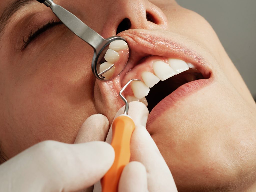
In one of every 10 people, and in one third of children with celiac disease, the enamel coating of the teeth appears defective, failing to protect the teeth properly. As a result, teeth become more sensitive to heat, cold and sour food, and they may decay faster. In most cases, the cause of the faulty enamel production is unknown.

Now, a study by Prof Jakub Abramson and his team at the Weizmann Institute of Science, published recently in Nature, may shed light on this problem by revealing a new children’s autoimmune disorder that hinders proper tooth enamel development. The disorder is common in people with a rare genetic syndrome and in children with celiac disease. These findings could help develop strategies for early detection and prevention of the disorder.

Tooth enamel is made up primarily of mineral crystals that are gradually deposited on protein scaffolds during enamel development. Once the crystals are in place, the protein scaffold is dismantled, leaving behind a thin, exceptionally hard layer of enamel. A strange phenomenon was identified in people with a rare genetic disorder known as APS-1: although the enamel layer of their milk teeth forms perfectly normally, something causes its faulty development in their permanent teeth. Since people with APS-1 suffer from a variety of autoimmune diseases, Abramson and his team hypothesised that the observed enamel defects may also be of an autoimmune nature

In autoimmune disease, to prevent T cells from triggering the immune system to attack body tissues, T cells developing in the thymus gland must be educated’ to discriminate between the body’s own proteins and those of foreign origin. To this end, T cells are presented with short segments of self-proteins that make up various tissues and organs in the body. When a ‘poorly educated’ T cell erroneously identifies a self-protein in the thymus as a target for attack, that T cell is labelled as dangerous and destroyed, so that it could not cause any damage after being released from the thymus.

This critical education step is impaired in APS-1 patients as a result of a mutation in a gene known as the autoimmune regulator (Aire). This gene is essential for the T cell education process: It produces a protein that is responsible for the collection of self-proteins presented to the T cells in the thymus. In their new study, scientists from Abramson’s lab in Weizmann’s Immunology and Regenerative Biology Department, led by research student Yael Gruper, sought to work out how mutations in the Aire gene lead to deficient tooth enamel production. The researchers discovered that, in the absence of Aire, proteins that play a key role in the development of enamel are not presented to the T cells in the thymus gland. As a result, T cells that are liable to identify these proteins as targets are released from the thymus, and they encourage the production of antibodies to the enamel proteins. But why do these autoantibodies damage permanent teeth and not baby teeth?

The answer to this question lies in the fact that milk teeth develop in the embryonic stage, when the immune system is not yet fully formed and cannot create autoantibodies. In contrast, the development of enamel on permanent teeth starts at birth and continues until around the age of six, when the immune system is sufficiently mature to thwart enamel development. Furthermore, the researchers found a correlation between high levels of antibodies to enamel proteins and the severity of the harm to enamel development in children with APS-1. This strengthens the assumption that the presence of enamel-specific autoantibodies in childhood can potentially lead to dental problems.

When the researchers looked into deficiencies in enamel development in people with other autoimmune diseases, they found a very similar phenomenon in children with celiac disease, a relatively common autoimmune disorder that affects around 1% of people in the West. When people with this disease are exposed to gluten, their immune system attacks and destroys the cellular layer lining the small intestine, leading to attacks on other self-proteins in the intestine.

In an attempt to understand how celiac disease, known to cause intestinal damage, may also cause damage to tooth enamel, the researchers first examined whether people with this disease have autoantibodies against enamel. They found that a large proportion of celiac patients have these autoantibodies, just as do people with APS-1. But the ‘education’ in the thymus gland of these patients seems normal, so why do they develop these antibodies? The researchers hypothesised that some proteins are found in both the intestine and the dental tissue and that these proteins play an important role in the development of tooth enamel. In this case, the antibodies that identify proteins in the intestine might move through the bloodstream to the dental tissue, where they could start to disrupt the enamel production process.

Since many celiac patients had previously been found to develop sensitivity to cow’s milk, the researchers decided to focus on the k-casein protein, a major component of dairy products. Strikingly, they found that the human equivalent of k-casein is one of the main components of the scaffold necessary for enamel formation. This led them to hypothesise that antibodies produced in the intestines of celiac patients in response to certain food antigens, such k-casein, may subsequently cause collateral damage to the development of enamel in the teeth, similarly to the way in which antibodies against gluten can eventually trigger autoimmunity against the intestine.

Indeed, they discovered that most of the children diagnosed with celiac had high levels of antibodies against k-casein from cows’ milk, which in many cases can also react against k-casein’s human equivalent expressed in the enamel matrix. This means that in theory, the same antibodies that are produced in the intestine against the milk protein could act against the human k-casein in the teeth.

These findings could have implications for the food industry. “Similarly to the lessons learned from gluten, we can assume that the consumption of large quantities of dairy products could lead to the production of antibodies against k-casein,” Abramson explains. “This protein increases the amount of cheese that can be produced from milk, so the dairy industry deliberately raises its concentration in cow’s milk. Our study, however, found that the milk k-casein is a potent immunogen, which may potentially trigger an immune response that can harm the body itself.”

Tooth enamel flaws are common, not just among people with celiac disease or APS-1. “Many people suffer from impaired tooth enamel development for unknown reasons,” Abramson says. “It is possible that the new disorder we discovered, along with the possibility of diagnosing it in a blood or saliva test, will give their condition a name. Most important, early diagnosis in children may enable preventive treatment in the future.”

Source: Weizmann Institute of Science