Tag: cancer

Why Some Cells Move Faster in Thicker Mediums
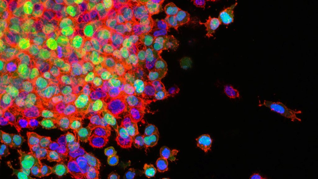
Lung cancer metastasising. Photo by National Cancer Institute on Unsplash

Researchers have discovered that, counterintuitively, certain cells move faster in thicker fluid – such as mucus as opposed to blood – because their ruffled edges sense the viscosity of their environment and adapt to increase their speed.

The researchers’ combined results in cancer and fibroblast cells suggest that the viscosity of a cell’s surrounding environment is an important contributor to disease. The findings, published in Nature Physics, may help explain tumour progression, scarring in mucus-filled lungs affected by cystic fibrosis, and the wound-healing process.

“This link between cell viscosity and attachment has never been demonstrated before,” noted Sergey Plotnikov, assistant professor at the University of Toronto and a co-corresponding author of the study. “We found that the thicker the surrounding environment, the stronger the cells adhere to the substrate and the faster they move – much like walking on an icy surface with shoes that have spikes, versus shoes with no grip at all.”

Understanding why cells behave in this surprising way is important because cancer tumours create a viscous environment, which means spreading cells can move into tumours faster than non-cancerous tissues. Since the researchers observed that cancer cells speed up in a thickened environment, they concluded that the development of ruffled edges in cancer cells may contribute to cancer spreading to other areas of the body.

Targeting the spreading response in fibroblasts, on the other hand, may reduce tissue damage in the mucus-filled lungs affected by cystic fibrosis. Because ruffled fibroblasts move quickly, they are the first type of cells to move through the mucus to the wound, contributing to scarring rather than healing. These results also imply that cell movement might be controlled by changing the viscosity of the lung’s mucus.

“By showing how cells respond to what’s around them, and by describing the physical properties of this area, we can learn what affects their behaviour and eventually how to influence it,” says Ernest Iu, PhD student at the University of Toronto and study co-author.

Plotnikov added, “For example, perhaps if you put a liquid as thick as honey into a wound, the cells will move deeper and faster into it, thereby healing it more effectively.”

Asst Prof Plotnikov and Iu used advanced microscopy techniques to measure the traction that cells exert to move, and changes in structural molecules inside the cells. They compared cancer and fibroblast cells, which have ruffled edges, to cells with smooth edges. They determined that ruffled cell edges sense the thickened environment, triggering a response that allows the cell to pull through the resistance – the ruffles flatten down, spread out and latch on to the surrounding surface.

The experiment originated at Johns Hopkins, where assistant professor Yun Chen, lead author of the study, and Matthew Pittman, PhD student and first author, were first examining the movement of cancer cells. Pittman created a viscous, mucus-like polymer solution, deposited it on different cell types, and saw that cancer cells moved faster than non-cancerous cells when migrating through the thick liquid. To further probe this behaviour, Asst Prof Chen collaborated with U of T’s Plotnikov, who specialises in the push and pull of cell movement.

Plotnikov was amazed at the change in speed going into thick, mucus-like liquid. “Normally, we’re looking at slow, subtle changes under the microscope, but we could see the cells moving twice as fast in real time, and spreading to double their original size,” he explained.

Typically, cell movement depends on myosin proteins, which help muscles contract. Asst Prof Plotnikov and Iu reasoned that stopping myosin would prevent cells from spreading, however were surprised when evidence showed the cells still sped up despite this action. They instead found that columns of the actin protein inside the cell, which contributes to muscle contraction, became more stable in response to the thick liquid, further pushing out the edge of the cell.   

The teams are now investigating how to slow the movement of ruffled cells through thickened environments, which may open the door to new treatments for people affected by cancer and cystic fibrosis.

Source: EurekAlert!

Cancer Cells Seek their ‘Goldilocks’ Zone of Tissue Softness

Source: National Cancer Institute on Unsplash

Research into the mechanisms of cell migration and the impact of tissue rigidity on cell positioning and steering has found that cancer cell migrate towards and opens new possibilities for stopping and directing it.

An international team of scientist have uncovered for the first time how tissue stiffness determines cell positioning and regulates all types of cell migration ranging from the neuronal growth cone turning to dissemination of malignant cancer cells in brain tumors and breast cancer.

Each cell in the body has a specific task and carefully determined position within a tissue. Cell positioning is regulated by many factors, including tissue rigidity. Cells are capable of probing and sensing their environment, and different cell types have different preferences for optimal conditions. A little bit like Goldilocks in the story trying out the different beds of the bear family and finding one bed to be too soft, the other one two hard and one to be just right. While this has been well-known for a long time, it has remained a mystery to researchers how cells are able to steer themselves to the optimal environment.

“The prevailing view among scientists was that all cell types prefer high-rigidity environments and migrate towards increasing stiffness. This process has been coined the term ‘durotaxis‘ – migration towards hard from Greek and Latin,” said Aleksi Isomursu, a doctoral researcher.

“I was visiting the University of Minnesota for a research project and noticed that brain cancer cells grown on engineered substrates with alternating stiffness show the opposite behaviour they turned towards soft,” Isomursu continues.

This observation launched an interdisciplinary research project involving cancer cell biology, computational modelling and engineering and involving researchers from three continents. As the outcome, the researchers uncovered the basic mechanism all cell types use to steer themselves towards their optimal environment.

These results will be important for future research in stopping and directing cancer cell migration.

“I experimented with different types of drugs and identified ones that could make brain cancer cells stop moving or change direction,” explains Postdoctoral Researcher Mathilde Mathieu.

Identification of the mechanism of cell steering provides explanations for many thus far mysterious steps in cancer dissemination, for example how cancer cells migrate out from the stiff core of a breast tumour.

“These findings have been gaining a lot of interest in researchers and we have even played around with the idea of launching a new term – ‘mollitaxis‘, migration towards soft,” says the Principal Investigator of the laboratory at the University of Turku, Professor Johanna Ivaska.

Source: University of Turku

Bariatric Surgery Reduces Cancer Risks with Obesity

Obesity
Image source: Pixabay CC0

A study published in JAMA shows that weight loss through bariatric surgery for adults with obesity was associated with a 32% lower risk of developing cancer and a 48% lower risk of cancer-related death compared with those who did not have the surgery.

Rising obesity numbers are being seen all over the world. The International Agency for Research on Cancer describes 13 types of cancer as obesity-associated cancers such as endometrial cancer, postmenopausal breast cancer, and cancers of the colon, liver, pancreas, ovary and thyroid.

Lead author of the study, Ali Aminian, MD, at Cleveland Clinic, said that bariatric surgery is currently the most effective treatment for obesity. “Patients can lose 20 to 40% of their body weight after surgery, and weight loss can be sustained over decades. The striking findings of this study indicate that the greater the weight loss, the lower the risk of cancer,” said Dr Aminian.

From 2004 and 2017, the SPLENDID (Surgical Procedures and Long-term Effectiveness in Neoplastic Disease Incidence and Death) study matched a group of 5053 adult patients with obesity who had bariatric surgery 1:5 to a control group of 25 265 patients with obesity who did not undergo the surgery.

After 10 years, 2.9% of patients in the bariatric surgery group and 4.9% of patients in the non-surgical group developed an obesity-associated cancer.

After 10 years, 0.8% of patients in the surgery group and 1.4% of patients in the non-surgical group died from cancer, indicating that bariatric surgery is associated with a 48% lower cancer mortality risk.

Researchers noted that the benefits of bariatric surgery were seen in a wide range of study participants in terms of age, sex and race. In addition, benefits were similarly observed after both gastric bypass and gastric sleeve operations.

“According to the American Cancer Society, obesity is second only to tobacco as a preventable cause of cancer in the United States,” said the study’s senior author, Steven Nissen, MD, Chief Academic Officer of the Heart, Vascular and Thoracic Institute. “This study provides the best possible evidence on the value of intentional weight loss to reduce cancer risk and mortality.”

Numerous studies have shown the health benefits of bariatric or weight-loss surgery in patients with obesity. The Cleveland Clinic-led STAMPEDE study showed that following bariatric surgery, significant weight loss and control of type 2 diabetes last over time. The SPLENDOR study showed that in patients with fatty liver, bariatric surgery decreases the risk of the progression of liver disease and serious heart complications.

The SPLENDID study adds important findings to the literature focused on the link between obesity and cancer. Given the growing epidemic of obesity worldwide, these findings have considerable public health implications.

“Based on the magnitude of benefit shown in our study, weight loss surgery can be considered in addition to other interventions that can help prevent cancer and reduce mortality,” said Jame Abraham, M.D., chairman of the Hematology and Medical Oncology Department at Cleveland Clinic. “Further research needs to be done to understand the underlying mechanisms responsible for reduced cancer risk following bariatric surgery.”

Source: Cleveland Clinic

Over-the-counter Drugs and Supplements Overlooked in Polypharmacy

Pills and tablets
Photo by Myriam Zilles on Unsplash

While patients usually report any medications they are on, over-the-counter drugs and supplements are not reported as often to the medical team, according to a study on polypharmacy published in The Oncologist, an overlooked situation that complicates the problem of polypharmacy, especially in cancer.

Polypharmacy can lead to harmful drug interactions, especially dangerous for cancer patients about to undergo therapy.

Even for those without cancer, multiple medication use has risks and is tricky to navigate because of the emotions involved, said Erika Ramsdale, MD, study leader.

“As doctors, we tell people to take medications but we don’t always do a great job of following up,” she said. “From the patient perspective, if it’s determined that a medication is no longer needed, it’s hard to stop taking it. There’s a sense of, ‘What will happen if I stop?’ or ‘Are you giving up on me?’ A lot of uncertainty and emotions are tied up in this issue.”

The more drugs and supplements a person takes, the higher the risk of inappropriate use and serious drug interactions, she said.

The fragmentation of healthcare across specialties complicates the issue. “Sometimes, there is no quarterback,” Dr Ramsdale said, which can result in “prescribing cascades,” where additional drugs are given to offset the adverse side effects of the original medications.

Researchers analysed medication use in a sample of 718 adults with a mean age of 77 who had stage 3 or 4 cancer and other health conditions. They screened for potentially inappropriate medications that have risks higher than benefits (known as PIMS), drug-drug interactions (DDI), and drug-cancer treatment interactions (DCI). Drug interaction can have consequences such as falls, functional decline, and death. Patients on multiple medications are also more likely to have anxiety or depression.

Among the 718 patients, 70% were at risk of drug-drug interactions and 67% were taking at least one drug that was potentially inappropriate.

In fact, 61% of the patients were taking five or more medications before starting chemotherapy – and nearly 15% were taking 10 or more medications.

Other findings from the study include:

  • Nearly 68% of the patients had serious health issues besides cancer, requiring associated medications. Most common was cardiovascular disease. When a person has cancer combined with other ailments, there is a greater risk of toxicity from cancer treatments due to polypharmacy.
  • Approximately 10% of hospital admissions for older adults are associated with hazardous drug interactions. Among older adults with cancer receiving chemotherapy, polypharmacy is associated with dramatic increases (up to 114%) in unplanned hospitalisations.
  • Cholesterol-lowering medications, minerals, and thyroid therapy are most commonly involved in potential drug interactions.
  • More than 25% of the medications used by the patients in the study were non-prescription—and these accounted for 40% of the potentially inappropriate medications detected by investigators.
  • Common non-prescription remedies included vitamins and minerals, anti-anemic preparations such as ferrous sulfate, and drugs for acid-related disorders and constipation.

“Older adults may incorrectly assume that over-the-counter medications are safe for them,” the authors wrote. “This study helps delineate the size and shape of a problem under-recognised by both providers and patients.”

It’s also an understudied problem, Dr Ramsdale said, and including over-the-counter medications sets Wilmot’s data apart from previous research; most polypharmacy studies among cancer patients look only at prescription drugs.

The study highlights an opportunity for education and problem-solving, such as deprescribing some drugs.

Deprescribing is the planned reduction of medications to avoid harm. Doctors take into account the risks-versus-benefits of each medication and the patient’s life expectancy. For example, statins that are taken for high cholesterol do not have an immediate effect. They are meant to be preventive and can take 10 years to have an impact. Therefore, if a patient is old and has incurable cancer, he or she may not need to take statins. (Discontinuing statins in this setting is supported by a landmark study, according to the Ramsdale publication.)

However, these conversations can be quite delicate, Ramsdale said. The goal is to promote better quality of life, and she is conducting a clinical trial to test the best way to intervene in cases of polypharmacy among older people with cancer.

Source: University of Rochester Medical Center

How Kaposi Sarcoma-associated Herpesvirus Evades the Immune System

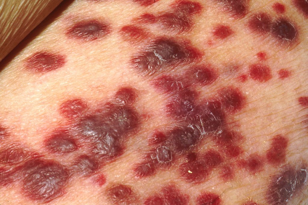
Kaposi sarcoma on the skin of an AIDS patient. Credit: National Cancer Institute

A study published in Cell Reports has identified a protein in the cancer cell’s nucleus as a critical agent keeping Kaposi sarcoma-associated herpesvirus (KSHV) dormant and hidden from the immune system. The virus, in the same family as Epstein-Barr virus, is linked to AIDS-related Castleman’s disease and cancers such as Kaposi sarcoma.

Up to 50% of the population in some parts of Africa are affected with KSHV, though not everyone with KSHV will develop Kaposi sarcoma. Those who do typically have a weakened immune system due to HIV infection, organ transplant, being older or other factors.

The introduction of antiretroviral therapy significantly reduced AIDS-related Kaposi sarcoma prevalence in Western countries; however, in sub-Saharan Africa, the disease continues to have a poor prognosis.

On entry into a human cell the virus causes a hidden infection in the nucleus: the virus simply latches onto parts of the cell’s chromosomes without replicating.

Researchers studied KSHV’s latent-lytic switch, a process in which the virus exits its dormancy state to replicate in the host cell. This replication phase, called the lytic cycle, ends with the disintegration of the cell and the release of the viruses, infecting neighbouring cells.

“The virus likes to stay silent as long as possible to avoid being detected by the body’s immune system,” said Professor Yoshihiro Izumiya, the study’s senior author.

The team sought to understand the mechanisms behind this latent-lytic switch and the role the host cell environment played in this process.

“Where the virus latches onto the host cell, how it manages to stay dormant, and what triggers its activation were very exciting and important puzzles to solve,” Prof Izumiya said.

The study identified where the virus genome could be found on the host genome.

Izumiya and his team profiled and analysed chromosomal interactions on three cancer cell lines naturally infected with KSHV, locating the virus’s preferred chromosome docking sites. The binding patterns, similar among the three cancer cell lines, showed a nuclear ecosystem that can attract and help keep the virus in its silent form.

The team also found that CHD4 (chromodomain helicase DNA binding protein 4) binds to the virus’s genomic elements. CHD4, a protein in the host cell’s chromosomes, suppresses the work of the gene responsible for viral replication. The study showed that CHD4 is a key regulator of the KSHV latency-lytic switch.

“The location where the virus genome attaches to the host chromosome is not random,” said Ashish Kumar, a postdoctoral researcher in Izumiya Lab and the paper’s first author. “Without having enriched CHD4 protein, the virus starts to replicate, kicking in a cell destructive mode. For the virus to select CHD4 among many other host proteins, CHD4 must play a unique and important role in host cells.”

Virology can help identify cellular proteins essential for cell homeostasis. Over millions of years, the virus’s genome developed to encode or assemble a small number of very efficient proteins, which strategically connect to host cell proteins to keep viral chromatin dormant and impact the host cell’s tumour suppression function.

“We used virology as an entry point to shed light on the function of CHD4 in gene regulation in general. During virus-host co-evolution, KSHV cleverly learned to hijack host proteins that can help keep the gene responsible for viral replication dormant.”

The researchers found a viral protein which could serve as the basis for a replication inhibitor. Since CHD4 is critical for cancer cell growth in a variety of cancers, they hope this virus-host interaction could inform cancer treatment research.

Source: University of California – Davis Health

Tumours Use Lactate to Bully Surrounding Cells into Helping Them

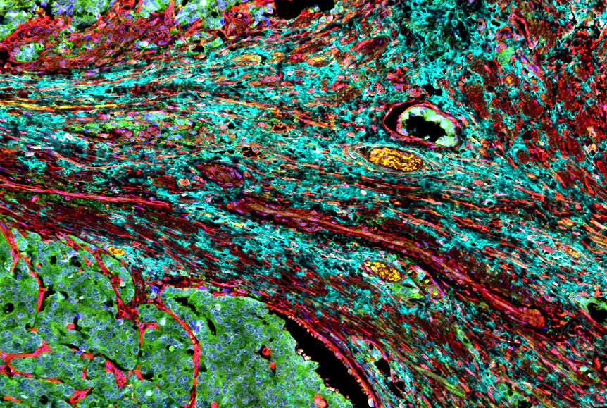
Cancer-associated fibroblasts surrounding a prostate tumour. Credit: Moscat/Diaz-Meco labs.

Some tumours can force neighbouring cells into supporting cancer growth by releasing lactate into their local environment, according to a study published in Cell Reports. The findings could lead to drugs that target that defence mechanism to help cancer patients ؘ– and also boost a current class of cancer drugs.

In the study, the researchers determined how developing tumours recruit nearby cells called fibroblasts to work as their enablers. Fibroblasts form part of the stroma, organs’ connective tissue, and normally have important repair and maintenance functions. But cancer-associated fibroblasts (CAFs) acquire properties that allow them to assist tumours in ways that make the tumours more malignant and harder to kill.

The researchers also discovered that PARP-1 inhibitors, a widely used class of cancer drugs, mimic one of the key steps in CAF recruitment, and can hamstring their own effectiveness by inadvertently switching local fibroblasts to this cancer-enabling mode.

“Future therapeutics that block this cancer-associated state of fibroblasts might be useful on their own or as a way to improve the effectiveness of PARP-1 inhibitors,” said study co-senior author Dr Maria Diaz-Meco.

Dr Diaz-Meco collaborated in the study with the laboratory of co-senior author Dr Jorge Moscat.

Developing tumours are known to often modify their local environments for their own survival and growth. Cancer-associated fibroblasts are a central component of the tumour microenvironment in prostate, lung, colon and other cancer types. Targeting these cells is therefore seen as a promising complementary approach to standard cancer treatment – and one that could work very broadly against cancers of different cellular and genetic origins.

“Cancer-associated fibroblasts support tumour growth by providing growth factors and essential metabolites to the tumour, by fending-off anti-tumour immune cells, and in many other ways,” Dr Moscat said. “The result is a tumour that is more malignant and treatment-resistant.”

Several years ago, the researchers discovered that a protein called p62, produced in fibroblasts, normally suppresses the CAF state, though many tumours find a way to restore this state by reducing fibroblast p62 production. In the new study, they showed that tumours achieve this by secreting high levels of an organic compound called lactate, also known as lactic acid.

Lactate is a normal by-product of certain energy-production processes in cells, which are often hyperactive in tumours. In experiments with prostate cancer cells, the researchers detailed the molecular chain of events by which tumour-secreted lactate disrupts the normal metabolism of fibroblasts, leading to reduced p62 gene activity and the activation of the tumour-enabling CAF state.

The finding is significant because it illuminates a major cancer-promoting pathway, which in principle can be targeted with future drugs as a standalone or add-on treatment strategy.

A second, surprising finding was that a key step leading from tumour lactate secretion to fibroblast p62 suppression turned out to be the inhibition of a DNA-repair enzyme called PARP-1, which has the same effect as PARP-1 inhibitors – suggesting that these drugs might be working partly against themselves by creating a more tumour-friendly microenvironment.

In vitro and animal testing confirmed that the PARP-1 inhibitor olaparib does reduce p62 in fibroblasts, and pushes them into the CAF state, in turn increase tumours’ resistance to the drug’s primary cancer-killing effect.

Thus, the researchers emphasised, future treatments that reprogram CAF cells to the non-cancer state or prevent their development might greatly enhance PARP-1 inhibitors’ anti-tumour effectiveness.

“We’re now studying several potential CAF-blocking therapeutics in our labs,” Dr Moscat said.

Source: Weill Cornell Medical College

Genetics is Only Part of the Cancer Puzzle

Killer T cells about to destroy cancer cell
Killer T cells about to destroy cancer cell (centre). Credit: NIH

Though cancer is a genetic disease, the environment and metabolism must also be considered – and could narrow down avenues for cancer treatment, according to a research review by a leading expert which appears in Metabolites.

Nearly all the theories about the causes of cancer that have emerged over the past several centuries can be sorted into three larger groups, said Professor David Wishart at the University of Alberta. The first is cancer as a genetic disease, focusing on the genome. The second is cancer as an environmental disease, focusing on the exposome, which includes everything the body is exposed to throughout life. The third is cancer as a metabolic disease, focusing on the metabolome, which is all the chemical byproducts of the process of metabolism.

Until now, there has been little research on the metabolic perspective, but it’s gaining the interest of more scientists, who are beginning to understand the metabolome’s role in cancer.

The genome, exposome and metabolome operate together in a feedback loop as cancer develops and spreads.

According to the data, heritable cancers account for only 5–10%of all cancers, Wishart said. The remaining 90–95% are initiated by factors in the exposome, which in turn trigger genetic mutations.

“That’s an important thing to consider, because it says that cancer isn’t inevitable.”

The metabolome is a crucial part of the process, as those genetically mutated cancer cells are sustained by the cancer-specific metabolome.

“Cancer is genetic, but often the mutation itself isn’t enough,” said Wishart. As cancer develops and spreads in the body, it creates its own environment and introduces certain metabolites. “It becomes a self-fuelled disease. And that’s where cancer as a metabolic disorder becomes really important.”

The multi-omics perspective considers the genome, exposome and metabolome in unison when thinking about cancer. It is showing promise for finding treatments and for overcoming the limitations of looking at only one of these factors.

Researchers focusing only on the genetic perspective, for example, are looking to address particular mutations. The problem is, Prof Wishart said, is that there are around 1000 genes that can become cancerous when mutated, at least two different mutations within these cells are usually for cancer to grow. Thus, a there are a million possible mutation pairs, so “it becomes hopeless” to narrow down the possibilities when seeking new treatments.

From the metabolic perspective, there are just four major metabolic types, said Prof Wishart. Therefore instead of searching for a treatment plan for a one in a million mutation combination, determining the patient’s cancer metabolic type can immediately guide doctors in deciding on the best treatment for their specific cancer.

“It really doesn’t make a difference where the cancer is – it’s something you’ve got to get rid of. It’s how it thrives or grows that matters,” said Prof Wishart. “It becomes a question of, ‘What’s the fuel that powers this engine?'”

However, health-care providers still need a mix of therapeutics for cancer, and he noted that a deeper understanding of the metabolome and its role in the cancer feedback loop is also critical to preventing cancer.

“If we understand the causes of cancer, then we can start highlighting the known causes, the lifestyle issues that introduce or increase our risk,” he said.

“From the prevention side, changing our metabolism through lifestyle adjustments will make a huge difference in the incidence of cancer.”

Source: University of Alberta

Pump up the Volume: Killing Cancer with Ultrasound

Sound waves
Image by Pawel Czerwinski on Unsplash

University of Michigan scientists have developed an ultrasound technology that uses high-powered pulses to break down liver tumours in rats, kill cancer cells and stimulate the immune system to destroy the remaining tumour and prevent metastasis.

The researchers reported in Cancers that, by destroying just 50% to 75% of liver tumour volume, the rats’ immune systems were able to clear away the rest, with no evidence of recurrence or metastases in more than 80% of animals.

“Even if we don’t target the entire tumour, we can still cause the tumour to regress and also reduce the risk of future metastasis,” said Professor Zhen Xu, corresponding author of the study.

The treatment was also found to spur the rats’ immune responses, possibly contributing to the eventual regression of the untargeted portion of the tumour and preventing further spread of the cancer.

The noninvasive technique, called histotripsy, focuses ultrasound waves to mechanically destroy target tissue with high precision. The relatively new technique is currently being used in a human liver cancer trial in the US and Europe.

Often, a tumour cannot be directly targeted for certain treatments due to the mass’ size, location or stage. To investigate the effects of partially destroying tumours with sound, this latest study targeted only a portion of each mass, leaving behind a viable intact tumour. It also allowed the team to demonstrate the technique in less ideal conditions.

“Histotripsy is a promising option that can overcome the limitations of currently available ablation modalities and provide safe and effective noninvasive liver tumour ablation,” said Tejaswi Worlikar, a doctoral student in biomedical engineering.

Liver cancer ranks among the top 10 causes of cancer related deaths worldwide, with poor prognosis despite multiple treatment options. Tumour recurrence and metastasis after initial treatment is common, demanding improved treatments.

The ultrasound approach comes without the side effects from present treatments such as radiation and chemotherapy.

“Our transducer, designed and built at U-M, delivers high amplitude microsecond-length ultrasound pulses – acoustic cavitation – to focus on the tumour specifically to break it up,” Prof Xu said. “Traditional ultrasound devices use lower amplitude pulses for imaging.”

The microsecond pulses create microbubbles within targeted tissues that rapidly expand and collapse, tearing up cancer cells and disrupting the tumour’s structure.

Source: University of Michigan

Exercise Slows Tumour Growth and Reduces Complications

Photo by Ketut Subiyanto on Pexels

In mouse studies, researchers found that exercising prior to developing cancer was associated with slower tumour growth and helped reduce the effects of a cachexia, a cancer complication also known as wasting syndrome.

Cachexia is a metabolic wasting disorder that affects up to 80% of patients with advanced cancer and is associated with about a third of all deaths from cancer. Cachexia is characterised by severe progressive muscle wasting, a decline in heart structure and function and an overall poorer quality of life.

“Most exercise, especially aerobic exercise, is easily accessible and affordable,” said Louisa Tichy, a graduate student in Traci Parry’s lab at the University of North Carolina at Greensboro. “Therefore, engaging in consistent aerobic exercise such as running is a cost-effective way to reduce the risk of cancer and cancer complications.”

Tichy presented the research at the American Society for Investigative Pathology annual meeting.

Previous research has shown that exercise could have anti-inflammatory effects and might positively impact cancer cachexia by slowing its development and preserving cardiac structure and function. However, very few studies have focused on preconditioning.

“Our preclinical study indicated that preconditioning – or exercise prior to tumour bearing – appears to play an important cardioprotective role during cancer cachexia by preserving cardiac structure and function,” said Tichy. “It also helped stunt tumour growth, even when animals did not exercise during the tumour-bearing period.”

For the new study, the researchers studied mice that exercised on a treadmill for eight weeks and non-exercising mice. After the eight weeks, the researchers induced cancer in some of the exercised mice and some sedentary mice while keeping some mice from both groups cancer-free to act as controls.

Mice with cancer and a sedentary lifestyle were found to have poorer heart function – as measured with echocardiography – than the mice that exercised prior to cancer induction. Also, exercising mice had a smaller tumour volume and a 60% smaller tumour mass than the sedentary mice.

“This data is crucial in identifying the significance of exercise and the best timing of exercise as a protective and preventative measure against the detrimental effects of cancer cachexia,” said Tichy.

Understanding how the underlying proteins and pathways are affected by cancer and exercise can be used to inform exercise interventions. The researchers say that devising safe and effective exercise interventions for cancer patients would require evaluating the best intensity, duration and timing of exercise in preclinical models before human tests.

Source: EurekAlert!

Not so Sweet: Artificial Sweeteners’ Cancer Risk

Fizzy drink cans
Source: Breakingpic on Pexels

An observational study with over 100 000 participants suggests that some artificial sweeteners are associated with increased cancer risk. The findings, published in PLOS Medicine, reflect other results from experimental studies.

The safety of artificial sweeteners has long been a subject of debate. To evaluate the potential carcinogenicity of artificial sweeteners, researchers analysed data from 102 865 French adults participating in the NutriNet-Santé study. The NutriNet-Santé study is an ongoing web-based cohort initiated in 2009 by the Nutritional Epidemiology Research Team (EREN). Participants enrolled voluntarily, and self-reported their medical history, sociodemographic, diet, lifestyle, and health data.

Researchers gathered data concerning artificial sweetener intake from 24-hour dietary records. After collecting cancer diagnosis information during follow-up, the researchers conducted statistical analyses to investigate the associations between artificial sweetener intakes and cancer risk. They also adjusted for a range of variables including age, sex, education, physical activity, medical history and dietary intake.

The researchers found that participants who consumed larger quantities of artificial sweeteners, particularly aspartame and acesulfame-K, had higher risk of overall cancer compared to non-consumers (hazard ratio 1.13). Higher risks were observed for breast cancer and obesity-related cancers.

In addition to being an observational study, there were a number of limitations; dietary intakes are self-reported. Selection bias may also have been a factor, as participants were more likely to be women, to have higher educational levels, and be more health-conscious. Additional research will be required to confirm the findings and clarify the underlying mechanisms.

According to the authors, “Our findings do not support the use of artificial sweeteners as safe alternatives for sugar in foods or beverages and provide important and novel information to address the controversies about their potential adverse health effects. While these results need to be replicated in other large-scale cohorts and underlying mechanisms clarified by experimental studies, they provide important and novel insights for the ongoing re-evaluation of food additive sweeteners by the European Food Safety Authority and other health agencies globally”.

First author Charlotte Debras added: “Results from the NutriNet-Santé cohort (n = 102 865) suggest that artificial sweeteners found in many food and beverage brands worldwide may be associated with increased cancer risk, in line with several experimental in vivo / in vitro studies. These findings provide novel information for the re-evaluation of these food additives by health agencies.”

Source: EurekAlert!