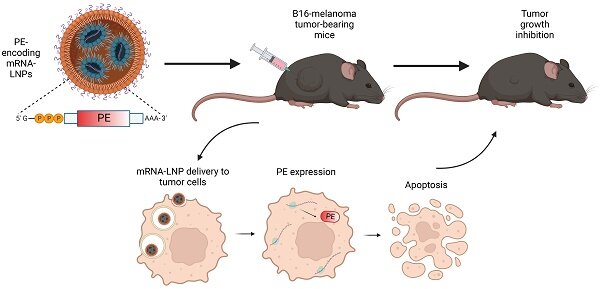
Tag: cancer treatment

Added Salt Found to Suppress Tumours in Mice

Source: Pixabay CC0

new study has found that adding salt to the diet of lab mice can suppress the growth of cancerous tumours.

Dietary salt reduction has been stressed by clinicians for many years, as research has shown that a high-sodium diet can result in inflammation, high blood pressure and an increased heart attack risk. Researchers from the Translational Health Science and Technology Institute wondered if the inflammation resulting from a high-salt diet could also confer positive health benefits, such as fighting cancerous tumours. 

To find out, the researchers fed two groups of mice with implanted melanoma tumours either a normal diet or a high-sodium diet (4.0% sodium chloride above normal diet) and then measured the differences in tumour suppression abilities between the two groups. They found that the mice on the high-sodium diet had an increase in Bifidobacterium probiotics, leading to an increase in natural killer cells that attack cancerous tumors. They also found an increased ability to inhibit PD-1 proteins which have been found to prevent T cells from attacking tumours.

On close examination, it was found that the high-sodium diet caused the gut barrier to be leakier, enabling the movement of Bifidobacteria from the gut to tumour locations. In addition, they found that once the Bifidobacteria arrived at a tumour, crosstalk between them and the immune cells engaged in attacking the tumour improved the success of the attack.

However, the researchers also found that a low-sodium diet worked in conjunction with several cancer-fighting drugs, showing an increased ability to reduce tumour growth. Since the researchers hypothesised that Bifidobacteria were responsible for the tumour immunity of a high-sodium diet, they performed faecal transplants from mice on a high-sodium diet to those on a normal diet and found that it also improved their ability to fight tumour growth.

The study was published in Science Advances.

Source: MedicalXpress

Cancer Follow-up Care Needs Improvement

With greater long-term cancer survival, previous standards of cancer follow-up care and support may no longer meet current needs.

The side-effects of anticancer medicines and impacts of the illness itself that sometimes persist after the end of treatment can hinder  a return to normal life after beating cancer. A study presented at the ESMO Congress 2021 showed that a significant proportion of survivors continue to suffer from burdensome symptoms for several years and reveal widespread dissatisfaction with the assistance provided.

Prof Dorothy Keefe, CEO of Australia’s national cancer agency, Cancer Australia, chair of the congress’s supportive and palliative care track, not involved in the study, underlined its importance in a context where survivorship research has lagged behind research on cancer treatment. “This is probably due to the increase in survival rates itself lagging behind the introduction of new therapies, but also to a lack of prioritisation compared to the need to develop a cure,” Keefe said, and highlighted the scale of the issue today: “We now have millions of cancer survivors in Australia, hundreds of millions around the world – and an ever-increasing number who could potentially have long-term side-effects.”

One of the most common symptoms experienced by patients and survivors alike is cancer-related fatigue (CRF), a persistent sense of exhaustion, not alleviated by sleep or rest and significantly interferes with the person’s usual functioning. The FiX study initially evaluated the patterns, severity and management of CRF among 2508 patients with 15 different types of cancer two years after the discovery of their illness. In a follow-up survey, 36 potential long-term problems, completed by participants around four years after diagnosis, almost 40% of survivors continued to report fatigue that they rated as a moderate or severe burden. As well as fatigue, over 40% of patients reported loss of physical capacity as a burden and over one third suffered from trouble sleeping, sexual problems, joint pains and anxiety.

Although there are recommendations for managing side-effects like CRF, study author Dr Martina Schmidt from the German Cancer Research Centre (DKFZ) in Heidelberg, Germany, drew attention to their lack of implementation and reported that more than one in three affected individuals in the study evaluated the support they were offered for fatigue as poor. “Despite increasing awareness of the effectiveness of mitigating measures like exercise to reduce fatigue, patients are still too often left alone to seek help for symptoms that cannot be directly addressed with medicines in the same way as something like pain, for which satisfaction with the support received was high in our study.”

Prof Keefe commented on the results, saying: “This research shows that a staggeringly high number of patients still suffer from significant health issues years after being declared disease-free. Their dissatisfaction with the care available is a wake-up call that we should be paying more attention to these individuals, trying to understand the mechanisms at play in order to identify interventions that could help them to better recover.”

According to Dr Schmidt, cancer follow-up care should therefore also incorporate more systematic screening for additional symptoms that can burden patients. “The first step should be to make sure that patients themselves are better informed about these potential issues early on, so they know that conditions like CRF are not only expected, but often manageable and that they should not wait for symptoms to disappear on their own,” she said.

Recognising that possible models of long-term support remain largely untested, Keefe further advocated that all patients should be provided with a survivorship care plan when they reach the end of their treatment. “Going forward, we need to develop these models of care in a way that minimises the burden on healthcare systems, implement them and research their impact so that we can come back in five years’ time and evaluate whether they have made a difference for cancer survivors,” she concluded.

Source: European Society for Medical Oncology

How Cancer Cells Develop Resistance to Chemotherapy

Source: National Cancer Institute on Unsplash

Researchers have found some answers as to why cancer cells can develop resistance to the cytotoxic drugs used in chemotherapy.

“We haven’t understood very much about how this resistance to chemotherapy develops and even less about how the microenvironment in cancer can affect the process,” said Kaisa Lehti, a professor at the Norwegian University of Science and Technology’s (NTNU) Department of Biomedical Laboratory Science.

Lehti has led this study into how cancerous tissues develop resistance to a particular form of chemotherapy, the results of which appear in Nature Communications.

If ovarian cancer is picked up early, almost all patients survive the first five years, while chances of survival are much worse if detected later. Finding effective treatment is therefore very important.

Platinum chemotherapy is one of the standard treatments for ovarian cancer, but cancer cells often develop resistance to this particular treatment. The reason lies in how the platinum-based cytotoxin itself can change the cancer cells and their environment.

Cytotoxin influences cancer cells and their environment
Lehti summed up the process: “The cytotoxin can change the way the cancer cells send and perceive signals and can modify the microenvironment around the cells.”

This change allows the cancer cells to withstand the damage caused by the cytotoxin—and can thus survive the chemotherapeutic attack. The researchers have found this key to the puzzle in a layer of tissue that often surrounds cancer cells.

“A fibrotic network of proteins, known as the extracellular matrix or ECM, surrounds the cancer cells, particularly the most aggressive ones,” said Lehti.

The fibrotic tissue, with the ECM network around the cancer cells, is mainly produced by normal connective tissue cells. But the cancer cells and connective tissue cells in the network can alter this tissue themselves.

“Previously, we haven’t known how the communication between the cancer cells and the extracellular matrix is affected by, or even itself influences, the development of cancer and its response to chemotherapy,” said Prof Lehti.

But it is now known that chemical and mechanical signals in the surrounding ECM tissue help cancer develop its ability to spread and to resist treatment.

“Certain signals from the ECM can critically change the cancer cells’ resistance to platinum-based cytotoxic drugs,” Prof Lehti explained.

In this way, the cytotoxin itself helps change both the microenvironment around the cancer cells and the ability of the cancer cells to sense their environment, and so resist the cytotoxin. By understanding this process, better therapies can be developed.

Source: MedicalXpress

Powering Up NK Cells With Magnetism

NK cells attack a cancer cell (centre). Source: NIH

Powering up natural killer cells with magnetic nanoparticles could enhance cancer immunotherapy, according to a Northwestern Medicine study published in ACS Nano.

This method could allow for the unleashing of natural killer (NK) cells on a variety of solid tumours, according to senior author Dong-Hyun Kim, PhD, associate professor at Northwestern.

“People have had trouble applying NK cells to solid tumours,” said Prof Kim. “If we can provide an easy path to modulate NK cells, perhaps this can become a useful therapy.”

Most cell-based immunotherapies involve T-cells, but these chimeric antigen receptor (CAR) T-cell therapies are costly and have a long incubation period and strong side effects.

On the other hand, NK cells belong to the innate immune system and are quicker to respond to pathogens. NK cell immunotherapy has been explored, according to Prof Kim, but that too has barriers.

“It’s pretty hard for these cells to penetrate inside the tumours which have thick barrier tissues,” Prof Kim said.

Magnetically activated NK cells
Boosting NK cell function with cytokines have proven unsuccessful and, like CAR T-cell therapy, have a high cost and lengthy manufacturing time. However, Prof Kim’s previous work with nanoparticles inspired a different approach.

Prof Kim and colleagues developed a magnetic nanocomplex that binds with NK cells and, when activated with an alternating magnetic field, exerts force on the exterior of the cell, promoting secretion of cytotoxic compounds. Testing this nanocomplex in animal models of hepatocellular carcinoma, the investigators found that magnetic activation increased the cancer-killing ability of NK cells when injected into solid tumours.

As a bonus, these nanoparticles show up with MRI, allowing for precise monitoring of NK cell distribution during and after injection.

“This creates a stronger NK cell, and can hopefully enhance the efficacy of the treatment,” Prof Kim said.

Source: Northwestern University

FLASH Radiation Treatment for Tumours a Step Closer

Heavy ion bombardment in FLASH radiation treatment could be the future of radiotherapy, with encouraging findings from a German lab.

The GSI Helmholtzzentrum für Schwerionenforschung and the future accelerator centre FAIR succeeded in performing a carbon ion FLASH experiment for the first time in their Phase 0 experiment. 

The scientists involved were able to achieve the very high dose rates required to irradiate tumours. The success was a collective effort of the GSI Biophysics Department and the accelerator crew on the GSI/FAIR campus in close collaboration with the German Cancer Research Center DKFZ and the Heidelberg Ion Therapy (HIT) center.

FLASH irradiation involves utra-short and ultra-high radiation, delivering the treatment dose in fractions of a second. Traditional radiation therapy, as well as proton or ion therapy, deliver smaller doses of radiation to a patient over an extended time period, whereas FLASH radiotherapy is thought to require only a few short irradiations, all lasting less than 100 milliseconds.

In the field of electron radiation, recent in vivo investigations have shown that the FLASH method’s ultra-high dose rate is less harmful to healthy tissue, but just as efficient as conventional dose-rate radiation to inhibit tumour growth. Such an effect has not yet been demonstrated for proton and for ion beam irradiation, which is the basis of the tumour therapy with carbon ions developed at GSI. There is still a lot of research to be done here. The results of the current experiment at GSI are now being evaluated and will contribute to new knowledge.

There have also been technical barriers to FLASH radiation. Until now, FLASH technique has only been applicable using electron and proton accelerators. While the required dose rates for electrons and protons can be achieved with a cyclotron (circular accelerator), this is more difficult with the synchrotrons required in heavy ion therapy, such as the SIS18 at GSI.

That is why the current FAIR Phase 0 experiment is a very crucial step: Thanks to the improvements at the existing GSI accelerator facility as part of the preparations for FAIR, the necessary dose rate in millisecond range can now also be achieved for carbon ions. However,  much development work remains to be done before this procedure is mature enough to be routinely used on patients in the field of electron radiation.

Professor Marco Durante, Head of the GSI Biophysics Research Department, was very pleased with this important milestone in  the development of FLASH irradiation:

“It is a forward-looking method that could significantly increase the therapeutic window in radiotherapy. I am very happy that the researchers and the accelerator team were able to demonstrate the possibility to create conditions with carbon beams that are necessary for FLASH therapy of tumors. If we can combine the great effect and precision of heavy ion therapy with FLASH irradiation while maintaining efficacy and causing little damage to healthy tissue, this could pave the way of a future radiation therapy several years from now.”

Professor Paolo Giubellino, The Scientific Managing Director of GSI and FAIR, expressed his delight at the results: “The combination of expertise in biophysics and medicine as well as engineering excellence allows the first world-class experiments FLASH irradiation with ion beams to be performed. This could result in important complements to existing radiation therapies. Applications in tumour therapy are one of the research areas that can benefit from the recent increased intensities of GSI accelerators. However, modern radiobiology will substantially benefit from beams with even higher intensities, such as we will have at the FAIR facility currently under construction. FLASH is a first example of these future directions of work”.

Source: GSI Helmholtzzentrum für Schwerionenforschung GmbH

Novel Approach Targets Pancreatic Cancers Which Depend on Mutant KRAS Gene

KRAS Protein Structure. RAS is a family of related proteins that is expressed in all animals. KRAS is one of three RAS genes found in humans. RAS genes are mutated in approximately one-third of all human cancers. Photo by National Cancer Institute on Unsplash

Researchers have identified a novel drug that effectively thwarts pancreatic tumours that are addicted to the cancer-causing mutant KRAS gene.

Because early detection of pancreatic cancer is difficult, it has a low survival rate, accounting for just over 3% of all new cancer cases in the US, but leading to nearly 8% of all cancer deaths, according to the National Cancer Institute.

The KRAS gene was recognised more that 25 years ago as the component of Kirsten sarcoma virus responsible for oncogenesis. Since then, mutations of KRAS have been described in a large proportion of solid tumors ranging from more than 90% of pancreatic carcinomas to 20% to 30% of pulmonary adenocarcinomas.

Through a pre-clinical study, Said Sebti, PhD, associate director for basic research at VCU Massey Cancer Center, identified a novel drug that effectively thwarts pancreatic tumors that are addicted to the cancer-causing mutant KRAS gene. 

“We discovered a link between hyperactivation of the CDK protein and mutant KRAS addiction, and we exploited this link preclinically to counter mutant KRAS-driven pancreatic cancer, warranting clinical investigation in patients afflicted with this deadly disease,“ said Dr Sebti, who is also the Lacy Family Chair in Cancer Research at Massey and a professor of pharmacology and toxicology at the VCU School of Medicine. “Our findings are highly significant as they revealed a new avenue to combat an aggressive form of pancreatic cancer with very poor prognosis due mainly to its resistance to conventional therapies.”

In 90 percent of pancreatic cancers, KRAS is mutated. Prior studies have shown that some tumours harbouring mutant KRAS are in fact addicted to the mutant gene, meaning they cannot survive or grow without it. Sebti set out to discover if there is a drug that can specifically kill those tumours with a mutant KRAS addiction.

Searching for a suitable drug

Dr Sebti and colleagues used a three-pronged approach to tackle this question.

First of all, they mapped the blueprint of pancreatic cancer cells through global phosphoproteomics, showing them how the addicted and non-addicted tumours differ at the phosphoprotein level. They found two proteins, CDK1 and CDK2, which signalled which cells were addicted to mutant KRAS.

Additionally, they analysed a comprehensive database from the Broad Institute of MIT and Harvard which contains genome-wide CRISPR gRNA screening datasets. They discovered that CDK1 and CDK2 as well as CDK7 and CDK9 proteins were associated with mutant KRAS-addicted tumors.

Finally, they evaluated 294 FDA drugs to selectively kill mutant KRAS-addicted cancer cells over non-KRAS-addicted cancer cells in the lab. They determined the most effective drug in preclinical experiments was AT7519, an inhibitor of CDK1, CDK2, CDK7 and CDK9.

“Using three entirely different approaches, the same conclusion presented itself clearly to us: pancreatic cancer patients whose tumors are addicted to mutant KRAS could benefit greatly from treatment with the CDK inhibitor AT7519,” Dr Sebti said.

To further validate these findings in fresh tumours taken from pancreatic cancer patients the researchers found that AT7519 suppressed the growth of xenograft cells from five mutant KRAS pancreatic cancer patients who relapsed on chemotherapy and/or radiation therapies.

Though AT7519 had previously been tested unsuccessfully in a number of clinical trials, none of the trials were for pancreatic cancer.

“If our findings are correct and translate in humans, then we should be able to see a positive response in pancreatic cancer patients whose tumors are addicted to mutant KRAS,” Dr Sebti said.

As well as pancreatic cancer, the study authors believe these findings may also have clinical implications for colorectal and non-small cell lung cancer patients with prevalent KRAS mutations.

Source: Virginia Commonwealth University

Journal information: Kazi, A., et al. (2021) Global Phosphoproteomics Reveal CDK Suppression as a Vulnerability to KRas Addiction in Pancreatic Cancer. Clinical Cancer Research. doi.org/10.1158/1078-0432.CCR-20-4781.

New Insights into How Kidney Cancer Cells Respond to Treatment

Photo by National Cancer Institute on Unsplash

Researchers from the University of Michigan Rogel Cancer Center have uncovered clues as to why kidney cancers respond so differently to treatment, opening up new tailored treatment options.

Not all kidney cancers behave the same, and some have wildly differing responses to immunotherapy or other treatments – resulting in wildly different outcomes for patients.

By sequencing the RNA of individual cells within multiple benign and cancerous kidney tumors, the researchers have identified the cells from which different subtypes of kidney cancer originate, the pathways involved and how the tumor microenvironment impacts cancer development and response to treatment.

The findings, published in PNAS, could help researchers better understand renal cell carcinoma development and guide oncologists in optimising therapies for each patient.

“Single cell RNA sequencing was key to allowing us to monitor gene expression patterns in each individual cell, revealing the mechanisms at play within the tumour microenvironment that can predict overall survival,” says study author Arul Chinnaiyan, MD, PhD, director of the Michigan Center for Translational Pathology and SP Hicks Professor of Pathology at Michigan Medicine.

Researchers produced gene expression atlases for normal kidney and renal cell carcinoma samples. They predicted the putative cell of origin for more than 10 subtypes of renal cell cancer. Their analysis also uncovered pathways and interactions within the tumour microenvironment that predicted if the tumour would respond to immunotherapy. These findings could help develop biomarkers to guide kidney cancer treatment.

“By understanding the cell type where a cancer originates, it may allow us to target more precise treatments for that cancer type as well as better understand response to therapy,” Dr Chinnaiyan said.

Source: University of Michigan

Journal information: “Single-cell analyses of renal cell cancers reveal insights into tumor microenvironment, cell of origin, and therapy response,” PNAS. DOI: 10.1073/pnas.2103240118

Olaparib Excels in Breast Cancer Trial

A clinical trial of olaparib has been shown to help keep certain early-stage, hard-to-treat breast cancers at bay after initial treatment in promising early findings.

The results were so promising they were published early, ahead of the American Society of Clinical Oncology’s annual meeting and published in the New England Journal of Medicine

Olaparib, sold under the name Lynparza, was found to help breast cancer patients with harmful mutations have a longer disease-free survival after their cancers had been treated with standard surgery and chemotherapy.

It was studied in patients with BRCA1 and BRCA2 gene mutations, which can not only predispose people to breast cancer if they don’t work properly, but who did not have a gene flaw that can be targeted by the drug Herceptin.

Most patients in the study also had tumours not fuelled by oestrogen or progesterone. Triple negative breast cancers are not fuelled by these two hormones nor by the gene Herceptin targets.

The new study tested Lynparza in 1836 women and men with early-stage disease who were given the drug or placebo pills for one year after surgery and chemotherapy. About 82% of participants had triple-negative breast cancer.

Independent monitors advised releasing the results after observing clear benefit from Lynparza. After three years, 86% of patients on it were alive without cancer recurrence compared to 77% in the placebo group.

The results suggest more patients should get their tumours tested for BRCA mutations to help guide treatment decisions, said ASCO president Dr Lori Pierce, a cancer radiation specialist at the University of Michigan.

Serious side effects were rare, and other less serious side effects included anaemia, fatigue and blood cell count abnormalities.

Lynparza, which is marketed by AstraZeneca and Merck, is already sold in the United States and elsewhere for treating metastatic breast cancers and for treating certain cancers of the ovaries, prostate and pancreas. It costs roughly US$14 000 per month, though what patients pay out of pocket varies depending on income, insurance and other factors.

Source: Medical Xpress

Lenvatinib Produces Impressive Results Against Tough Tumours

Image by doodlartdotcom from Pixabay

Lenvatinib, a multitargeted tyrosine kinase inhibitor (TKI) induced a strong tumour response in patients with advanced gastrointestinal or pancreatic tumours, according to results from a phase II trial.

The study focused on previously treated advanced gastroenteropancreatic neuroendocrine tumors (GEP-NETs). An overall response rate (ORR) of 29.9% was seen in the trial, with a particularly high ORR — 44.2% — in patients with pancreatic NETs. 

“This study provides novel evidence for the efficacy of lenvatinib in patients with disease progression following treatment with other targeted agents, suggesting the potential value in the treatment of advanced GEP-NETs,” wrote Jaume Capdevila, MD, PhD, of Vall Hebron University Hospital in Barcelona, and colleagues.

TKIs are a group of pharmacologic agents that disrupt the signal transduction pathways of protein kinases by several modes of inhibition. Since sunitinib maleate (Sutent), another multitargeted TKI, was approved ten years ago, investigators have been evaluating newer-generation TKIs that target VEGF receptors (VEGFRs), among other receptors, both in pancreatic and non-pancreatic NETs.

Lenvatinib targets VEGFR 1-3, fibroblast growth factor receptors (FGFR) 1-4, and platelet-derived growth factor receptor alpha.

The researchers noted that studies have demonstrated its particular effectiveness against FGFR-1, which is a key driver of resistance to antiangiogenic drugs, “suggesting that it could potentially also reverse primary and acquired resistance to anti-VEGFR treatments or to other targeted agents.”

A total of 111 patients were enrolled in the study; 55 had histologically confirmed grade 1-2 pancreatic NETs, while 56 had gastrointestinal NETs. Patients were administered 24-mg lenvatinib once daily until disease progression or treatment intolerance. Median follow-up was 23 months.

The ORR was 16.4% for patients with gastrointestinal NETs, and median duration of response was 19.9 months for patients with pancreatic NETs and 33 months for gastrointestinal NETs. The median progression-free survival (PFS) for both groups was 15.7 months.

These results compare well with PFS outcomes reported in phase III trials, including those evaluating sunitinib and surufatinib, the authors noted.

“Interestingly, the ORR in pancreatic NETs was 44%, a rate not seen before with targeted agents,” Jonathan Strosberg, MD, head of the neuroendocrine tumor division at Moffitt Cancer Center in Tampa, told MedPage Today.

Dr Strosberg, who was not involved with this research, noted that the study group had been heavily treated beforehand, and that 29% had received prior sunitinib. “In contrast, the response rates with other TKIs have been <20% in this population, even in less heavily treated populations. The ORR for gastrointestinal NETs was more modest, but still impressive,” he added.

The most common grade 3/4 adverse events was hypertension (22.7%), while a majority of patients needed either a dose reduction or a pause.

“This suggests that lower starting doses might be considered in this population, and that particularly close monitoring of blood pressure is necessary,” said Dr Strosberg.

The study results “suggest that lenvatinib is more than just a ‘me-too’ competitor to sunitinib,” he noted. “It actually seems to have superior activity, potentially due to its ability to target both the VEGF and FGF receptors. Moreover, it appears to have activity in patients who have progressed on sunitinib. Randomized phase III studies with this drug are warranted, both for pancreatic and GI/lung NETs.”

Source: MedPage Today

Journal information: Capdevila J, et al “Lenvatinib in patients with advanced grade 1/2 pancreatic and gastrointestinal neuroendocrine tumors: results of the phase II TALENT trial (GETNE1509)” J Clin Oncol 2021; DOI: 10.1200/JCO.20.03368.

Nanoparticles Deliver Chemotherapy to Cancer Cell’s Doorsteps

Fanciful depiction of nanoparticles. Photo by Landon Arnold on Unsplash

New research has developed a nanoparticle system that can deliver large, unwieldy protein-based chemotherapy drugs right to the doorsteps of cancer cells.

Some cancer treatments make use of antibodies’ ability to recognise specific cancer cells in order to target those cells with small active agents, but have not been able to deliver larger protein-based drugs.

Research published in the journal Angewandte Chemie shows how, using a new protein transport system, proteins can arrive at their target intact, protected from destructive proteases by polymer brushes.

Two problems keep coming up when scientists try to develop new anticancer drugs. Firstly, an active agent needs to be able to kill the body’s cells at the root of the cancer, and secondly it should be active in target cancer cells rather than in healthy cells. To this end, some medical researchers are  trying to implement a cargo package as a method of delivery. The active agent stays protected and packaged until it reaches the target location, while antibodies that only attach to cancer cells help with “finding the right address”. 

These antibodies recognise specific receptor structures on the outer membrane of cancer cells, attaching to these structures with the cell absorbing the active agent. However, this strategy is unsuccessful when the active agents are large proteins. 

These large proteins are usually water soluble, and unable to penetrate the cell membrane. The body’s own protease enzymes throw in another complication, because they break down the protein cargoes before they can reach their target location.

Sankaran Thayumanavan and colleagues at the University of Massachusetts in Amherst, USA, have now developed a protected nanosized cargo package, which meets both requirements of targeted delivery and keeping the cargo intact. For the container, they use miniscule beads made of silicon dioxide with a diameter of just 200 nanometres. The surface of these beads is coated with brush-like polymer strands made of polyethylene glycol (PEG) that can be doubly functionalised, giving tiny “brush beads”. This is termed a protein-antibody conjugate (PAC).

With simple click chemistry, the researchers attach the desired active-agent protein and antibodies to the polymer bristles. The finished bead-shaped packages have antibodies on the outermost layer, with the proteins safely concealed in the forest of polymer strands.

Besides the ability to transport water-soluble proteins, this PAC also possessed another advantage: a possible high protein-antibody ratio. The researchers said that, at least in theory, over 10 000 proteins could be transported per (expensive) antibody using the researchers’ PACs, compared to the maximum of four active agents per antibody in previous antibody-drug combinations.

The team tested their system on various cell cultures with different antibodies and test proteins. The test was a success; the PACs delivered their deadly cargoes to their cellular targets as planned.
The team is now going to figure out if and how the packages can be shielded from macrophages. They are optimistic about this because the PEG functionalities and the surface antibodies are designed for a quick delivery while minimising clearance by macrophages.

Source: News-Medical.Net

Journal information: Liu, B., et al. (2021) Protein–Antibody Conjugates (PACs): A Plug‐and‐Play Strategy for Covalent Conjugation and Targeted Intracellular Delivery of Pristine Proteins. Angewandte Chemie International Edition. doi.org/10.1002/anie.202103106.