A recent double-blinded, peer-reviewed analysis published in Cancer Letters revealed that an experimental test for pancreatic cancer correctly identified 71% of lab samples compared to only 44% correctly identified by the current gold-standard test.
An experimental blood test for pancreatic cancer that was developed by teams led by VAI Professor Brian Haab, PhD, and Randall E. Brand, MD, a physician-scientist and professor of medicine at the University of Pittsburgh, created the test. This evaluation by a commercial laboratory is an important milestone toward making the test available for patients.
Before the new test can be used by doctors to diagnose cancer, it must undergo clinical validation. During this process, a CLIA-accredited diagnostics laboratory adapts the experimental test into a version that reliably works under the strict conditions in a clinical lab. CLIA is a rigorous federal standard that ensures lab quality.
“Validation studies are essential for transforming a test developed in an academic lab into one that is used to diagnose real people,” Haab said. “For a person being evaluated for pancreatic cancer, the stakes are high. Validation studies ensure that new tests work as intended.”
The new test works by detecting two sugars — CA199.STRA and CA19-9 — that are produced by pancreatic cancer cells and escape into the bloodstream. CA19-9 is the current gold-standard biomarker for pancreatic cancer. Haab’s lab identified CA199.STRA as a cancer biomarker and developed the technology to detect it.
The new test also greatly reduced the number of false negatives while maintaining a low false positive rate, according to the recent analysis. Low rates of false positives and false negatives are important because they reflect the test’s ability to correctly identify the presence or absence of cancer.
Clinical validation of the test will be conducted by ReligenDx, a CLIA-accredited diagnostics lab based in Pennsylvania. The process is expected to take two years.
If successful in clinical validation, Haab envisions the test being used in two main ways: 1. Catching pancreatic cancer more quickly in people at high risk of the disease, which would enable earlier treatment and 2. Monitoring progression and treatment response in people diagnosed with pancreatic cancer.
A blood test, performed when metastatic prostate cancer is first diagnosed, can predict which patients are likely to respond to treatment and survive the longest. It can help providers decide which patients should receive standard treatment versus who might stand to benefit from riskier, more aggressive new drug trials. The research, which forms part of a Phase III clinical trial, was just published in JAMA Network Open.
Once prostate cancer has metastasised and is no longer curable, systemic treatments are used to prolong survival as much as possible. Biomarkers that predict how patients will respond could allow for better personalisation of treatments, but they are few and far between.
A new study found that measuring circulating tumour cells (CTCs), rare cancer cells shed from tumours into the blood, is a reliable way to predict later treatment response and survival prospects. CTCs have been studied in prostate cancer before, but only in its later stages.
“No one, until now, has looked at whether CTC counts can be used right at the beginning, when a man first presents with metastatic prostate cancer, to tell us whether he’s going to live a long or short time, or whether or not he will progress with therapies,” said Amir Goldkorn, MD, lead author of the study and associate director of translational sciences at the USC Norris Comprehensive Cancer Center at the Keck School of Medicine of USC.
The research leveraged CellSearch (Menarini, Inc.), an FDA-cleared liquid biopsy technology at the Norris Comprehensive Cancer Center, to detect and measure CTCs in blood samples. Patients with more CTCs had shorter median survival lengths and a greater risk of death during the study period. Those with more CTCs also had less “progression-free survival,” which refers to the length of time when a patient’s disease is controlled by treatment without getting worse.
“You couldn’t tell these men apart when they walked through the door,” said Goldkorn, who is also a professor of medicine at the Keck School of Medicine. “All of their other variables and prognostic factors were seemingly the same, and yet they had very, very different outcomes over time.”
The researchers say that the CellSearch blood test, which is already widely available from commercial providers, can help quickly identify patients who are unlikely to respond to standard treatment options. Those men could benefit from a more intensive approach to therapy, including clinical trials of new drugs that may have more side effects but could improve survival in these high-risk patients.
Counting CTCs
The research was part of a phase 3 clinical trial of the NCI-funded SWOG Cancer Research Network, a group of more than 1300 institutions around the country that collaborate to study various cancers. Baseline blood samples from 503 patients with metastatic prostate cancer, who were participating in a new drug trial, were sent to the Keck School of Medicine team for analysis.
To analyze the blood samples, the researchers used the CellSearch platform at the Norris Comprehensive Cancer Center’s Liquid Biopsy Research Core, a facility that Goldkorn founded and directs. CellSearch uses immunomagnetic beads, antibodies attached to small magnetic particles, which bind to CTCs in the blood and pull them out to be detected and counted by specialised equipment.
Patients with five or more CTCs in their blood sample had the worst outcomes. Compared to patients with zero CTCs, they were 3.22 times as likely to die during the study period and 2.46 times as likely to have their cancer progress. They were only 0.26 times as likely to achieve a complete prostate-specific antigen (PSA) response, meaning they responded poorly to treatment.
Men with five or more CTCs had a median survival length of 27.9 months following the blood test, compared to 56.2 months for men with one to four CTCs and at least 78 months for men with zero CTCs. (Many patients in the latter group survived past the date of publication, so the median survival length could not yet be calculated.)
The bottom line: more CTCs meant that patients survived for less time, progressed much more quickly and were unlikely to respond to standard treatments.
Candidates for clinical trials
The new study shows that measuring CTC counts at the start of therapy can predict long-term survival rates, even in men who go on to receive many treatments for metastatic prostate cancer over a years-long period. That means the test can help identify men early on for trials of new and potentially more aggressive therapies.
“We want to enrich these clinical trials with men who need all that extra help – who really would benefit from three drugs versus just two, or from being on a new chemotherapy drug, even though it may have more side effects,” Goldkorn said.
Goldkorn and his team are now testing a new blood test that measures not just CTC counts, but also the molecular composition of CTCs and tumour DNA circulating in the blood, as well as other factors. Their goal is to create biomarkers with even more predictive power, which may ultimately help match patients with specific treatment options.
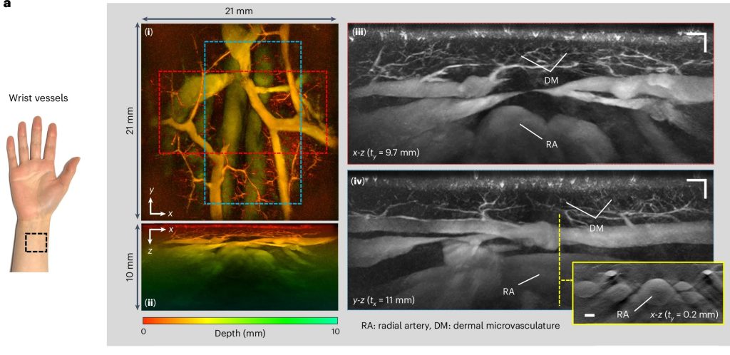
A new hand-held scanner developed by UCL researchers and tested in a series of clinical trials on UCLH patients can generate highly detailed 3D photoacoustic images in just seconds, paving the way for their use in a clinical setting for the first time and offering the potential for earlier disease diagnosis.
In the study, published in Nature Biomedical Engineering, the UCL and UCLH team show their technology can deliver photoacoustic tomography (PAT) imaging scans to doctors in real time, providing them with accurate and intricate images of blood vessels, helping inform patient care.
Photoacoustic tomography imaging uses laser-generated ultrasound waves to visualise subtle changes (an early marker of disease) in the sub-millimetre-scale veins and arteries up to 15mm deep in human tissues.
However, up until now, existing PAT technology has been too slow to produce high-enough quality 3D images for use by clinicians.
The older PAT scanners took more than five minutes to take an image – by reducing that time to a few seconds or less, image quality is much improved and far more suitable for people who are frail or poorly.
The researchers say the new scanner could help to diagnose cancer, cardiovascular disease and arthritis in three to five years’ time, subject to further testing.
In this study, the team tested the scanner during pre-clinical tests on 10 UCLH patients with type-2 diabetes, rheumatoid arthritis or breast cancer, along with seven healthy volunteers. They also compared the PAT scans to regular clinical scans taken at UCLH. Larger scale trials of the device are ongoing at UCLH and UCL.
In three patients with type-2 diabetes, the scanner was able to produce detailed 3D images of the microvasculature in the feet, highlighting deformities and structural changes in the vessels. The scanner was also used to visualise the skin inflammation linked to breast cancer.
UCLH consultant radiologist Andrew Plumb, a senior author of the study and Chief Investigator of the clinical PAT studies, said: “One of the complications often suffered by people with diabetes is low blood flow in the extremities, such as the feet and lower legs, due to damage to the tiny blood vessels in these areas. But until now we haven’t been able to see exactly what is happening to cause this damage or characterise how it develops.
“In one of our patients, we could see smooth, uniform vessels in the left foot and deformed, squiggly vessels in the same region of the right foot, indicative of problems that may lead to tissue damage in future. Photoacoustic imaging could give us much more detailed information to facilitate early diagnosis, as well as better understand disease progression more generally.” Dr Plumb is also Associate Professor of Medical Imaging at UCL.
Patients were identified and recruited from a number of clinics at UCLH, including consultant rheumatologist Madhura Castelino, consultant interventional radiologist Conrad von Stempel and research staff Katerina Soteriou and Antonia Yeung who co-ordinated safe, timely scanning at UCLH and UCL on the new PAT scanner.
UCL Professor of Biomedical Photoacoustics Paul Beard, corresponding author, said: “We’ve come a long way with photoacoustic imaging in recent years, but there were still barriers to using it in the clinic.
“The breakthrough in this study is the acceleration in the time it takes to acquire images, which is between 100 and 1000 times faster than previous scanners.
“This speed avoids motion-induced blurring, providing highly-detailed images of a quality that no other scanner can provide. It also means that rather than taking five minutes or longer, images can be acquired in real time, making it possible to visualise dynamic physiological events.
“These technical advances make the system suitable for clinical use for the first time, allowing us to look at aspects of human biology and disease that we haven’t been able to before.
“Now more research is needed with larger groups of patients to confirm our findings.”
Professor Beard added that a key potential use for the new scanner was to assess inflammatory arthritis, which requires scanning all 20 finger joints in both hands. With the new scanner, this can be done in a few minutes – older PAT scanners take nearly an hour, which is too long for elderly, frail patients, he said.
Cancer incidence trends in 2021 largely returned to what they were before the COVID pandemic, according to a study by researchers at the National Institutes of Health (NIH). However, there was little evidence of a rebound in incidence that would account for the decline in diagnoses in 2020, when screening and other medical care was disrupted, according to findings published in the Journal of the National Cancer Institute. One exception was breast cancer, where the researchers did see an uptick in diagnoses of advanced-stage disease in 2021.
A previous study showed that new cancer diagnoses fell abruptly in early 2020, as did the volume of pathology reports, suggesting that many cancers were not being diagnosed in a timely manner. To determine whether these missed diagnoses were caught in 2021, possibly as more advanced cancers, researchers from NIH’s National Cancer Institute (NCI) compared observed cancer incidence rates for 2021 with those expected from pre-pandemic trends using data from NCI’s Surveillance, Epidemiology, and End Results Program.
A full recovery in cancer incidence should appear as an increase over pre-pandemic levels (also known as a rebound) to account for the missed diagnoses. The researchers looked at cancer overall, as well as five major cancer types that vary in how they are typically detected: through screening (female breast and prostate cancer), due to symptoms (lung and bronchus and pancreatic cancer), or incidentally during other medical procedures (thyroid cancer).
Cancer incidence rates overall and for most specific cancers approached pre-pandemic levels, with no significant rebound to account for the 2020 decline. However, in addition to an uptick in new diagnoses of advanced breast cancer in 2021, the data also provided some evidence of an increase in diagnoses of advanced pancreatic cancer. Also, new diagnoses of thyroid cancers in 2021 were still below pre-pandemic levels.
The researchers concluded that 2021 was a transition year that was still affected by new variants and new waves of COVID-19 cases, which continued to impact medical care. They said the findings highlight the need for ongoing monitoring to understand the long-term impacts of the pandemic on cancer diagnoses and outcomes.
An analysis of Swedish data, where the definition of triple negative breast cancer (TNBC) differs from that used internationally, brings additional insights to on ongoing discussion in the scientific community. The study was presented at the 2023 European Society for Medical Oncology (ESMO) meeting and is now published in Lancet Regional Health – Europe.
The Swedish definition of TNBC differs from the international version in that it also includes tumours with low expression of the Oestrogen Receptor (ER) biomarker, ie in 1–9% of tumour cells. Internationally, ER-low breast cancer is classified as hormone-sensitive and treated differently from TNBC patients. This is despite previous studies demonstrating that the majority of ER-low tumours are molecularly similar to ER-zero, the latter completely without expression of ER, and meta-analyses that show no survival benefit from endocrine therapy in ER-low tumours.
The Swedish population-based study included all women diagnosed with TNBC in Sweden during 2008–2020 using the National Quality Register for Breast Cancer. Patient and tumour characteristics, treatment and survival in patients with low ER expression was compared to patients with no ER tumour expression.
The study identified and included 5655, and 560 patients (10%) were defined as ER-low and 5095 (90%) as ER-zero. The data demonstrated there are only small differences in tumour characteristics, no differences in response to neoadjuvant chemotherapy and no significant differences in prognosis.
“The international cut-off for ER-positivity and thus the definition of TNBC as only completely ER-negative is now increasingly questioned. ER-low tumours behave like ER-zero tumours and should be treated as such. On the basis of real-world data, the Swedish cutoff for hormone receptor positivity appears to be more clinically relevant. A changed international definition would give patients with ER-low expressing breast cancer the same treatment options as in TNBC, within studies and in clinical routine,” says study leader Dr Irma Fredriksson.
The study was carried out in collaboration with the pharmaceutical company MSD.
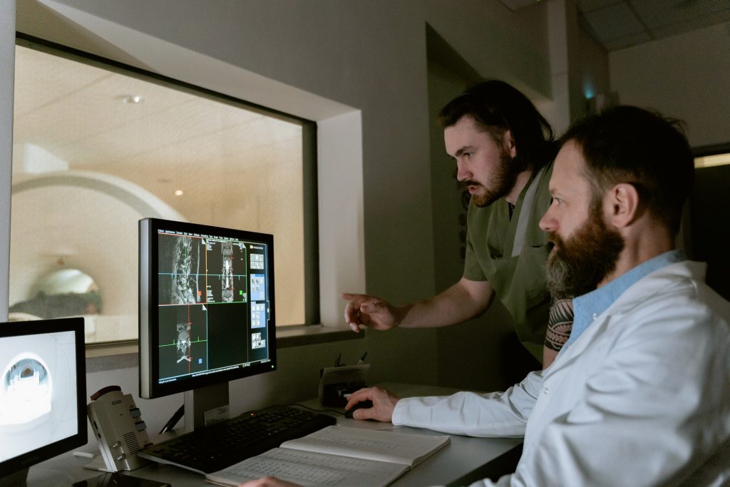
Diffuse gliomas are malignant brain tumours that cannot be optimally examined by means of conventional MRI imaging. So-called amino acid PET (positron emission tomography) scans are better able to image the activity and spread of gliomas. An international team of researchers from the RANO Working Group have drawn up the first ever international criteria for the standardised imaging of gliomas using amino acid PET. It has published its results in the journal The Lancet Oncology.
PET uses a radioactive tracer to measure metabolic processes in the body. Amino acid PET is used in the diagnosis of diffuse gliomas, with tracers that work on a protein basis (amino acids) and accumulate in brain tumours.
The Response Assessment in Neuro-Oncology (RANO) Working Group is an international, multidisciplinary consortium founded to develop standardised new response criteria for clinical studies relating to brain tumours.
Under the joint leadership of nuclear physician Nathalie Albert from LMU and oncologist Professor Matthias Preusser from the Medical University of Vienna, the RANO group has developed new criteria for assessing the success of therapies for diffuse gliomas.
Nathalie Albert explains: “PET imaging with radioactively labelled amino acids has proven extremely valuable in neuro-oncology and permits reliable representation of the activity and extension of gliomas. Although amino acid PET has been used for years, it had not been evaluated in a structured manner before now. In contrast to MRI-based diagnostics, there have been no criteria for interpreting these PET images.” According to the researchers, the new criteria allow PET to be used in clinical studies and everyday clinical practice and create a foundation for future research and the comparison of treatments for improved therapies.
New criteria for PET examinations of brain tumours
Diffuse gliomas are malignant brain tumorus that cannot be optimally examined by means of conventional MRI imaging. So-called amino acid PET scans are better able to image the activity and spread of gliomas.
These malignant brain tumours develop out of glial cells and are generally aggressive and difficult to treat.
The RANO group has developed criteria that permit evaluation of the success of treatment using PET. Called PET RANO 1.0, these PET-based criteria open up new possibilities for the standardised assessment of diffuse gliomas.
Diagnoses of early, curable stages of prostate cancer are being missed because national guidelines and media health campaigns in the UK focus on urinary symptoms despite a lack of scientific evidence, according to University of Cambridge researchers.
Prostate cancer is the most common type of cancer in men. And while 78% of men diagnosed with this cancer survive for over ten years, this proportion has barely changed over the past decade in the UK, largely because the disease is detected at a relatively late stage. In England, for example, nearly half of all prostate cancers are picked up at stage three of four (stage four being the latest stage).
Despite no evidence of a link between urinary symptoms and prostate cancer, national guidelines, health advice and public health campaigns continue to promote this link. In a review published in BMC Medicine, Cambridge researchers argue that not only is this unhelpful, but it may even deter men from coming forward for early testing and detection of a potentially treatable cancer.
“When most people think of the symptoms of prostate cancer, they think of problems with peeing or needing to pee more frequently, particularly during the night,” said Vincent Gnanapragasam, Professor of Urology at the University of Cambridge. “This misperception has lasted for decades, despite very little evidence, and it’s potentially preventing us picking up cases at an early stage.”
Prostate enlargement can cause the urinary problems often included in public health messaging, but evidence suggests that this is rarely due to malignant prostate tumours. Rather, research suggests that the prostate is smaller in cases of prostate cancer. A recent study – the UK PROTECT trial – even went as far as to say that a lack of urinary symptoms may in fact be an indicator of a higher likelihood of cancer.
Screening programmes are one way that cancers are often detected at an early stage, but in the case of prostate cancer, some argue that such programmes risk overwhelming health services and leading to men being treated for relatively benign disease.
Testing for prostate cancer involves a blood test that looks for a protein known as a prostate-specific antigen (PSA) that is made only by the prostate gland; however, it is not always accurate. PSA density is significantly more accurate than PSA alone in predicting a positive biopsy and is used in everyday clinical practice.
The researchers point to evidence that there is a misconception that prostate cancer is always symptomatic: a previous study found that 86% of the public associated prostate cancer with symptoms, but only 1% were aware that it could be asymptomatic.
“We urgently need to recognise that the information currently given to the public risks giving men a false sense of security if they don’t have any urinary symptoms,” said Prof Gnanapragasam.
“We need to emphasise that prostate cancer can be a silent or asymptomatic disease, particularly in its curable stages. Waiting out for urinary symptoms may mean missing opportunities to catch the disease when it’s treatable.
“Men shouldn’t be afraid to speak to their GP about getting tested, and about the value of a PSA test, especially if they have a history of prostate cancer in their family or have other risk factors such as being of Black or mixed Black ethnicity.”
The researchers say they are not advocating for an immediate screening programme, and acknowledge that changes in messaging could mean more men approaching their GPs for a PSA test, potentially resulting in unnecessary investigations and treatment. However, they argue that there are ways to reduce the risk of this happening. These include the use of algorithms to assess an individual’s risk and whether they need to be referred to a specialist, and for those who are referred, MRI scans could help rule out ‘indolent’ (mild) disease or negative findings, reducing the risks of an unnecessary biopsy.
“We’re calling on organisations such as the NHS, as well as patient charities and the media, to review the current public messaging,” said Prof Gnanapragasam.
“If men were aware that just because they have no symptoms doesn’t necessarily mean they are cancer free, then more might take up offers for tests. This could mean more tumours identified at an earlier stage and reduce the numbers of men experiencing late presentation with incurable disease.”
Researchers at the University of Waterloo have developed a new form of magnetic resonance imaging (MRI) that makes cancerous tissue glow in medical images. This innovation could enable more accurate detection and tracking of cancer over time.
“Our studies show this new technology has promising potential to improve cancer screening, prognosis and treatment planning,” said first author Professor Alexander Wong.
Irregular packing of cells leads to differences in the way water molecules move in cancerous tissue compared to healthy tissue. The new technology, called synthetic correlated diffusion imaging, highlights these differences by capturing, synthesising and mixing MRI signals at different gradient pulse strengths and timings.
In the largest study of its kind, the researchers collaborated with medical experts at the Lunenfeld-Tanenbaum Research Institute, several Toronto hospitals and the Ontario Institute for Cancer Research to apply the technology to a cohort of 200 patients with prostate cancer.
The synthetic correlated diffusion imaging was found to be better at delineating significant cancerous tissue than current imaging technique, making it a potentially powerful addition to the toolbox for doctors and radiologists.
“Prostate cancer is the second most common cancer in men worldwide and the most frequently diagnosed cancer among men in more developed countries,” said Prof Wong. “That’s why we targeted it first in our research.
“We also have very promising results for breast cancer screening, detection, and treatment planning. This could be a game-changer for many kinds of cancer imaging and clinical decision support.”
In an encouraging sign, the RAND corporation reports that despite COVID, cancer screenings in the United States rebounded in the wake of the first wave.
There has been concern since the COVID pandemic was keeping people from going in for routine cancer screening, resulting in more undetected cancers to progress unchecked and an increase in cancer deaths. As some 600 000 Americans were expected to die of cancer in 2020, any impact on screening is a considerable health concern.
In a statement, Ryan McBain, PhD, of the RAND Corp. in Santa Monica, California, said: “These are the first findings to show that, despite real fears about the consequences of drop off in cancer screens, health facilities figured out how to pick this back up after the initial pandemic restrictions. Our study shows that health systems were able to recalibrate resources and protocols in a relatively short interval to deliver these important services.”
In Spain, cancer diagnoses were down 38% in the first half of 2020, according to one study. The pandemic’s impact on cancer screening that required in-person examinations, such as mammography and colonoscopy, were a particular concern, McBain and co-authors noted. Moreover, little was known about the magnitude of the decline in screening rates or longer-term trends in screen.
Using data on Castlight Health beneficiaries from January 15 to July 31, 2020, the researchers calculated weekly screening rates per 10 000 eligible beneficiaries. Before the declaration of national emergency in the US on March 13, screening mammography weekly rate was 87.8 per 10 000, falling to a low of 6.9 per 10 000 in April. Thereafter, screening rates began a steady recovery, to 88.2 per 10 000 at the end of July.
Over the same time period, weekly colonoscopy screenings fell from 15.1 to 0.9 per 10 000, before rebounding to a weekly median of 12.6 per 10 000 by July 31, 2020.
Multivariable regression analyses confirmed the significant declines in screening mammography and colonoscopy. A larger decline in colonoscopy was observed in high-income counties. Otherwise, the analyses showed no significant demographic variations.
Laura Makaroff, DO, of the American Cancer Society in Atlanta, said that the pandemic’s ultimate impact on cancer screening and cancer care is still largely unknown.
“We have seen similar data showing some rebound in cancer screening rates last summer and fall, but even those turnarounds show an approximate 30% decrease in cancer screening compared with pre-pandemic rates,” she said to MedPage Today in an email. “We also don’t yet know the full impact of the late fall and winter surges on disruptions in cancer screening and diagnosis.”
“The COVID-19 pandemic has had numerous consequences secondary to the disease itself, including reduced access to care for other illnesses,” Dr Makaroff added. “While these measures were necessary, delays in cancer screening, diagnosis, and treatment due to reduced healthcare access will likely result in a short-term drop in cancer diagnoses followed by increases in late-stage diagnoses and preventable cancer deaths.
“The full impact of the COVID-19 pandemic on cancer prevention and early detection will not be known until population-based nationwide data become available in the years to come,” she said.
Journal information: McBain RK, et al “Decline and rebound in routine cancer screening rates during the COVID-19 pandemic” J Intern Med 2021; DOI: 10.1007/s11606-021-06660-5.