Tag: breast cancer

For Large Breast Sizes, Prone Positioning is Less Toxic for Radiotherapy

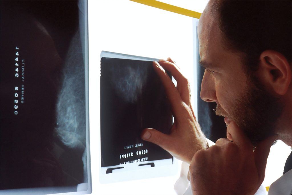
Woman receiving mammogram
A woman receiving a mammogram, Source: National Cancer Institute

For women with large breast sizes, receiving radiotherapy with prone positioning is less toxic than while supine, according to a study published in JAMA Oncology.

Patients treated in the supine position had significantly higher rates of moist desquamation anywhere in the breast compared with those treated in the prone position (39.6% vs 26.9%).

“Treatment in the prone position has several dosimetric advantages for these patients,” the researchers explained. “It allows for more homogeneous dose distribution owing to the smaller separation when compared with the supine position, which decreases deposition of higher doses in the inframammary fold and axilla.”

Fewer toxic effects of the skin were seen when patients were treated with hypofractionated radiotherapy compared with extended fractionation, they added.

“Prone radiotherapy appears to be an excellent option for patients with large breast size and right-sided breast cancer, and may benefit many women with left-sided breast cancer with large breast size if acceptable cardiac avoidance is feasible,” observed Mayo Clinic’s Dean Shumway, MD, and Cedars-Sinai Medical Center’s Katelyn Atkins, MD, PhD, in an accompanying editorial. “In summary, prone positioning for whole-breast radiotherapy represents a valuable addition to the armamentarium of treatment techniques to reduce the adverse effects associated with whole-breast radiotherapy.”

Of the 357 women (mean age 61 years) included, 182 were treated in the supine position and 175 were treated in the prone position.

From April 2013 until June 2016, 167 patients received 50 Gy in 25 fractions (extended fractionation) with or without boost (range 10-16 Gy). After the trial was amended in June 2016, the majority of patients (93.2%) received the hypofractionation regimen of 42.5 Gy in 16 fractions.

The researchers also found that the supine position was associated with more grade 3 desquamation compared with the prone position (15.4% vs 8.0%; OR 2.09, 95% CI 1.62-2.69, P<0.001).

In addition, when broken down by treatment with either extended fractionation or hypofractionation, extended fractionation was associated with more:

  • Toxic effects (43.3% vs 23.2%)
  • Grade 3 desquamation (17.2% vs 6.3%)
  • Pain (9.4% vs 3.4%)

“These differences were primarily driven by the rates of toxic effects in patients treated in the supine position,” the authors noted.

Specifically, in patients treated in the supine position, extended fractionation was associated with increased desquamation compared with hypofractionation (51.1% vs 27.8%), and grade 3 desquamation (23.9% vs 6.7%).

Extended fractionation was also associated with increased toxicity in patients treated in the prone position, although the link was less pronounced. Desquamation occurred in 35.2% of patients treated with extended fractionation versus 18.4% of patients treated with hypofractionation (OR 2.41), while grade 3 desquamation occurred in 10.2% versus 5.7% of patients (OR 1.87).

No differences in quality of life as measured by global health status, breast symptoms, or pain scales between the supine and prone groups were seen, the researchers noted.

How Breast Cancer Cells Sabotage Insulin Production to Fuel Themselves

A breast cancer cell
Scanning Electron Micrograph of a breast cancer cell. Credit: NIH

Breast cancer and diabetes have long been suspected to have some kind of relationship, but now new research in Nature Cell Biology reveals how breast cancer cells sabotage insulin production to fuel their own cravings for glucose.

Diabetes risk begins to increase two years after a breast cancer diagnosis, and by 10 years post-diagnosis, the risk is 20% higher in breast cancer survivors than in age-matched women without breast cancer.

But these epidemiological linkages are not clear-cut or definitive, and some studies have found no associations at all. In the paper, a research team describe a possible biological mechanism connecting the two diseases, in which breast cancer suppresses the production of insulin, resulting in diabetes, and the impairment of blood sugar control promotes tumour growth.

“No disease is an island because no cell lives alone,” said corresponding study author Shizhen Emily Wang, PhD, professor of pathology at UC San Diego School of Medicine. “In this study, we describe how breast cancer cells impair the function of pancreatic islets to make them produce less insulin than needed, leading to higher blood glucose levels in breast cancer patients compared to females without cancer.”

The researchers name the culprit as extracellular vesicles (EV), which carry DNA, RNA, proteins, fats and other materials between cells, a sort of cargo communication system.

The cancer cells were found to be secreting microRNA-122 into the vesicles. When vesicles reach the pancreas, Prof Wang said, they can enter the islet cells, offload their miR-122 cargo and damage the islets’ critical function in maintaining a normal blood glucose level.

“Cancer cells have a sweet tooth,” Prof Wang said. “They use more glucose than healthy cells in order to fuel tumor growth, and this has been the basis for PET scans in cancer detection. By increasing blood glucose that can be easily used by cancer cells, breast tumors make their own favorite food and, meanwhile, deprive this essential nutrient from normal cells.”

Feeding mice slow-releasing insulin pellets or an SGLT2 inhibitor restored normal control of glucose in the presence of a breast tumour, in turn suppressed the tumour’s growth.

“These findings support a greater need for diabetes screening and prevention among breast cancer patients and survivors,” remarked Prof Wang, noting that a miR-122 inhibitor is currently in clinical trial as a potential treatment for chronic hepatitis C. It has been found to be effective in restoring normal insulin production and suppressing tumour growth in mouse models of breast cancer.

“These miR-122 inhibitors, which happen to be the first miRNA-based drugs to enter clinical trials, might have a new use in breast cancer therapy,” Prof Wang posited.

Source: University of California – San Diego

Why Breast Cancer Metastases Spread to the Bones

A breast cancer cell
Scanning Electron Micrograph of a breast cancer cell. Credit: NIH

A team of biologists has discovered a factor that increases cellular plasticity can explain why, in breast cancer, metastases spread to the bones. Their findings appear in the journal Nature Communications.

The organs affected by cancer metastases depend in part on their tissue of origin – in the case of breast cancer, they usually form in the bones. No cure for metastatic breast cancer exists yet, and it is associated with a poor prognosis with a 5-year survival rate of 26%. However, treatments can improve and extend the lives of patients.

From the primary site of a tumour, cancer cells can invade their microenvironment and then circulate via blood and lymphatic vessels to distant healthy tissue to form metastases. In the case of metastatic breast cancer, the cancer cells primarily colonise the bones, but can also be found in other organs such as the liver, lungs or brain.

Plasticity of tumour cells

Although the mechanisms behind the different stages of the metastatic process are not yet fully understood, cellular plasticity plays an important role: tumour cells that become metastatic change their shape and become mobile.

Using mice, the researchers investigated the potential role of the protein ZEB1, known to increase cell plasticity, in breast cancer cell migration.

“Unlike in women, mice transplanted with human breast cancer cells develop metastasis to the lungs, not the bones. We therefore sought to identify factors capable of inducing metastasis in bone tissue and in particular tested the effect of the factor ZEB1.,” explained researcher Nastaran Mohammadi Ghahhari, first author of the study

Directing metastasis to bone

In in vitro migration and invasion experiments, the scientists found that cancer cells expressing ZEB1 moved to bone tissue, unlike cancer cells that did not express it. These results were later confirmed when human breast cancer cells were transplanted into the mammary glands of mice. If the cancer cells did not express ZEB1, metastasis occurred primarily in the lungs. In contrast, when ZEB1 was present, metastases also developed in the bones, as is the case in women.

‘‘We can therefore assume that this factor is expressed during tumour formation and that it directs cells that have acquired metastatic characteristics to the bones,’’ explained Didier Picard, the study’s last author. These findings confirm the importance of plasticity in metastases, and could help lead to new therapies.

Source: University of Geneva

Key Factors in Hospitalisation after Breast Reconstruction Surgery

Photo by Jafar Ahmed on Unsplash

Factors such as anaemia and anticoagulants have more impact on hospitalisation time after breast reconstruction than “common” risk factors according to a new study published in the Journal of Clinical Medicine.

The study investigated the impact of different factors on postoperative blood loss and drainage fluid volume, two factors which can lengthen hospitalisation time of patients after breast reconstruction after breast cancer surgery. The findings of the study allow for an improved risk assessment and planning of reconstructive breast surgery to offer patients personalised and improved treatment.

Partial or total mastectomy is often necessary in breast cancer surgery, and reconstructive breast surgery lessens the psychological stress on the patient. Fast wound healing after surgical breast reconstruction is crucial to not delay subsequent cancer treatments. Factors influencing the length of hospital stay (LOS) or wound healing are therefore particularly significant in cancer treatment. This study identified previously unrecognised risk factors.

Blood loss and drainage fluid volumes after breast reconstruction due to breast cancer were recorded, parameters which are closely linked to the healing process and LOS .Lower loss equals earlier patient discharge and early start of subsequent treatment. “We analysed factors that might affect blood loss and drainage fluid volumes after surgery – but can be identified before the surgery,” explained lead author Dr Tonatiuh Flores, plastic surgeon. “These factors included age, body mass index and smoking status – factors that are known to have a strong impact on the course of disease.” Additionally, haemoglobin levels and possible antithrombotic prophylaxis were reviewed – two parameters that are particularly significant in oncological treatment.

Surprising results emerged from the evaluation of a total of 257 breast reconstructions in 195 patients. Professor Konstantin Bergmeister, senior author of the study explained that “the classic risk factors did not significantly influence postoperative blood loss and drainage fluid output. Haemoglobin levels and anticoagulant concentration, however, did.” The analysis revealed a close relation between low haemoglobin values or anaemia and fluid loss after reconstructive breast surgery. Co-author Prof. Klaus Schroegendorfer, elaborated on this: “Especially breast cancer patients often show perioperative anaemia, caused by the frequently required neoadjuvant chemotherapy which can affect blood values, in particular haemoglobin.”

There were similar findings regarding low molecular heparin used in cancer patients as antithrombotic prophylaxis. Patients receiving heparin tended to have increased drainage fluid output after surgery, though the effect was not as strong as with perioperative anaemia.

The study authors recommend that, to cut LOS and continue the necessary cancer treatment after reconstructive breast surgery in cancer patients as early as possible, patients should preoperatively be screened for anaemia and administration of low molecular heparin should be adapted to the patients’ risk. In correlation to the results, follow-up treatment can be improved, patients can be discharged earlier and cancer treatment can be continued.

Source: Karl Landsteiner University of Health Sciences

Cardamonin may Have Anticancer Properties

Breast cancer cells. Image source: National Cancer Institute on Unsplash

The natural compound cardamonin present in the spice cardamom and other plants could have therapeutic potential for triple-negative breast cancer, according to a new study using human cancer cells. The findings also show that the compound targets a gene that helps cancer cells elude the immune system.

Around 10–15% of breast cancers are triple-negative, which means they lack receptors for oestrogen or progesterone and don’t make excess amounts of a protein called HER2. These tumours are difficult to treat because they don’t respond to the hormone-based therapies used for other types of breast cancer. They also tend to be more aggressive and have a higher mortality rate than other breast cancers.

“It has been challenging to develop a targeted therapy for triple-negative breast cancer that is safe and effective at the same time,” said Assistant Professor Patricia Mendonca, PhD, of Florida A&M University. “Because of this, there is a critical need to investigate medicinal plants as a new way to combat this cancer.”

The research was presented at the American Society for Investigative Pathology annual meeting.

“The fact that cardamonin has been used for centuries as a spice and, more recently, as a supplement shows that its intake is safe and may bring health benefits,” said A/Prof Mendonca. “Our research shows that cardamonin holds potential for improving cancer therapy without as many side effects as other chemotherapeutic agents.”

For the new study, the researchers investigated how cardamonin affected the expression of the programmed cell death ligand 1 (PD-L1) gene, which is found in tumour cells. PD-L1 is overexpressed during breast cancer progression and plays a critical role in helping breast cancer cells evade the body’s immune system.

The researchers used two genetically different triple-negative breast cancer cell lines – one derived from women with African American ancestry and the other from women of European origin (Caucasian). They found that cardamonin treatment caused a dose-dependent decrease in cell viability in both cell lines. It also reduced PD-L1 expression in the Caucasian cell line but not the African American cell line, indicating that cells from different races may respond differently to cardamonin because of genetic variations among races. 

“This is the first study to describe cardamonin’s inhibitory effect on the expression of PD-L1, which is relevant for the treatment of triple-negative breast cancer,” said Mendonca. “These findings add support to other research that has shown differences in the tumour microenvironment between African and non-African Americans.”

Source: EurekAlert!

To Properly Use AI to Analyse Breast Cancers, Look to Past Mistakes

Source: National Cancer Institute

Doctors writing in an editorial in JAMA Health Forum caution that while using AI to analyse breast cancer tumours has the potential to improve healthcare efficiency and outcomes, similar technological leaps have previously led to higher rates of false-positive tests and over-treatment.

The editorial wasco-written by Joann G. Elmore, MD, MPH, professor of medicine at the David Geffen School of Medicine at UCLA, and Christoph I. Lee, MD, MS, MBA, a professor of radiology at the University of Washington School of Medicine.

“Without a more robust approach to the evaluation and implementation of AI, given the unabated adoption of emergent technology in clinical practice, we are failing to learn from our past mistakes in mammography,” the authors wrote.

One of those “past mistakes in mammography,” the authors said, was adjunct computer-aided detection (CAD) tools, which grew rapidly in popularity in the field of breast cancer screening starting more than two decades ago. CAD was approved by the FDA in 1998, and by 2016 more than 92% of U.S. imaging facilities were using the technology to interpret mammograms and hunt for tumours. However, CAD did not improve mammography accuracy., according to the evidence. “CAD tools are associated with increased false positive rates, leading to overdiagnosis of ductal carcinoma in situ and unnecessary diagnostic testing,” the authors wrote. The US Medicare system stopped paying for CAD in 2018, but by then the tools had run up more than $400 million a year in wasted health costs.

“The premature adoption of CAD is a premonitory symptom of the wholehearted embrace of emergent technologies prior to fully understanding their impact on patient outcomes,” Drs Elmore and Lee wrote. “As AI algorithms are increasingly receiving FDA clearance and becoming commercially available with ROC curves similar to what we observed prior to CAD clearance and adoption, how can we prevent history from repeating itself?”

The doctors suggest a number of safeguards to avoid “repeating past mistakes” such as tying reimbursement to proven efficacy.

Source: UCLA Health

New Recommendations for Earlier Breast Cancer MRI Screening

This screening MRI detected a very small cancer (circled) in the patient’s breast.
Credit: Dr. Kathyrn Lowry

Annual MRI screenings starting at ages 30 to 35 may slash breast-cancer mortality by more than 50% among women with genetic changes in three genes, according to a study published in JAMA Oncology.

The pathogenic variants are in the ATM, CHEK2 and PALB2 genes – which collectively are as prevalent as the much-reported BRCA1/2 gene mutations. The study authors state that their findings support earlier MRI screening in these women.

“Screening guidelines have been difficult to develop for these women because there haven’t been clinical trials to inform when to start and how to screen,” said lead author Dr Kathryn Lowry.

The work was a collaboration of the Cancer Intervention and Surveillance Modeling Network (CISNET), the Cancer Risk Estimates Related to Susceptibility (CARRIERS) consortium, and the Breast Cancer Surveillance Consortium.

To arrive at their model, the researchers input age-specific risk estimates from CARRIERS involving some 64 000 women and recent published data for screening performance.

“For women with pathogenic variants in these genes, our modeling analysis predicted a lifetime risk of developing breast cancer at 21% to 40%, depending on the variant,” Dr Lowry said. “We project that starting annual MRI screening at age 30 to 35, with annual mammography starting at age 40, will reduce cancer mortality for these populations of women by more than 50%.”

The simulations compared the combined performance of mammography and MRI against mammography alone, and projected that annual MRI conferred significant additional benefit to these populations.

“We also found that starting mammograms earlier than age 40 did not have a meaningful benefit but increased false-positive screens,” Dr Lowry added.

Results from CISNET models have informed past guidelines, including the 2009 and 2016 U.S. Preventive Services Task Force recommendations for breast cancer screening in average-risk women.

“Modelling is a powerful tool to synthesise and extend clinical trial and national cohort data to estimate the benefits and harms of different cancer control strategies at population levels,” said senior author Dr Jeanne Mandelblatt.

The study projected about four false-positive screening results and one to two benign biopsies per woman over a 40-year screening span, the authors noted.

To get any benefit from genetic susceptibility-based screening guidelines, a woman would have to know beforehand that she carries the gene, yet most often a genetic test panel is done after a positive cancer result – too late for any benefit.

“People understand very well the value of testing for variants in BRCA1 and BRCA2, the most common breast cancer predisposition genes. These results show that testing other genes, like ATM, CHEK2, and PALB2, can also lead to improved outcomes,” said senior author Dr Mark Robson.

The researchers hope their analysis will aid the National Comprehensive Cancer Network (NCCN), the American Cancer Society and other organizations that issue guidance for medical oncologists and radiologists.

“Overall what we’re proposing is slightly earlier screening than what the current guidelines suggest for some women with these variants,” said senior author Professor Allison Kurian. “For example, current NCCN guidelines recommend starting at age 30 for women with PALB2, and at 40 for ATM and CHEK2. Our results suggest that starting MRI at age 30 to 35 appears beneficial for women with any of the three variants.”

Source: University of Washington

Researchers Halt Aspirin Trial to Prevent Breast Cancer Recurrence

Source: National Cancer Institute

A large randomised trial was halted after preliminary analysis found that taking aspirin after treatment for breast cancer did not reduce the risk of disease recurrence.

Laboratory studies had previously shown that aspirin and nonsteroidal anti-inflammatory drugs (NSAIDs) reduced breast cancer growth and invasion. Non-steroidal anti-inflammatory drugs (NSAIDs) display anticancer activity through the inhibition of the COX-2 enzyme, triggering processes such as apoptosis, a reduction in proliferation and inhibition of carcinogenesis.  Several observational studies have shown a reduced risk of breast cancer mortality among regular aspirin users. 

There was a 25% higher risk of invasive recurrence in patients who took aspirin for a median of 18 months, but not statistically different from placebo (P = 0.1258). The aspirin group had an excess of all disease-related events, including death, local and distant recurrence/progression, and new primary tumours.

The results are in line with similar trials that ended while the Aspirin after Breast Cancer (ABC) trial was ongoing, Wendy Y. Chen, MD, of Dana-Farber Cancer Institute in Boston, said during a presentation at the American Society of Clinical Oncology (ASCO) Plenary Series.

“In this double-blind, placebo-controlled randomised trial, there was no benefit of aspirin 300 milligrams daily in terms of breast cancer invasive disease-free survival,” reported Dr Chen. “Although follow-up was short, the futility bound was clearly crossed. We had reached 50% of the events, and there was a numerically higher number of events in the aspirin arm. Therefore, it was unlikely that even with further follow-up there wouldn’t be any benefit associated with aspirin.”

“Although inflammation may still play a key role in cancer, it’s important to remember that aspirin may have different effects in other cancers, such as colon, or in different settings, such as primary versus secondary prevention,” she added.

Though the trial was well designed, enrolled the right population and with adequate dosing. the trial was stopped early for futility, commented Angela DeMichele, MD, of the Abramson Cancer Center at the University of Pennsylvania.

“The direction and magnitude [of the difference in events] highly preclude the possibility that there would have been a benefit with more follow-up,” said Dr DeMichele. “Although it was not statistically significant, we cannot rule out the possibility of a potential increase in breast cancer recurrence from the use of aspirin.”

“For patients and providers at this time, aspirin should not be used simply to prevent breast cancer recurrence,” she continued. “For those situations in which there are other options, decisions about aspirin use for other indications should definitely include an individualised risk/benefit discussion between physician and patient.”

The results underscore the need for prospective, randomised clinical trials to validate the effects of interventions from observational studies, she concluded.

The ABC trial involved patients under 70 with HER2-negative, high-risk breast cancer. The study randomised 3021 participants to 300 mg of aspirin daily or matching placebo for 5 years, with the primary endpoint being invasive disease-free survival. 

Dr Chen further noted that three clinical trials of aspirin or NSAID treatment ended while the ABC trial was ongoing. The Canadian-led MA.27 trial of an aromatase inhibitor plus celecoxib ended due to toxicity in the celecoxib arm. The randomised REACT trial of celecoxib in HER2-negative breast cancer showed no difference in disease-free survival after more than 6 years of follow-up.

The ASPREE trial tested low-dose aspirin on all-cause mortality in healthy older patients, and results showed a trend to increased all-cause mortality and significantly higher cancer mortality in the aspirin arm. 
During the post-presentation discussion, an audience member asked whether the results definitively ruled out a late benefit of aspirin, given that most patients had HR-positive disease wherein late relapse is not uncommon.

“It’s always frustrating when a study is closed early, and it was done in this case after we had reached 50% of the expected benefits,” said Chen. “There was an increase [in clinical events]. Not a statistically significant increase, but it was bordering on statistical significance. In order for aspirin to have a benefit, it would mean that in the second half, there would need to be a significantly decreased risk. It would basically need to flip and that would be biologically difficult to imagine.”

“I think it’s fair to say that this study doesn’t say definitively that there’s harm, but as for the likelihood of a benefit of aspirin, that would be extremely unlikely,” she said.

Source: MedPage Today

Metformin Ineffective in Most Breast Cancers

Breast cancer cells. Image source: National Cancer Institute on Unsplash

Researchers have found that the diabetes drug metformin, once hoped to hold enormous promise in treating breast cancer, does not prevent or stop the spread of the most common forms of the disease but may still have potential in HER2-positive breast cancer.

The randomised, double-blind trial enrolled 3600 patients who received two pills a day of either placebo or metformin. Overall, researchers found the addition of metformin to standard breast cancer treatments did not improve outcomes in the two most common types of breast cancer, hormone receptor-positive or negative.

“The results tell us that metformin is not effective against the most common types of breast cancer and any off-label use of this drug for the treatment of these common types of breast cancer should be stopped,” said Pamela Goodwin, a professor in the department of medicine at the University of Toronto’s Temerty Faculty of Medicine.

Prof Goodwin presented the findings at the 2021 San Antonio Breast Cancer Symposium.

While metformin was found not to be effective in treating the most common forms of breast cancer, there was evidence that use of metformin for five years might lead to a reduction in deaths from HER2-positive breast cancer, a less aggressive subtype which makes up about 20% of all breast cancers.

“Metformin is not beneficial for use in most common breast cancers, but in the cases of HER2 positive breast cancer, our findings suggest it may be beneficial,” said Prof Goodwin. “These results need to be replicated in future research before metformin is used as a breast cancer treatment, however, it could provide an additional treatment option for HER2-positive breast cancer,” she added

Previous studies suggested metformin may also reduce the risk of development and increase survival of some cancers, including breast cancer.

Metformin was theorised to slow breast cancer growth by improving patient metabolism, notably insulin levels, leading to reduced cancer cell growth, or that it might impact cancer cells directly.

Next steps would be to prospectively test the impact of metformin in patients with HER2-positive breast cancer in a randomised clinical trial. 

Source: University of Toronto

Anti-diabetes Drug under Development May Also Treat Breast Cancer

Source: NCI

A new study has shown that a small molecule inhibitor drug, with the unwieldy designation of PF05175157, originally developed to treat diabetes by Pfizer, may help in the treatment of breast cancer by blocking a key enzyme. 

The findings from the Yale Cancer Center-led study were reported at the 2021 San Antonio Breast Cancer Symposium in San Antonio, Texas.

“Our research shows the preclinical, anti-cancer activity using PF05175157 may lead us to bring this drug back into the clinic to help treat patients with breast cancer,” said lead study author Julia Foldi, MD, PhD, a clinical fellow at Yale Cancer Center and Smilow Cancer Hospital. “More studies are needed, but our initial data looks very promising.”

Cancer cells are characterised by altered metabolism. In this study, the Yale team identified new metabolic vulnerabilities in cancer cells that are based on a loss of enzyme diversity. They found that an enzyme called acetyl-CoA-carboxylase-1 (ACC1), is critical for the survival of breast cancer cells. The ACC1 enzyme is the key initial step in fatty acid synthesis. Fatty acids are building blocks of the various types of lipids and fat that are the critical ingredients of cell membranes and play an important role in energy generation in cells. The team’s analysis demonstrated that blocking ACC1 using PF05175157 can inhibit the growth of breast cancer cells grown in mice and also in patient-derived cancer models.

“We are currently testing this drug in combination with other approved breast cancer drugs to see if it could improve their activity, with the hope to bring the most promising combinations to the clinic to help patients with breast cancer,” added Lajos Pusztai, MD, DPhil, Professor of Medicine (Medical Oncology), Director of Breast Cancer Translational Research at Yale Cancer Center, and senior author of the study.

Source: Yale Cancer Center