Tag: breast cancer

New Trial Flips the Script for Hormonal Treatment of Breast Cancer

Photo by National Cancer Institute

For decades, hormonal treatment of breast cancer has been going in one direction: blocking oestrogen. Now, a global study has discovered there may be another, less toxic way to defeat the most common form of breast cancer. The results, published in The Lancet Oncology, showed that the androgen receptor (AR) agonist enobosarm, is effective against oestrogen receptor-positive (ER+) breast cancer, which constitutes up to 80% of all breast cancer cases.

“The effectiveness of enobosarm lies in its ability to activate the AR and trigger a natural defence mechanism in breast tissue, thereby slowing the growth of ER+ breast cancer, which relies on the hormone oestrogen to grow and spread,” said senior co-author Professor Wayne Tilley, Director of the Dame Roma Mitchell Cancer Research Laboratories at the University of Adelaide.

“This clinical study is supported by our pre-clinical research, previously published in Nature Medicine, which established that the AR is a tumour suppressor in both normal breast tissue and ER+ breast cancer.”

Along with investigators from the University of Adelaide and Dana-Farber Cancer Institute (DFCI) in Boston, USA, the international study also included researchers from the University of Liverpool in the UK and other experts around the world.

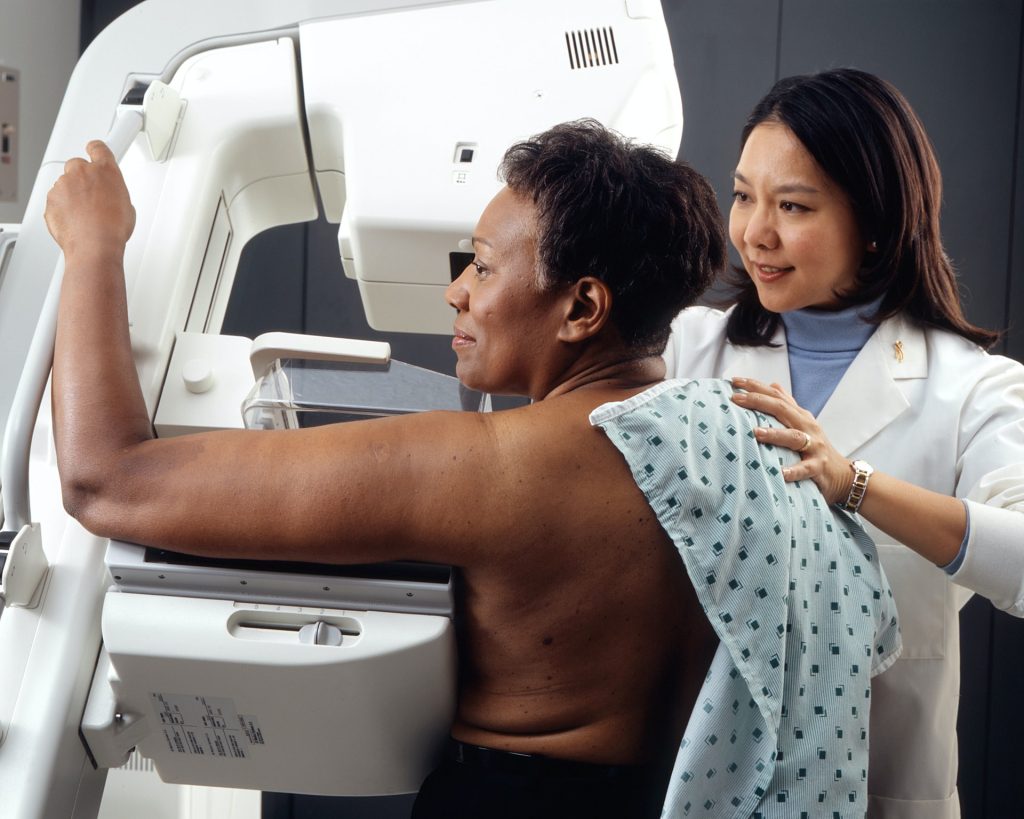
The team assessed enobosarm’s efficacy and safety in 136 postmenopausal women with advanced or metastatic ER-positive, HER2-negative breast cancer.

Enobosarm showed significant anti-tumour activity and was well-tolerated by patients, without adversely affecting their quality of life or causing masculinising symptoms.

This discovery represents the first advancement in hormonal treatment of ER+ breast cancer in decades and offers a promising new oral treatment strategy for the most prevalent form of breast cancer.

The new hormonal strategy differs from the existing standard-of-care hormonal treatments, which have been around for decades and involve suppressing oestrogen activity in the body or inhibiting the ER.

Although successful initially, treatments targeting ER can cause severe side effects and treatment-resistant progression of the disease is common.

“Our findings are very promising. They demonstrate that stimulating the androgen receptor pathway with enobosarm can be beneficial,” said senior co-author and study Principal Investigator Dr Beth Overmoyer from DFCI.

“This is the first time a non-oestrogen receptor hormonal treatment approach has been shown to be clinically advantageous in ER+ breast cancer. The study supports further investigation of enobosarm in earlier stages of breast cancer as well as in combination with targeted therapies, such as ribociclib, a CDK 4/6 inhibitor.”

estrogen to grow and spread,” said senior co-author Professor Wayne Tilley, Director of the Dame Roma Mitchell Cancer Research Laboratories at the University of Adelaide.

“The data strongly encourages more clinical trials for AR-stimulating drugs in treating AR-positive and ER-positive breast cancer. The fact that this drug is well-tolerated also opens possibilities for its use in breast cancer prevention,” said co-author Dr Stephen Birrell, a clinical affiliate of the University of Adelaide.

Source: University of Adelaide

ER+ Tumours Driving Surge in Breast Cancer Diagnoses among Younger Women

Photo by National Cancer Institute on Unsplash

Diagnoses of breast cancer have increased steadily in women under age 50 over the past two decades, with steeper increases in more recent years, according to a study published in JAMA Network Open. The surge is driven largely by increases in the number of women diagnosed with oestrogen-receptor positive (ER+) tumours.

While overall trends show increases, however, some decreases have occurred in specific tumour types and among specific groups of women. Such changes in disease rates in young women observed over time – analysed by age, race, tumour type, tumour stage and other factors – may offer clues to possible prevention strategies.

“For most women, regular breast cancer screening does not begin until at least age 40, so younger women diagnosed with breast cancer tend to have later-stage tumours, when the disease is more advanced and more difficult to treat,” said senior author Adetunji T. Toriola, MD, PhD, a professor of surgery and co-leader of the Cancer Prevention and Control Program at Siteman Cancer Center, based at Barnes-Jewish Hospital and Washington University School of Medicine. “This research offers a way to begin identifying the factors driving these increasing rates, with the goal of finding ways to slow or reverse them. It also could help identify young women who are at high risk of developing early-onset breast cancer, so that we can design interventions to evaluate in clinical trials to see if we can lower that risk.”

The research team analysed data from over 217 000 U.S. women diagnosed with any type of breast cancer from 2000 through 2019. In 2000, the incidence of breast cancer among women ages 20 to 49 was about 64 cases per 100 000 people. Over the next 16 years, that rate slowly went up, increasing at about 0.24% per year. By 2016, the rate had reached about 66 cases per 100 000. But after 2016, for reasons researchers do not yet understand, the trend line made a steep uphill turn, suddenly increasing at 3.76% per year. By 2019 the rate had reached 74 cases per 100 000.

An additional intriguing aspect of the data is that the increase in breast cancer incidence is due almost entirely to an increase in tumours that are ER+ according to Toriola, who is also a William H. Danforth Washington University Physician-Scientist Scholar. These tumours have proteins on their surfaces that bind to oestrogen, which fuels tumour growth. In fact, the incidence of tumours without the oestrogen receptor decreased over the 20 years of data analysed in the study.

“We need to understand what is driving the specific increase in oestrogen-receptor positive tumours,” Toriola said. “We also hope to learn from the decrease in oestrogen-receptor negative tumours. If we can understand what is driving that rate down, perhaps we can apply it in efforts to reduce or prevent other breast tumour types.”

The researchers also found higher rates of breast cancer among Black women, especially among those ages 20 to 29. Black women in this age group have a 53% increased risk of breast cancer compared with white women of the same age group. A higher risk for Black women also continues from ages 30 to 39, but the increased risk is smaller, at about 15% greater risk compared with white women of the same age range. Then, from ages 40 to 49, the rate for Black women drops below that of white women.

Toriola said his group is evaluating breast tumour tissue from cancer patients of different ages and races to see if there are molecular differences that could shed light on what is driving cancer to develop more in young Black women. Of note, Hispanic women in the study had the lowest incidence of breast cancer of any group.

The researchers also showed an increase in diagnoses of stage 1 and stage 4 tumours, and a decrease in diagnoses of stage 2 and stage 3 tumours. Toriola said such data suggest that improvements in screening over the past two decades, and perhaps greater awareness of family history and genetic risk factors for breast cancer, have led to many tumours being caught earlier. But it also suggests that when stage 1 tumours are missed in younger women, the tumours tend not to be found until they reach stage 4.

The researchers also found differences in breast cancer risk by year of birth. Toriola said the most dramatic difference was a greater than 20% increased risk of breast cancer among women born in 1990 compared with women born in 1955.

“We are hopeful this study will offer clues to prevention strategies that will be effective in younger women, especially younger Black women, who are at particularly high risk of developing breast cancer before age 40,” Toriola said.

Source: Washington University School of Medicine in St. Louis

Choice of Breast Cancer Surgery Facilities may Drive Inequality

Photo by Michelle Leman on Pexels

Including patients as partners for making decisions about their medical treatments is an important aspect of patient-centred care. A new study from England examined choices that patients with breast cancer make when considering where to have surgery for their condition and assessed how policies that offer such choices might affect inequalities in the health care system. The findings are published in CANCER.

For the study, investigators analysed data from the National Health Service (NHS), the publicly funded health care system in the United Kingdom that offers patients with cancer the opportunity to select any hospital providing cancer treatment, and identified all women diagnosed with breast cancer from 2016 to 2018 who had breast-conserving surgery or a mastectomy.

Records showed that 22 622 of 69,153 patients undergoing breast-conserving surgery (32.7%) and 7179 of 23 536 patients undergoing mastectomy (30.5%) bypassed their nearest hospital to receive surgery farther away from home. Women who were younger, without additional medical conditions, of white ethnic background, or lived in rural areas were more likely to travel to more distant hospitals.

Patients were more likely to be treated at hospitals classified as specialist breast reconstruction centres even if they personally were not undergoing breast reconstruction after surgery. Patients who had a mastectomy and immediate breast reconstruction were more likely to travel to hospitals that had surgeons with a strong media reputation for breast cancer surgery, and patients were less likely to travel to hospitals with shorter surgical waiting times. Patients did not seem to make choices based on hospitals’ research activity, quality rating, breast re-operation rates (to remove additional cancer cells that were missed), or status as a multidisciplinary cancer centre (where patients can receive all their care at one location).

The investigators noted that this separation – elderly patients, those with comorbidities, and those from ethnic minority backgrounds receiving care at their local hospital, while others travel to other hospitals and specialist centres – could further drive inequalities in access to quality care.

“As marginalized groups already face barriers to high-quality care, it is important for policy makers to consider measures that mitigate against the risks of increasing inequalities in access and outcomes, by for example providing free transport, accommodation, or even protection against loss of income,” said co-author Lu Han, PhD, of the London School of Hygiene & Tropical Medicine. “Moreover, patients prefer to access information on the quality of breast cancer care of the hospitals in their region at the start of the management pathway when a diagnosis is sought. Such information should be easy to understand and presented in a format that can support the trade-offs that patients have to make.”

Source: Wiley

New Combination Salvages Immunotherapy Drug for Breast Cancer

Immunotherapy in combination with chemotherapy has become an important therapeutic treatment option in some patients with metastatic breast cancer. Which patients will benefit the most, however, remains unclear; current biomarkers such as PD-L1 that are used to predict response are mediocre at best. Vanderbilt researchers led a clinical trial combining atezolizumab, an immunotherapy, in combination with chemotherapy in patients with metastatic triple-negative breast cancer to both evaluate the efficacy of the treatment combination and to understand biomarkers of response to immunotherapy.

Atezolizumab became the first approved immunotherapy for breast cancer when the Food and Drug Administration granted it accelerated approval in 2019, but two years later, its maker voluntarily withdrew the indication after additional data from a follow-up clinical trial failed to corroborate its efficacy. Atezolizumab had been approved for metastatic PD-L1-positive triple-negative breast cancer in combination with the chemotherapy nab-paclitaxel. Results from Vanderbilt’s clinical trial, published in JAMA Oncology, indicate that this immunotherapy does have a clinically meaningful benefit with a different chemotherapy partner and the correlative analyses provide insight to which patients will respond.

The clinical trial combined atezolizumab with carboplatin – a chemotherapy that works differently than nab-paclitaxel. The new combination significantly improved progression-free and overall survival of patients with metastatic triple-negative breast cancer. Atezolizumab with carboplatin lengthened progression-free survival from a median of 2.2 months to 4.1 months. Overall survival increased from a median of 8.6 months for the control group, who received carboplatin alone, to 12.6 months for those who received the combination therapy.

The phase 2 randomized clinical trial was conducted at six cancer centers through the Translational Breast Cancer Research Consortium and involved 106 patients of diverse ethnicities.

“Triple-negative breast cancer is difficult to treat because we don’t have a clear target, and understanding the underlying factors that affect response to a treatment is key. This study is so important because we were able to collect biopsies in all of the participants and really understand factors that affect response,” said Vandana Abramson, MD, the Donna S. Hall Professor in Cancer Research and co-leader of the Breast Cancer Research Program at Vanderbilt-Ingram Cancer Center.

The researchers hypothesised that atezolizumab would have superior efficacy to carboplatin because the chemotherapy is a platinum agent, which causes structural DNA changes and generates neoantigens that may stimulate an immune response. Nab-paclitaxel chemotherapy works differently: it is a microtubule-stabilising agent that stops cancer cell division.

“The tremendous knowledge gained from our multidisciplinary analyses of the patients and their tumours will continue to be important for clinical decision-making. After our first description of the triple-negative breast cancer subtypes over 10 years ago, more recently, we refined the subtypes further into four, which were analysed in this study: two basal-like subtypes, a mesenchymal subtype and a lumen androgen receptor-expressing subtype. When we refined the triple-negative breast cancer subtypes, we revealed an immune-modulatory descriptor or correlation.

“This JAMA Oncology study and others continue to confirm that lymphocytes, as measured by the immune-modulatory correlation, have predictive value for better relapse-free survival for triple-negative patients. Further, this study provides evidence that the luminal androgen receptor subtype is more like oestrogen receptor-positive (or ER+) disease. Prior studies investigating immunotherapy in breast cancers have shown that patients with ER+ disease have less benefit from immunotherapy, and we found that to be the case with patients with luminal androgen-positive tumours in this trial,” said Jennifer Pietenpol, PhD, the study’s corresponding author.

Interestingly, patients with higher body mass indexes and uncontrolled blood glucose levels had greater benefit from atezolizumab with carboplatin. The researchers noted that these patients may have more immune cells upon which anti-PD1/PD-L1 therapies can act. A lower risk of disease progression was also associated with high mutation burden and increased tumour-infiltrating lymphocytes.

“In this study, we observed that patients received benefit with atezolizumab even if the tumours were PD-L1 negative. We also show that, like prior clinical trials in melanoma and renal and lung cancers, tumours with high mutation burdens and the presence of immune cells within or around the tumour receive greater benefit from immunotherapy. This makes sense because each mutation has the potential to be recognised as non-self by the immune system, increasing the probability of immune cells already positioned around the tumour to recognise and target the cancer,” said Brian Lehmann, PhD, Research Associate Professor of Medicine and lead correlative scientist on the study.

“One surprising finding was the trend toward greater benefit for patients with higher body mass indexes and patients with uncontrolled blood glucose at prediabetic and diabetic levels while on the study. Both obesity and diabetes are linked to systemic inflammation, and the increased benefit may be attributed to higher adipose tissue composition in the breast and augmented by metabolic syndrome conditions such as Type 2 diabetes. Further studies are necessary to validate these findings and delineate the effects of blood glucose and obesity on immunotherapy,”

The combination therapy was generally well-tolerated, and toxic effects were consistent with previous reports for atezolizumab. The most common drug complications on the combination arm of the clinical trial were low blood platelet counts, anaemia, lymphocytopenia, nausea, fatigue and increased liver enzymes. The participants identified as 69% white, 19% African American, 10% unknown and 1% Asian.

Source: Vanderbilt University Medical Center

A New Robotic ‘Hand’ that can Carry out Clinical Breast Examination

The device. Credit: George Jenkinson

University of Bristol researchers have created a robotic hand that could carry out Clinical Breast Examinations (CBE). The device is able to apply very specific forces over a range similar to forces used by human examiners and can detect lumps using sensor technology at larger depths than before.

This could revolutionise how women monitor their breast health by giving them access to safe electronic CBEs, located in easily accessible places, such as pharmacies and health centres, which provide accurate results. The technology is described in the journal Sensors.

Precision, repeatability and accuracy are of paramount importance in these tactile medical examinations to ensure favourable patient outcomes. A range of automatic and semi-automatic devices have been proposed to aid with optimising this task, particularly for difficult to detect and hard to reach situations such as during minimally invasive surgery.

The research team included a mix of postgraduate and undergraduate researchers, supervised by Dr Antonia Tzemanaki from Bristol Robotics Laboratory. Lead author George Jenkinson explained: “There are conflicting ideas about how useful carrying out Clinical Breast Examinations (CBE) are for the health outcomes of the population.

“It’s generally agreed upon that if it is well performed, then it can be a very useful and low risk diagnostic technique.

“There have been a few attempts in the past to use technology to improve the standard to which healthcare professionals can perform a CBE by having a robot or electronic device physically palpate breast tissue. But the last decade or so of technological advances in manipulation and sensor technology mean that we are now in a better position to do this.

“The first question that we want to answer as part of this is whether a specialised manipulator can be demonstrated to have the dexterity necessary to palpate a realistic breast size and shape.”

The team created their manipulator using 3D printing and other Computerised Numerical Control techniques and employed a combination of laboratory experiments and simulated experiments on a fake (silicone) breast and its digital twin, both modelled on a volunteer at the Simulation and Modelling in Medicine and Surgery research group at Imperial College London.

The simulations allowed the team to perform thousands of palpations and test lots of hypothetical scenarios such as calculating the difference in efficiency when using two, three, or four sensors at the same time. In the lab, they were able to carry out the experiments on the silicone breast to demonstrate the simulations were accurate and to experimentally discover the forces for the real equipment.

George added: “We hope that the research can contribute to and complement the arsenal of techniques used to diagnose breast cancer, and to generate a large amount of data associated with it that may be useful in trying to identify large scale trends that could help diagnose breast cancer early.

“One advantage that some doctors have mentioned anecdotally is that this could provide a low-risk way to objectively record health data. This could be used, for example, to compare successive examinations more easily, or as part of the information packet sent to a specialist if a patient is referred for further examination.”

As a next step, the team will combine CBE techniques learned from professionals with AI, and fully equip the manipulator with sensors to determine the effectiveness of the whole system at identifying potential cancer risks.

The ultimate goal is that the device and sensors will have the capability to detect lumps more accurately and deeper than it is possible only from applying human touch. It could also be combined with other existing techniques, such as ultrasound examination.

“So far we have laid all of the groundwork,” said George. “We have shown that our robotic system has the dexterity necessary to carry out a clinical breast examination – we hope that in the future this could be a real help in diagnosing cancers early.”

Source: The University of Bristol

Addition of a Statin Reduces Treatments Needed to Shrink Breast Cancer Tumour

Colourised scanning electron micrograph of a breast cancer cell. Credit: NIH

A novel therapeutic approach that combines human epidermal growth receptor factor 2 (HER2)-targeted therapies with the cholesterol-lowering drug lovastatin can reduce the number of cancer treatments required to prevent tumour growth. Monitored by immuno-PET scans, this combination therapy has the potential to personalise treatment for cancer patients and spare them from harmful side effects. This research was published in The Journal of Nuclear Medicine.

Antibody-drug conjugates (ADCs) have become an eminent cancer treatment because of their ability to precisely target tumours with potent efficacy. HER2-ADC therapies have been effective in treating breast, lung, bladder, and stomach cancers. Although usually well-tolerated, multiple doses of the drugs can result in severe side effects, including low blood counts, liver damage, and lung damage. Strategies that reduce toxic side effects caused by ADCs and predictive biomarkers of ADC toxicity are currently an unmet clinical need.

“In this study, we sought to determine whether a single dose of HER2-ADCs could be administered in combination with lovastatin (which temporarily elevates cell-surface HER2) to achieve therapeutic efficacy similar to that of a multiple dose regime,” said Patricia Pereira, PhD, assistant professor at the Washington University School of Medicine. “We also used HER2-targeted immuno-PET to monitor changes in HER2 expression after ADC therapy.”

Researchers injected mice with cultured gastric cancer cells and patient-derived gastric cancer cells. When tumours grew sufficiently, the mice were divided into groups and received various treatment schedules (no treatment, multiple doses of ADC, multiple doses of ADC with lovastatin, single dose of ADC, or single dose of ADC with lovastatin). Immuno-PET was used to investigate the dosing regimen and the efficacy of the treatment schedules.

A single dose of ADC therapy combined with lovastatin was found to reduce tumour volume at rates similar to those resulting from multiple doses of ADC in a preclinical setting. The study results showed that immuno-PET can noninvasively monitor HER2 tumour levels after treatment with HER2-targeted ADC therapies.

“This preclinical work is significant because it has the potential to improve therapy for patients with HER2-positive cancers,” noted Pereira. “It not only simplifies treatment by exploring single-dose schedules of antibody-drug conjugates but can also reduce side effects by minimizing the number of doses required. Additionally, it personalises therapy using molecular imaging, enhancing treatment efficacy.”

She continued, “The findings suggest a future where molecular imaging techniques play a critical role in guiding drug development and cancer treatment decisions, particularly as various ADCs are being tested and approved for cancer treatment. Currently, there is no perfect way to select tumours or monitor their response to ADCs. This research indicates that molecular imaging can bridge this gap by providing real-time insights into therapy response.”

Source: Society of Nuclear Medicine and Molecular Imaging

High Rates of Hyperglycaemia with Alpelisib Treatment for Breast Cancer

Photo by National Cancer Institute on Unsplash

New research has uncovered high rates of hyperglycaemia among breast cancer patients being treated with the oral medication alpelisib. The researchers say that patients receiving this medication should have their blood sugar levels monitored and managed well before treatment with alpelisib. The results are published by Wiley online in CANCER, a peer-reviewed journal of the American Cancer Society.

Alpelisib targets the phosphoinositide 3-kinase (PI3K) protein that is involved in cell growth and when mutated can fuel cancer. In 2019, the US Food and Drug Administration approved the use of this drug in combination with fulvestrant, an oestrogen receptor blocker, for certain cases of metastatic breast cancer that have mutations in the gene that codes for a PI3K subunit.

Unfortunately, targeting PI3K can lead to hyperglycaemia as a side effect which, if severe, can result in dehydration or kidney damage and can require hospitalisation. Sherry Shen, MD, of Memorial Sloan Kettering Cancer Center, and her colleagues set out to describe the incidence, risk factors, and treatment patterns of alpelisib-associated hyperglycaemia in patients with metastatic breast cancer treated in a clinical trial or as standard care at their institution.

Among 147 patients treated with alpelisib as standard care, the rate of hyperglycaemia was 80.3%, and the rate of serious hyperglycaemia was 40.2%. Among 100 patients who were treated during a clinical trial, rates were lower (34.0% any grade and 13.0% serious hyperglycaemia). The median time to onset of hyperglycaemia after initiating alpelisib was 16 days. An initially elevated haemoglobin A1c, an indicator of high blood sugar such as in prediabetes or diabetes, was a risk factor for later developing hyperglycaemia.

Among patients who developed hyperglycemia, 66.4% received treatment, most commonly with the diabetes drug metformin.

“If a patient is identified to have a PI3KCA mutation and thus eligible for treatment with alpelisib, we should be checking haemoglobin A1c level and partnering with the patient’s primary care physician and/or endocrinologist to optimise their blood sugar levels,” said Dr Shen. “This needs to be done months before initiating alpelisib, because once alpelisib is started, hyperglycaemia usually develops within the first two weeks of treatment. Being pre-emptive about improving glycaemic status and treating prediabetes/diabetes will hopefully lower the patient’s risk of developing hyperglycaemia and thus, lower their risk of needing to discontinue a drug that could be effective for their cancer.”

Senior author Neil M. Iyengar, MD noted that optimising a patient’s blood sugar levels often involves changes to dietary and exercise patterns, and potentially introducing certain medications. “Improving metabolic risk factors through lifestyle interventions may also improve dose delivery of alpelisib, and ongoing clinical trials by our group and other groups are testing whether metabolic interventions such as the ketogenic diet or newer medications used to treat diabetes could also improve the treatment efficacy of cancer therapies that target the PI3K pathway,” he said.

Source: Wiley

Scientists Create Protein that Blocks Breast Cancer Metastasis

In a paper published in the journal Biomolecules, UK and Chinese researchers report their creation of a biomedical compound that has the potential to stop breast cancer metastasis.

The scientists from the Chemistry and Biochemistry Departments at the University of Liverpool and Nanjing Medical School in China have discovered a possible way to block proteins produced by cancer cells that promote metastasis – the chief impediment to successful cancer treatment.

Prof Philip Rudland from the University of Liverpool explained: “As a general rule, cancer that has spread is treated with chemotherapy, but this treatment can rarely be given without severely harming or becoming toxic to the patient. The importance of our work was to identify a specific and important target to attack, without toxic side effects.”

The University’s research team have in the past discovered that specific proteins are involved in the metastatic process; these proteins are different from those involved in the production of the primary tumour. One such example is a protein called ‘S100A4’, and is the protein chosen by the research team to target for the identification of chemical inhibitors of metastasis, using model systems of cells from the highly metastatic and incurable hormone receptor-free breast cancer.

Using these model systems, researchers at the University’s Department of Biochemistry discovered a novel compound that can specifically block the interaction of this metastasis-inducing protein S100A4 with its target inside the cell. Researchers in the Department of Chemistry then synthesised a simpler chemical and connected it to a warhead which stimulates cells’ normal protein-degrading machinery. This compound now works at very low doses to inhibit properties associated with metastasis, an improvement of over 20 000-fold on the original unarmed inhibitor, with virtually no toxic side effects. Moreover, in collaboration with Chinese researchers at Nanjing Medical School, they have shown that this compound inhibits metastasis in similar metastatic tumours in mice, suggesting a potential therapeutic role.

Dr Gemma Nixon, Senior Lecturer in Medicinal Chemistry at the University of Liverpool said: “This is an exciting breakthrough in our research. We now hope to take the next steps, and repeat this study in a large group of animals with similar metastatic cancers so that the efficacy and stability of the compounds can be thoroughly investigated and if necessary improved by further design and syntheses, prior to any clinical trials.”

“Significantly, this particular protein we’re investigating occurs in many different cancers, which could mean this approach may be valid for many other commonly occurring human cancers.”

Source: University of Liverpool

Study Improves Accuracy for Breast Cancer Genetic Markers in Ashkenazi Jewish Women

Photo by Robert Thiemann on Unsplash

A new study by researchers in the UK and Israel has investigated how to improve breast cancer genetic tests for Ashkenazi Jewish women. By analysing genetic samples from women from Ashkenazi Jewish backgrounds, they were able to correctly adjust the risk estimates from commercially available genetic screening tests, giving a more accurate result. They detail their technique in the journal Genetics in Medicine.

New forms of genetic tests can tell women their personal risk of developing breast cancer. However, previous research has shown they are not accurate for many Black, Asian or Ashkenazi Jewish women, or women with a mixed ethnic background.

The study focuses on tiny genetic variations called Single Nucleotide Polymorphisms (SNPs) – which, depending on the unique combination of them, can increase or decrease the risk of breast cancer.

This information is used to produce a Polygenic Risk Score (PRS), which can inform women whether they are at low, average, or high risk of developing breast cancer in the next 10 years. PRS are becoming more widely available through commercial companies and research studies on the NHS breast screening programme.

Current PRSs were developed from large-scale genome studies which predominantly collected genetic data from mainstream White European populations. As a result, the accuracy of a PRS for an individual will depend on how closely their genetic material resemble those of the people whose data was used to develop the risk score.

This means that while commercially available PRS can accurately predict breast cancer risk for mainstream White European women, they often exaggerate this risk for Black, Asian or Ashkenazi Jewish women, or women with a mixed ethnic background.

In this new study, researchers compared two available PRS based on two SNPs – SNP142 and the commercial SNP78 – and analysed their accuracy for women of Ashkenazi Jewish ancestry.

The findings showed that these PRS tests inaccurately predicted Ashkenazi Jewish women to be at higher risk of developing breast cancer.

After adjusting the test for Ashkenazi Jewish ancestry, the researchers were able to generate a more accurate prediction of breast cancer risk for these women.

The research team used genetic information from Ashkenazi Jewish women in both Manchester and Israel, with data from the Predicting the Risk of Cancer at Screening (PROCAS) study conducted in Greater Manchester, a Manchester regional genetics database, and the Breast Cancer in Northern Israel (BCINIS) study.

This research was led by Professor Gareth Evans, a leading expert in breast cancer genetics and SNPs testing and NIHR Manchester BRC Cancer Prevention and Early Detection Theme Lead.

Professor Evans said: “Polygenic Risk Scores (PRS) are a major component of accurate breast cancer risk prediction and have great potential to improve personalised screening methods. However, it is clear from our findings that you cannot simply apply current PRS developed using genetic data from individuals of white European ancestry to those from Ashkenazi Jewish backgrounds.

“A test result which exaggerates a woman’s risk of the disease could lead to undue stress or concern and unnecessary screening and preventative measures that they don’t need. Future PRS for Ashkenazi Jewish women should be based on their genetic data to provide a more accurate risk prediction.

“This study is an important step forward in our continued research into breast cancer genetic testing for people of different ethnic backgrounds to improve equity. More accurate and personalised PRS are required to avoid further increasing health inequalities and so patients can receive high-quality screening, care, and treatments.”

Source: University of Manchester

How Accurate is Supplemental Ultrasound in Breast Cancer Screening Failures?

Photo by National Cancer Institute on Unsplash

Dense breast tissue, which contains a higher proportion of fibrous tissue than fat, is a risk factor for breast cancer and also makes it more difficult to identify cancer on a mammogram. Many US states have enacted laws that require women with dense breasts to be notified after a mammogram, so that they can choose to undergo supplemental ultrasound screening to improve cancer detection. A recent study published by Wiley online in CANCER, a peer-reviewed journal of the American Cancer Society, evaluated the results of such additional screening to determine its benefits and harms to patients.

Although supplemental ultrasound screening may detect breast cancers missed by mammography, it requires additional imaging and may lead to unnecessary breast biopsies among women who do not have breast cancer. Therefore, it is important to use supplemental ultrasound only in women at high risk of mammography screening failure – in other words, women who develop breast cancer after a mammogram shows no signs of malignancy.

Brian Sprague, PhD, of the University of Vermont Cancer Center, and his colleagues evaluated 38 166 supplemental ultrasounds and 825 360 screening mammograms without supplemental ultrasounds during 2014–2020 at 32 US imaging facilities within three regional registries of the Breast Cancer Surveillance Consortium.

The team found that 95.3% of supplemental ultrasounds were performed in women with dense breasts. In comparison, 41.8% of mammograms without additional screening were performed in women with dense breasts.

Among women with dense breasts, a high risk of interval invasive breast cancer was present in 23.7% of women who underwent ultrasounds, compared with 18.5% of women who had mammograms without additional imaging.

The findings indicate that ultrasound screening was highly targeted to women with dense breasts, but only a modest proportion of these women were at high risk of mammography screening failure. A similar proportion of women who received only mammograms were at high risk of mammography screening failure.

“Among women with dense breasts, there was very little targeting of ultrasound screening to women who were at the highest risk of a mammography screening failure. Rather, women with dense breasts undergoing ultrasound screening had similar risk profiles to women undergoing mammography screening alone,” said Dr Sprague. “In other words, many women at low risk of breast cancer despite having dense breasts underwent ultrasound screening, while many other women at high risk of breast cancer underwent mammography alone with no supplemental screening.”

Clinicians can consider other breast cancer risk factors beyond breast density to identify women who may be appropriate for supplemental ultrasound screening. Publicly available risk calculators from the Breast Cancer Surveillance Consortium are available that also consider age, family history, and other factors (https://www.bcsc-research.org/tools).

Source: Wiley