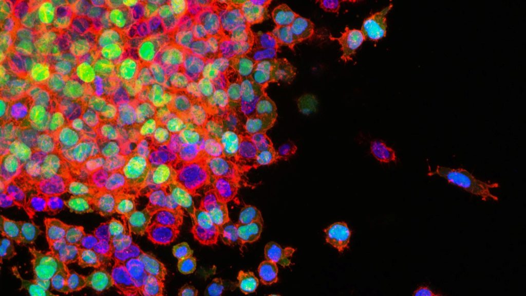
A new study led by researchers at Moffitt Cancer Center shows that asymptomatic brain metastasis is more common in stage 4 breast cancer patients than previously believed. The study, published in Neuro-Oncology, suggests that doctors may need to rethink current screening guidelines for detecting brain metastasis in patients without symptoms.
Researchers examined 101 asymptomatic patients diagnosed with stage 4 breast cancer, including triple-negative, HER2-positive and hormone receptor-positive/HER2-negative breast cancer. These patients underwent MRI scans to check for brain metastasis, with a follow-up MRI six months later if the initial scan showed no signs of cancer spread.
Of the patients who completed the initial MRI, 14% had brain metastasis. The rates by subtype were:
18% in triple-negative breast cancer
15% in HER2-positive breast cancer
10% in hormone receptor-positive/HER2-negative breast cancer
After the second MRI, the number of patients with brain metastasis grew to about 25% in each subtype. Following diagnosis, patients went on to receive early treatment for their brain metastases, including changes in systemic therapy and local therapies.
“Our study suggests that asymptomatic brain metastasis is quite common in stage 4 breast cancer,” said Kamran Ahmed, MD, associate member and section chief for Breast Radiation Oncology at Moffitt and principal investigator of the study. “Although larger studies are needed to confirm our findings, given the improvements in systemic and local therapies for breast cancer brain metastasis, the time may be appropriate to reconsider current guidelines that recommend against routine MRI surveillance in late stage breast cancer.”
Lung cancer metastasis. Credit: National Cancer Institute
In new research, a team led by University of Cincinnati researchers has identified a potential new way to make radiation more effective and improve outcomes for patients with lung cancer that has spread to the brain. The study, led by first author Debanjan Bhattacharya, PhD, appears in the journal Cancers, and uses a benzodiazepine analogue.
According to the American Cancer Society, lung cancer is the leading cause of cancer death in the United States, accounting for about one in five cancer deaths. Non-small cell lung cancer (NSCLC) is the most prevalent type of lung cancer, making up approximately 80% to 85% of all lung cancer cases.
Up to 40% of lung cancer patients develop brain metastases during the course of the disease, and these patients on average survive between eight and 10 months following diagnosis.
Current standard of care treatments for lung cancer that spreads to the brain include surgical removal and stereotactic brain radiosurgery (using precisely focused radiation beams to treat tumours) as well as whole brain irradiation in patients with more than 10 metastatic brain lesions.
“Lung cancer brain metastasis is usually incurable, and whole brain radiation treatment is palliative, as radiation limits therapy due to toxicity,” said Bhattacharya, research instructor in the Department of Neurology and Rehabilitation Medicine in UC’s College of Medicine. “Managing potential side effects and overcoming resistance to radiation are major challenges when treating brain metastases from lung cancer. This highlights the importance of new treatments which are less toxic and can improve the efficacy of radiation therapy, are less expensive, and can improve the quality of life in patients.”
Research focus
Bhattacharya and his colleagues at UC focused on AM-101, a synthetic analogue, meaning it has a close resemblance to the original compound, in the class of benzodiazepine drugs. It was first developed by James Cook, a medicinal chemist at the University of Wisconsin-Milwaukee. Prior to this study, AM-101’s effect in non-small cell lung cancer was unknown.
AM-101 is a particularly useful drug in the context of brain metastases in NSCLC, Bhattacharya said, as benzodiazepines are known to be able to pass through the blood-brain barrier that protects the brain from potential harmful invaders that can also block some drugs from reaching their target in the brain.
Research results
The team found that AM-101 activated GABA(A) receptors located in the NSCLC cells and lung cancer brain metastatic cells. This activation triggers the “self-eating” process of autophagy where the cell recycles and degrades unwanted cellular parts.
Specifically, the study showed that activating GABA(A) receptors increases the expression and clustering of GABARAP and Nix (an autophagy receptor), which boosts the autophagy process in lung cancer cells. This enhanced “self-eating” process of autophagy makes lung cancer cells more sensitive to radiation treatment.
Using animal models of lung cancer brain metastases, the team found AM-101 makes radiation treatment more effective and significantly improves survival. Additionally, the drug was found to slow down the growth of the primary NSCLC cells and brain metastases.
In addition to making radiation more effective, adding AM-101 to radiation treatments could allow for lower radiation doses, which could reduce side effects and toxicity for patients, Bhattacharya said. The team is now working toward opening Phase 1 clinical trials testing the combination of AM-101 and radiation both in lung cancer within the lungs and lung cancer that has spread to the brain.
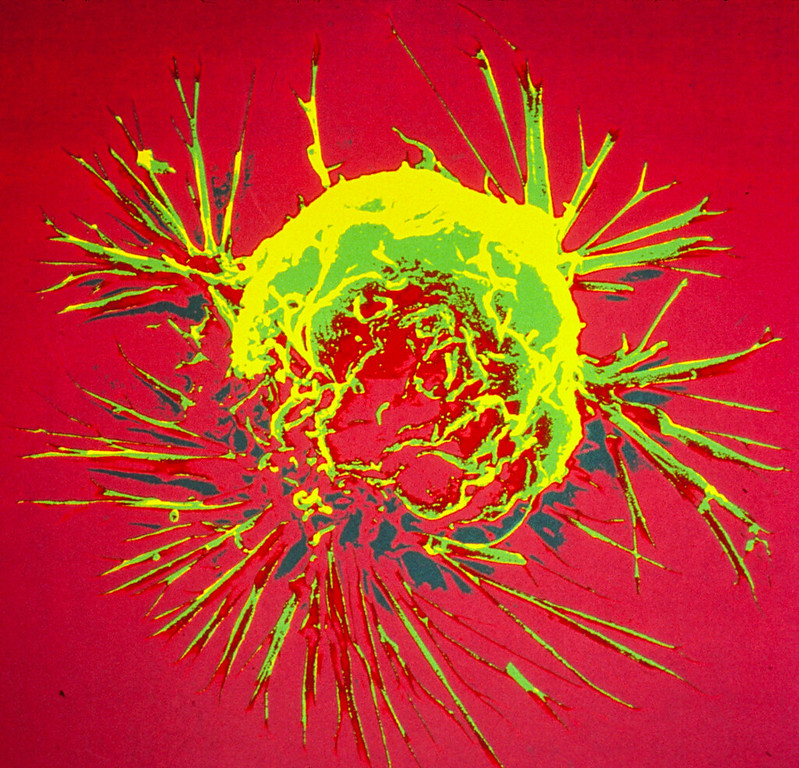
Colourised scanning electron micrograph of a breast cancer cell. Credit: NIH
A study led by researchers from the University of Arizona Cancer Center at UArizona Health Sciences identified a biological mechanism that could lead to more effective treatments for breast cancer that has metastasised to the brain.
By studying the metabolic differences between primary breast cancer cells and those that metastasise to the brain, they determined that autophagy was significantly upregulated in brain metastases. Autophagy is a cellular recycling process that cancer cells can use to stay alive when faced with stressful conditions such as those triggered by anticancer drugs.
“The prognosis for individuals with brain metastases from breast cancer is extremely unfavourable, and the management of breast cancer metastases in the brain remains a formidable challenge,” said senior author Jennifer Carew, PhD. “We were able to disrupt breast cancer cells’ ability to form brain metastases by impairing the autophagy pathway.”
In the study published in Clinical and Translational Medicine, the researchers first showed that targeting the key autophagy regulating gene ATG7 significantly reduced the ability of breast cancer cells to form brain metastases in mouse models.
With the goal of developing a strategy to bring this discovery to patients, the research team investigated whether hydroxychloroquine, a Food and Drug Administration-approved drug, could potentially be used to treat breast cancer brain metastases. Hydroxychloroquine inhibits autophagy at a later point in the pathway and, importantly, readily crosses the blood-brain barrier.
“Most drugs do not efficiently cross the blood-brain barrier, and that is one of the key reasons why brain metastases are so difficult to treat,” said Carew, who is a professor of medicine at UArizona.
The research team combined hydroxychloroquine with lapatinib, which is FDA-approved to treat breast cancer. They showed that this drug combination successfully reduced the number and size of breast cancer brain metastases in mouse models.
Hydroxychloroquine has been combined with a number of other anticancer agents in early phase clinical trials, but this is the first time researchers have studied its effectiveness when combined with lapatinib for breast cancer therapy.
Carew said the team was amazed by how significantly they were able to diminish the ability of breast cancer cells to form brain metastases by targeting a single pathway.
“Cancer cells, unfortunately, have evolved so many ways that make it difficult for us to stop their growth or kill them,” Carew said. “It is always somewhat surprising when you see how changing only one thing can have an impact.”
“Our group and others have shown that activation of autophagy makes it harder for many different types of cancer therapies to kill cancer cells and this promotes drug resistance,” said first author Steffan Nawrocki, PhD, UArizona professor. “Because hydroxychloroquine and lapatinib are already FDA approved, we can advance this drug combination quickly into a clinical trial for patients with breast cancer brain metastases.”
Brain metastases are the most prevalent adult central nervous system tumours, with 20% to 30% of cases resulting from breast cancer patients, particularly those with triple negative and HER2 amplified disease. Managing breast cancer metastases in the brain is challenging, with only 20% of patients with breast cancer brain metastases surviving beyond five years.
Lung cancer metastasis. Credit: National Cancer Institute
The largest review of papers for brain metastases of lung cancer has found abnormalities in their genetic mutations and for which licensed drugs could be clinically trialled to find out if they could treat the disease. The research led by the University of Bristol and published in Neuro-Oncology Advances also uncovered differences in those mutations between smokers and non-smokers.
Brain metastases most commonly occur from lung and breast cancer, and in the majority of cases are fatal. The genetic mutations in primary lung cancers have been widely studied, but less is known about the changes in the cancer once it has metastasised to the brain.
The research team wanted to find out the genetic changes in brain metastasis from non-small cell lung cancer (NSCLC) and whether there are drugs already available that could potentially be offered to these patients.
The researchers carried out a review from 72 papers of genetic mutations in brain metastasis of NSCLC from 2346 patients’ data on demographics, smoking status, genomic data, matched primary NSCLC, and PD-L1 – a protein found on cancer cells.
The study found the most commonly mutated genes were EGFR, TP53, KRAS, CDKN2A, and STK11.
Common missense mutations – mutations that lead to a single amino acid change in the protein coded by the gene – included EGFR L858R and KRAS G12C
In certain cases the genetic mutations were different in the brain metastasis from the primary lung cancer.
There were also differences in the genetic mutations in smokers versus patients who had never smoked. Brain metastases of smokers versus non-smokers had different missense mutations in TP53 and EGFR, except for L858R and T790M in EGFR, which were seen in both subgroups.
The research team found from the top ten commonly mutated genes which had primary NSCLC data, 37% of the specific mutations assessed were different between primary NSCLC and brain metastases.
The researchers suggest Medicines and Healthcare products Regulatory Agency-approved drugs already licensed could potentially be tested to treat the disease in clinical trials.
The genetic landscape of the different subtypes of NSCLC is well known. TP53 and LRP1B mutations are common to all NSCLC subtypes, but certain subtypes also have specific alterations.
Lung adenocarcinoma is the most common type of lung cancer and has higher frequencies of KRAS, EGFR, KEAP1, STK11, MET, and BRAF somatic mutations – changes that have accumulated in the cancer genome.
Some studies suggested that the genomic landscape of NSCLC in smokers vs non-smokers differ independent of subtype.
One study found EGFR mutations, ROS1 and ALK fusions to be more prevalent in non-smokers, whereas KRAS, TP53, BRAF, JAK2, JAK3 and mismatch repair gene mutations were more commonly mutated in smokers.
Kathreena Kurian, Professor of Neuropathology and Honorary Consultant at North Bristol NHS Trust, Head of the Brain Tumour Research Centre at the University of Bristol and co-author of the paper, said: “Our research recommends that all patients should have their brain metastasis examined for mutations in addition to their primary lung cancer because they may be different.
“This evidence could form the backbone for new clinical trials for patients with brain metastasis in non-small cell lung cancer using drugs that are already available.”
The team suggest the next steps for the research would be to consider whole genome sequencing on brain metastasis to look for other types of mutations, such as, common insertions/deletions for which drugs are already available.
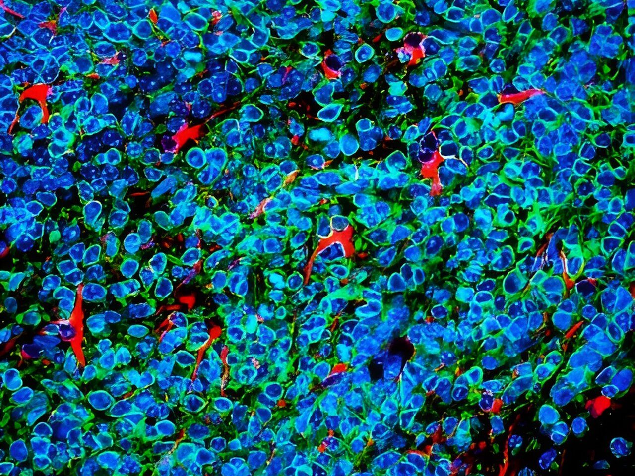
Small cell lung cancer cells (green and blue) that metastasised to the brain in a laboratory mouse recruit brain cells called astrocytes (red) for their protection. Credit: Fangfei Qu
Lung cancer cells that metastasise to the brain survive by convincing brain cells called astrocytes that they are baby neurons in need of protection, according to a study by researchers at Stanford Medicinepublished in Nature Cell Biology.
The cancer cells carry out their subterfuge by secreting a chemical signal prevalent in the developing human brain, the researchers found. This signal draws astrocytes to the tumour, encouraging them to secrete other factors that promote the cancer cells’ survival. Blocking that signal may be one way to slow or stop the growth of brain metastases of small cell lung cancer, which account for about 10% to 15% of all lung cancers, the researchers believe.
In the adult brain, astrocytes play a critical role in maintaining nerve function and connectivity. They are also important during brain development, when they facilitate connections between developing neurons.
The researchers studied laboratory mice, human tissue samples and human mini-brains, or organoids, grown in a lab dish to dissect the unique relationship between the cancer cells and their ‘big sister’ astrocytes, which hover nearby and shower them with protective factors.
“Small cell lung cancers are known for their ability to metastasise to the brain and thrive in an environment that is not normally conducive to tumour growth,” said professor of paediatrics and of genetics Julien Sage, PhD. “Our study suggests that these cancer cells reprogram the brain microenvironment by recruiting astrocytes for their protection.”
Professor Sage is the senior author of the study, while postdoctoral scholar Fangfei Qu, PhD, is the lead author.
Invasion of the brain
Small cell lung cancer excels at metastasising to the brain – about 15% to 20% of people already have clusters of cancer cells in their brains when their lung tumours are first diagnosed. As the cancer progresses, about 40% to 50% of patients will develop brain metastases. The problem is so prevalent, and the clinical outcome so dire, that clinicians recommend cranial radiation even before brain metastases have been found.
How and why small cell lung cancer has such an affinity for the brain has been something of a mystery. Brain metastases are rarely biopsied or removed because doing so has not been shown to affect a patient’s survival, and brain surgery is so invasive. Using laboratory mice is also of little help since small cell lung cancers in those animals rarely develop metastases in the brain, perhaps due to subtle biological differences between species.
Small cell lung cancers have another distinguishing feature – they are neuroendocrine cancers, meaning they arise from cells with similarities to both neurons and hormone-producing cells. Neuroendocrine cells link the nervous system with the endocrine system throughout the body, including in the lung.
Sage and his colleagues wondered whether neuronal-associated proteins on the surface of small cell lung cancer cells give them a leg up when the cells first begin to infiltrate the brain.
“We know the brain is full of neurons,” Sage said. “Maybe that’s why these cancer cells with some neuronal traits are happy in the brain and are accepted into that environment.”
Qu and Sage developed a way to inject mouse small cell lung cancer cells grown in the laboratory into the brains of mice to spark the development of brain tumours. They saw that astrocytes, a subtype of glial cell, flocked to the infant tumours and began to churn out proteins critical during brain development, including factors that stimulate nerve growth.
A plethora of astrocytes
A similar call happens in human brains, they noted: Brain tissue samples from people who had died of metastatic small cell lung cancer, shared by professor of pathology and paper co-author Christina Kong, MD, had many more protective astrocytes in the interior of the tumours than did metastases of melanoma, breast cancer and another type of lung cancer called adenocarcinoma.
Qu worked with assistant professor of paediatrics and co-author Anca Pasca, MD, to fuse aggregates of small cell lung cancer, lung adenocarcinoma or breast cancer cells with what are called cortical organoids – in vitro-grown clumps of brain cells including neurons and astrocytes that begin to mimic the organisation and connectivity of a human cortex. Within 10 days, many more protective astrocytes had infiltrated the small cell lung cancer pseudo-tumours than the adenocarcinoma or breast cancer.
“This showed us that the astrocytes actively move toward the small cell lung cancer cells, rather than simply being engulfed by the growing tumour,” Sage said. “What’s more exciting, though, is that these organoids, or mini-brains, realistically model the developing human brain. So, we’re no longer relying on a mouse model. It’s a perfect system to study brain metastases.”
Further research showed that the small cell lung cancer cells summon protective astrocytes by secreting a protein called Reelin that mediates the migration of neuronal and glial cells during brain development. Triggering Reelin expression in mouse breast cancer cells injected into the brain significantly increased the number of astrocytes in the resulting tumours in the mice, and the tumours were larger than in control animals injected with cells with low Reelin expression.
The apparent reliance of the cancer cells on chemical signals and responses specific to the developing brain may give clues for the development of future therapies, Sage believes.
“Some of these signals may not be as relevant or as highly expressed in the adult brain,” Sage said. “As a result, perhaps they could still be targeted to slow or prevent brain metastases without harming a normal brain. This might be an important window of opportunity for therapy.”
At 18 Spanish hospitals, when a patient with brain metastasis undergoes surgery, they can donate a tiny part of their brain to the first repository of brain metastasis living samples in the world, based at the Spanish National Cancer Research Centre (CNIO). A world-first collection, it was created to accelerate the search for therapies against brain metastasis, a disease that affects up to 30% of patients with systemic cancer.
The creators of this repository, called RENACER (Spanish acronym for the National Brain Metastasis Network), are two CNIO researchers, Manuel Valiente, head of the Brain Metastasis Group, and Eva Ortega-Paíno, director of the Biobank. They explained the advantages of the collection in the journal Trends in Cancer. In just three years RENACER has compiled samples from more than 150 patients.
The truly unique feature of RENACER, which makes it a valuable tool for the international scientific community, is that it contains living samples, conserved in cultures that enable the cells to continue behaving in a similar way as they were in the body.
A living biobank that enables organotypic cultures
“We have built a ‘living’ biobank” write Valiente and Ortega-Paíno. And this characteristic can be “transformative, not only for research but also for clinical trial design, especially when focused on unmet clinical needs, such as brain metastasis”.
The fact that the cells are living allows them, for example, to study their response to specific drugs. RENACER paves the way to create avatars for each patient in order to identify the best therapeutic options in an individualised way.
“Research contracts have been already signed to exploit patient-derived organotypic cultures (PDOCs) as avatars, thus providing the possibility to generate biomarkers of sensitivity or resistance to specific drugs” the authors explain.
The hospitals involved with RENACER work as a network to pass on research findings to patients as quickly as possible. In fact, thanks to this network, there are already two clinical trials underway, which will determine the capacity of two biomarkers to discriminate cases in which radiotherapy – a technique with side effects – will be effective.
From the operating theatre to the biobank in hours
The requirement for cells to be “alive” is not easy to achieve, since it involves a sophisticated logistics chain. The samples are taken from the operating theatre in a special container, in their culture medium, at a temperature of between 4 and 8 degrees centigrade.
They must reach the CNIO Biobank, in Madrid, in less than 24 hours. There, they are processed, organotypic cultures are created, and they are divided into proportional parts that are stored as samples for future investigations. They are also analysed using various techniques and sequenced, to extract as much information as possible from them. All the data are put into a database that is open to the international scientific community.
“It is pivotal to empower patients”
“This is happening just a few years after the project was launched,” said Valiente. “It’s a strategy that helps to improve knowledge as well as diagnosis and treatment options, but also brings all the people involved closer together: patients, core researchers, chemical re searchers, healthcare professionals, and the biobank.”
Patients, “[because they act] as donors during a difficult brain metastasis neurosurgery, play a crucial role and we strongly believe that it is pivotal to empower them,” the researchers explain. GEPAC (Spanish Group of Patients with Cancer) is also involved with RENACER.