Tag: brain connectivity

Researchers 3D-print Functional Human Brain Tissue

AI-generated image illustrating 3-D tissue printing

A team of scientists has developed the first 3D-printed brain tissue that can grow and function like typical brain tissue. This has important implications for scientists studying the brain and working on treatments for a broad range of neurological and neurodevelopmental disorders, such as Alzheimer’s and Parkinson’s disease.

“This could be a hugely powerful model to help us understand how brain cells and parts of the brain communicate in humans,” says Su-Chun Zhang, professor of neuroscience and neurology at UW-Madison’s Waisman Center. “It could change the way we look at stem cell biology, neuroscience, and the pathogenesis of many neurological and psychiatric disorders.”

Printing methods have limited the success of previous attempts to print brain tissue, according to Zhang and Yuanwei Yan, a scientist in Zhang’s lab. The group behind the new 3D-printing process described their method today in the journal Cell Stem Cell.

Instead of using the traditional 3D-printing approach, stacking layers vertically, the researchers went horizontally. They situated brain cells, neurons grown from induced pluripotent stem cells, in a softer “bio-ink” gel than previous attempts had employed.

“The tissue still has enough structure to hold together but it is soft enough to allow the neurons to grow into each other and start talking to each other,” Zhang says.

The cells are laid next to each other like pencils laid next to each other on a tabletop.

“Our tissue stays relatively thin and this makes it easy for the neurons to get enough oxygen and enough nutrients from the growth media,” Yan says.

The results speak for themselves – which is to say, the cells can speak to each other. The printed cells reach through the medium to form connections inside each printed layer as well as across layers, forming networks comparable to human brains. The neurons communicate, send signals, interact with each other through neurotransmitters, and even form proper networks with support cells that were added to the printed tissue.

“We printed the cerebral cortex and the striatum and what we found was quite striking,” Zhang says. “Even when we printed different cells belonging to different parts of the brain, they were still able to talk to each other in a very special and specific way.”

The printing technique offers precision – control over the types and arrangement of cells – not found in brain organoids, miniature organs used to study brains. The organoids grow with less organisation and control.

“Our lab is very special in that we are able to produce pretty much any type of neurons at any time. Then we can piece them together at almost any time and in whatever way we like,” Zhang says. “Because we can print the tissue by design, we can have a defined system to look at how our human brain network operates. We can look very specifically at how the nerve cells talk to each other under certain conditions because we can print exactly what we want.”

That specificity provides flexibility. The printed brain tissue could be used to study signaling between cells in Down syndrome, interactions between healthy tissue and neighboring tissue affected by Alzheimer’s, testing new drug candidates, or even watching the brain grow.

“In the past, we have often looked at one thing at a time, which means we often miss some critical components. Our brain operates in networks. We want to print brain tissue this way because cells do not operate by themselves. They talk to each other. This is how our brain works and it has to be studied all together like this to truly understand it,” Zhang says. “Our brain tissue could be used to study almost every major aspect of what many people at the Waisman Center are working on. It can be used to look at the molecular mechanisms underlying brain development, human development, developmental disabilities, neurodegenerative disorders, and more.”

The new printing technique should also be accessible to many labs. It does not require special bio-printing equipment or culturing methods to keep the tissue healthy, and can be studied in depth with microscopes, standard imaging techniques and electrodes already common in the field.

The researchers would like to explore the potential of specialization, though, further improving their bio-ink and refining their equipment to allow for specific orientations of cells within their printed tissue..

“Right now, our printer is a benchtop commercialised one,” Yan says. “We can make some specialised improvements to help us print specific types of brain tissue on-demand.”

Source: University of Wisconsin-Madison

Antidepressants Impact Prefrontal Cortex Development

Photo by William Fortunato on Pexels

A new study published in Nature Communications suggests that use of antidepressants can impact early post-natal brain development, potentially contributing to the development of mental health disorders. The study, led by researchers at the University of Colorado Anschutz Medical Campus, focused on the effect of fluoxetine, commonly used in medications such as Prozac and Sarafem for treating depression and perinatal depression, on the developing prefrontal cortex of mice.

Since fluoxetine works by increasing the levels of serotonin in the brain, the researchers looked at the impact serotonin has on prefrontal cortex development.

“While it is known that serotonin plays a role in the brain development, the mechanisms responsible for this influence, specifically in the prefrontal cortex, have been unclear, ” said lead author Won Chan Oh, PhD, assistant professor in the Department of Pharmacology at CU Anschutz.

Changes in gestational and early postnatal serotonin levels can arise from many causes including maternal deprivation or abuse, diets high or low in tryptophan, or the use of medications such as selective serotonin reuptake inhibitors (SSRIs) that can readily cross the placenta or be passed to offspring through breast feeding. Disbalances of 5-HT during brain development are associated with increased risk of neurodevelopmental disorders such as autism spectrum disorder and long-lasting behavioural deficits, but the underlying mechanisms remain elusive.

Oh and his student, Roberto Ogelman, a neuroscience PhD candidate, found serotonin directly influences nascent and immature excitatory synaptic connections in the prefrontal cortex, which if disrupted or dysregulated during early development can contribute to various mental health disorders.

“Our research uncovers the specific processes at the synaptic level that explain how serotonin contributes to the development of this important brain region during early-life fluoxetine exposure,” adds Oh. “We are the first to provide experimental evidence of the direct impact of serotonin on the developing prefrontal cortex in mice.”

To study the effect, the researchers looked at the impact of deficiency and surplus of serotonin on brain development in mice. They discovered that serotonin is not just involved in overall brain function but also has a specific role in influencing how individual connections between neurons change and adapt, contributing to the brain’s ability to learn and adjust.

“Understanding this correlation has the potential to help with early intervention and the development of new therapeutics for neurodevelopmental disorders involving serotonin dysregulation,” said Oh.

The researchers plan to continue studying the impact of fluoxetine, next examining its impact on a developing brain later in life.

Source: Colorado University Anschutz Medical Campus

Scans of Brain Connectivity in Veterans Yield Objective Pain Measures

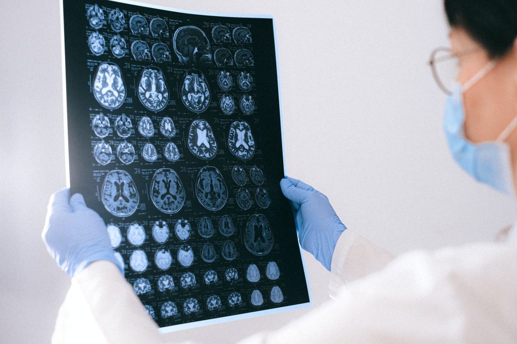
MRI images of the brain
Photo by Anna Shvets on Pexels

A brain connectivity study of military veterans discovered three unique brain subtypes potentially indicating high, medium, and low susceptibility to pain and trauma symptoms. This could constitute an objective measurement of pain and trauma susceptibility, possibly leading to personalised treatments and new therapies based on neural connectivity patterns.  

Comorbidity Goes Unexplored

“Chronic pain is a major public health concern, especially among veterans,” said first author Prof Irina Strigo. “Moreover, chronic pain sufferers almost never present with a single disorder but often with multiple co-morbidities, such as trauma, posttraumatic stress, and depression.”

It is already understood that both pain and trauma can affect brain connections, but this had not been studied in the context of comorbid trauma and pain. Much pain and trauma research also relies on subjective measurements, such as questionnaires, rather than objective measurements like brain scans. This study, published in Frontiers in Pain Research, addresses these problems.

Theresearchers studied a group of 57 veterans with both chronic back pain and trauma, who had quite varied symptoms in terms of pain and trauma severity. Functional MRI scans of the veterans’ brains showed the strength of connections between brain regions involved in pain and trauma. The researchers then used a statistical technique to automatically group the veterans based on their brain connection signatures, regardless of their self-reported pain and trauma levels.

Based on the veterans’ brain activity, they were sorted into three groups. Strikingly, these divisions were comparable to the severity of the veterans’ symptoms, and they fell into a low, medium, or high symptom group.

The team hypothesised that the pattern of brain connections found in the low symptom group allowed veterans to avoid some of the emotional fallout from pain and trauma, and also included natural pain reduction capabilities. Conversely, the high symptom group demonstrated brain connection patterns that may have increased their chances of anxiety and catastrophising when experiencing pain.

Interestingly, based on self-reported pain and trauma symptoms, the medium symptom group was largely similar to the low symptom group. However, the medium symptom group showed differences in their brain connectivity signature, which suggested that they were better at focusing on other things when experiencing pain, reducing its impact.

Putting the findings into future practice

“Despite the fact that the majority of subjects within each subgroup had a co-morbid diagnosis of pain and trauma, their brain connections differed,” said Prof Strigo.

“In other words, despite demographic and diagnostic similarities, we found neurobiologically distinct groups with different mechanisms for managing pain and trauma. Neurobiological-based subgroups can provide insights into how these individuals will respond to brain stimulation and psychopharmacological treatments.”

Thus far, it’s not known whether these neural hallmarks represent a vulnerability to trauma and pain or a consequence of these conditions. The technique does however provide an objective and unbiased hallmark of pain and trauma susceptibility or resilience, not reliant on subjective measures such as the surveys. In fact, subjective measurements of pain in this study would not differentiate between the low and medium groups.

Techniques using objective measures like brain connectivity appear more sensitive and could provide a clearer overall picture of someone’s resilience or susceptibility to pain and trauma, thereby guiding personalised treatment and paving the way for new treatments.

Source: Frontiers