Tag: brain-computer interface

Person with Tetraplegia Pilots Drone with Brain-computer Interface

Photo by Thomas Bjornstad on Unsplash

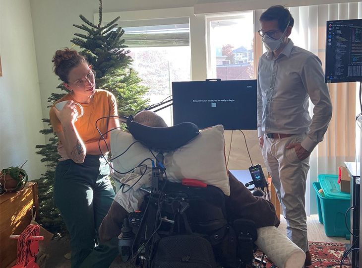
A brain-computer interface, surgically placed in a research participant with tetraplegia, paralysis in all four limbs, provided an unprecedented level of control over a virtual quadcopter – just by thinking about moving his unresponsive fingers.

The technology divides the hand into three parts: the thumb and two pairs of fingers (index and middle, ring and small). Each part can move both vertically and horizontally. As the participant thinks about moving the three groups, at times simultaneously, the virtual quadcopter responds, manoeuvring through a virtual obstacle course.

It’s an exciting next step in providing those with paralysis the chance to enjoy games with friends while also demonstrating the potential for performing remote work.

“This is a greater degree of functionality than anything previously based on finger movements,” said Matthew Willsey, U-M assistant professor of neurosurgery and biomedical engineering, and first author of a new research paper in Nature Medicine. The testing that produced the paper was conducted while Willsey was a researcher at Stanford University, where most of his collaborators are located.

While there are noninvasive approaches to allow enhanced video gaming such as using electroencephalography to take signals from the surface of the user’s head, EEG signals combine contributions from large regions of the brain. The authors believe that to restore highly functional fine motor control, electrodes need to be placed closer to the neurons. The study notes a sixfold improvement in the user’s quadcopter flight performance by reading signals directly from motor neurons vs. EEG.

To prepare the interface, patients undergo a surgical procedure in which electrodes are placed in the brain’s motor cortex. The electrodes are wired to a pedestal that is anchored to the skull and exits the skin, which allows a connection to a computer.

“It takes the signals created in the motor cortex that occur simply when the participant tries to move their fingers and uses an artificial neural network to interpret what the intentions are to control virtual fingers in the simulation,” Willsey said. “Then we send a signal to control a virtual quadcopter.”

The quadcopter is on a serpentine path around rings that hang in midair over a virtual basketball court. The fingers of the hand are curled in with a line indicating a neutral point for the fingers. Four vectors point away from the thumb: up, down, right and left.
A screenshot of the game display shows the quadcopter following a green path around the rings. The inset shows a hand avatar. The neural implant records from nearby neurons and algorithms determine the intended movements for the hand avatar. The finger positions are then used to control the virtual quadcopter. Image credit: Nature Medicine

The research, conducted as part of the BrainGate2 clinical trials, focused on how these neural signals could be coupled with machine learning to provide new options for external device control for people with neurological injuries or disease. The participant first began working with the research team at Stanford in 2016, several years after a spinal cord injury left him unable to use his arms or legs. He was interested in contributing to the work and had a particular interest in flying.

“The quadcopter simulation was not an arbitrary choice, the research participant had a passion for flying,” said Donald Avansino, co-author and computer scientist at Stanford University. “While also fulfilling the participant’s desire for flight, the platform also showcased the control of multiple fingers.”

Co-author Nishal Shah, incoming professor of electrical and computer engineering at Rice University, explained, “controlling fingers is a stepping stone; the ultimate goal is whole body movement restoration.”

Jaimie Henderson, a Stanford professor of neurosurgery and co-author of the study, said the work’s importance goes beyond games. It allows for human connection.

“People tend to focus on restoration of the sorts of functions that are basic necessities – eating, dressing, mobility – and those are all important,” he said. “But oftentimes, other equally important aspects of life get short shrift, like recreation or connection with peers. People want to play games and interact with their friends.”

A person who can connect with a computer and manipulate a virtual vehicle simply by thinking, he says, could eventually be capable of much more.

“Being able to move multiple virtual fingers with brain control, you can have multifactor control schemes for all kinds of things,” Henderson said. “That could mean anything, from operating CAD software to composing music.”

Source: University of Michigan

New Brain–computer Interface Allows Man with ALS to ‘Speak’ Again

A new brain-computer interface (BCI) developed at UC Davis Health translates brain signals into speech with up to 97% accuracy – the most accurate system of its kind. The researchers implanted sensors in the brain of a man with severely impaired speech due to amyotrophic lateral sclerosis (ALS). The man was able to communicate his intended speech within minutes of activating the system.

A study about this work was published in the New England Journal of Medicine.

ALS, also known as Lou Gehrig’s disease, affects the nerve cells that control movement throughout the body. The disease leads to a gradual loss of the ability to stand, walk and use one’s hands. It can also cause a person to lose control of the muscles used to speak, leading to a loss of understandable speech.

The new technology is being developed to restore communication for people who can’t speak due to paralysis or neurological conditions like ALS. It can interpret brain signals when the user tries to speak and turns them into text that is ‘spoken’ aloud by the computer.

“Our BCI technology helped a man with paralysis to communicate with friends, families and caregivers,” said UC Davis neurosurgeon David Brandman. “Our paper demonstrates the most accurate speech neuroprosthesis (device) ever reported.”

Brandman is the co-principal investigator and co-senior author of this study. He is an assistant professor in the UC Davis Department of Neurological Surgery and co-director of the UC Davis Neuroprosthetics Lab.

The new BCI breaks the communication barrier

When someone tries to speak, the new BCI device transforms their brain activity into text on a computer screen. The computer can then read the text out loud.

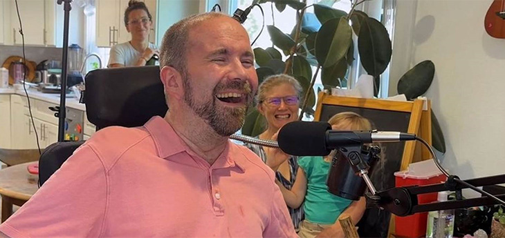
To develop the system, the team enrolled Casey Harrell, a 45-year-old man with ALS, in the BrainGate clinical trial. At the time of his enrolment, Harrell had weakness in his arms and legs (tetraparesis). His speech was very hard to understand (dysarthria) and required others to help interpret for him.

In July 2023, Brandman implanted the investigational BCI device. He placed four microelectrode arrays into the left precentral gyrus, a brain region responsible for coordinating speech. The arrays are designed to record the brain activity from 256 cortical electrodes.

“We’re really detecting their attempt to move their muscles and talk,” explained neuroscientist Sergey Stavisky. Stavisky is an assistant professor in the Department of Neurological Surgery. He is the co-director of the UC Davis Neuroprosthetics Lab and co-principal investigator of the study. “We are recording from the part of the brain that’s trying to send these commands to the muscles. And we are basically listening into that, and we’re translating those patterns of brain activity into a phoneme – like a syllable or the unit of speech – and then the words they’re trying to say.”

Casey Harrell with his personal assistant Emma Alaimo and UC Davis neuroscientist Sergey Stavisky

Faster training, better results

Despite recent advances in BCI technology, efforts to enable communication have been slow and prone to errors. This is because the machine-learning programs that interpreted brain signals required a large amount of time and data to perform.

“Previous speech BCI systems had frequent word errors. This made it difficult for the user to be understood consistently and was a barrier to communication,” Brandman explained. “Our objective was to develop a system that empowered someone to be understood whenever they wanted to speak.”

Harrell used the system in both prompted and spontaneous conversational settings. In both cases, speech decoding happened in real time, with continuous system updates to keep it working accurately.

The decoded words were shown on a screen. Amazingly, they were read aloud in a voice that sounded like Harrell’s before he had ALS. The voice was composed using software trained with existing audio samples of his pre-ALS voice.

At the first speech data training session, the system took 30 minutes to achieve 99.6% word accuracy with a 50-word vocabulary.

“The first time we tried the system, he cried with joy as the words he was trying to say correctly appeared on-screen. We all did,” Stavisky said.

In the second session, the size of the potential vocabulary increased to 125 000 words. With just an additional 1.4 hours of training data, the BCI achieved a 90.2% word accuracy with this greatly expanded vocabulary. After continued data collection, the BCI has maintained 97.5% accuracy.

“At this point, we can decode what Casey is trying to say correctly about 97% of the time, which is better than many commercially available smartphone applications that try to interpret a person’s voice,” Brandman said. “This technology is transformative because it provides hope for people who want to speak but can’t. I hope that technology like this speech BCI will help future patients speak with their family and friends.”

The study reports on 84 data collection sessions over 32 weeks. In total, Harrell used the speech BCI in self-paced conversations for over 248 hours to communicate in person and over video chat.

“Not being able to communicate is so frustrating and demoralising. It is like you are trapped,” Harrell said. “Something like this technology will help people back into life and society.”

“It has been immensely rewarding to see Casey regain his ability to speak with his family and friends through this technology,” said the study’s lead author, Nicholas Card. Card is a postdoctoral scholar in the UC Davis Department of Neurological Surgery.

“Casey and our other BrainGate participants are truly extraordinary. They deserve tremendous credit for joining these early clinical trials. They do this not because they’re hoping to gain any personal benefit, but to help us develop a system that will restore communication and mobility for other people with paralysis,” said co-author and BrainGate trial sponsor-investigator Leigh Hochberg. Hochberg is a neurologist and neuroscientist at Massachusetts General Hospital, Brown University and the VA Providence Healthcare System.

Brandman is the site-responsible principal investigator of the BrainGate2 clinical trial. The trial is enrolling participants. To learn more about the study, visit braingate.org or contact braingate@ucdavis.edu.

Source: UC Davis Health

Visual Cortex Stimulation Boosts Brain-computer Interface

Deep brain stimulation illustration. Credit: NIH

Brain-computer interfaces, or BCIs, promise life-changing benefits for people suffering from a range of neurological conditions, but implementation is for both the invasive and noninvasive methods is challenging. Researchers led by Bin He at Carnegie Mellon University used an innovative electroencephalogram (EEG) wearable. They successfully integrated a novel focused ultrasound stimulation to realise bidirectional BCI that both encodes and decodes brain waves using machine learning in a study with 25 human subjects.

This work, published in Nature Communicationsopens up a new avenue to significantly enhance not only the signal quality, but also, overall nonivasive BCI performance by stimulating targeted neural circuits.

Noninvasive BCI is lauded for its merits of being cheap, safe, and virtually applicable to everyone, but because signals are recorded over the scalp versus inside the brain, low signal quality presents some limitations. The He group is exploring ways to improve the effectiveness of noninvasive BCIs and, over time, has used deep learning approaches to decode what an individual was thinking and then facilitate control of a cursor or robotic arm.

In their latest research, the He group demonstrated that through precision noninvasive neuromodulation using focused ultrasound, the performance of a BCI could be improved for communication.

“This paper reports a breakthrough in noninvasive BCIs by integrating a novel focused ultrasound stimulation to realise bidirectional BCI functionality,” explained Bin He, professor of biomedical engineering at Carnegie Mellon University. “Using a communication prosthetic, 25 human subjects spelled out phrases like ‘Carnegie Mellon’ using a BCI speller. Our findings showed that the addition of focused ultrasound neuromodulation significantly boosted the performance of EEG-based BCI. It also elevated theta neural oscillation that enhanced attention and led to enhanced BCI performance.”

For context, a BCI speller is a 6×6 visual motion aide containing the entire alphabet that is commonly used by nonspeakers to communicate. In He’s study, subjects donned an EEG cap and just by looking at the letters, were able to generate EEG signals to spell the desired words. When a focused ultrasound beam was applied externally to the V5 area (part of the visual cortex) of the brain, the performance of the noninvasive BCI greatly improved among subjects. The neuromodulation-integrated BCI actively altered the engagement of neural circuits to maximize the BCI performance, compared to previous uses, which consisted of pure processing and decoding recorded signals.

Following this discovery, the He lab is further investigating the merits and applications of focused ultrasound neuromodulation to the brain, beyond the visual system, to enhance noninvasive BCIs. They also aim to develop more compact-focused ultrasound neuromodulation device for better integration with EEG-based BCIs, and to integrate AI to continue to enhance the overall system performance.

“This is my lifelong interest, and I will never give up,” emphasized He. “Working to improve noninvasive technology is difficult, but I strongly believe that if we can find a way to make it work, everyone will benefit. I will keep working, and someday, noninvasive lifesaving technology will be available for every household.

Source: College of Engineering, Carnegie Mellon University

‘Digital Bridging’ Enables Paraplegic Man to Walk Again

Study participant Gert-Jan Oskam walking with the brain-spine interface. Credit: Swiss Federal Institute of Technology in Lausanne

A 40 year-old man, Gert-Jan Oskam, has regained the ability to walk independently after being paralysed from a spinal cord injury with the use of a new brain-spine interface. The ‘digital bridging’ technology, developed at the Swiss Federal Institute of Technology in Lausanne and described in Nature, consists of implants and a computer to translate brain signals of the intention to move into stimulations that move the legs accordingly..

This BSI system could be calibrated in minutes, and remained stable for one year, including use at home. The BSI enabled the participant to exert natural control over the movements of his legs to stand, walk, climb stairs and even traverse complex terrains.

In addition to the digital bridging, neurorehabilitation supported by the BSI improved neurological recovery. The participant regained the ability to walk with crutches overground even when the BSI was switched off. This digital bridge establishes a framework to restore natural control of movement after paralysis.

The system consists of a pair cortical of sensors, each an array with 64 electrodes housed in 5cm-diameter titanium discs. These discs are implanted snugly in the skull to pick up brain activity. They transmit the data wirelessly to a personalised headset, which also provides power for the sensors. The headset then sends the data to a portable processing unit (which may be carried in a backpack). Using specialised software, it uses this brain signal data to generates real-time predictions of motor intentions. These decoded intentions are translated into stimulation commands and sent on to another implant, a paddle array of 16 electrodes implanted next to the spinal cord, delivering current to the targeted dorsal root entry zones.

Neurosurgical implantation procedure

Oskam had sustained an incomplete cervical (C5/C6) spinal cord injury during a biking accident 10 years previously. He had already participated in a neurological recovery programme, the STIMO trial, which had used neurostimulation to get him to the stage where he could walk with the aid of a front-wheel walker. The neurorehabilitation from the trial also enabled him to use his hip flexors and lift his legs against gravity, but recovery had plateaued for the three years prior to his participation in the present study.

For the BSI to function, the researchers needed to locate neural features related to the intention to move the legs. To pinpoint the cortical regions associated with the intention to move, they used CT scans and magnetoencephalography. Taking into account anatomical restraints, they then decided on the positions of the implants.

Under general anaesthesia, surgeons performed a bicoronal incision of the scalp to allow two circular-shaped craniotomies over the planned locations of the left and right hemispheres. They then replaced the bone flaps with the two implantable recording devices, before closing the scalp.

The paddle lead had already been emplaced over the dorsal root entry zones of the lumbar spinal cord during the STIMO clinical trial. Its optimal positioning was identified using high-resolution structural imaging of the spine, and its final position was decided during the surgery based on electrophysiological recordings. The implantable pulse generator was inserted subcutaneously in the abdomen. Oskam was able to return home 24 hours after each procedure.

Elon Musk’s Neuralink Brain-computer Interface Receives Human Testing Approval

Elon Musk’s company Neuralink had finally received approval for human testing of its brain-computer interface (BCI). After initially rejecting the application, the US Food and Drug Administration finally gave the company the go-ahead on Thursday.

Neuralink, which aims to develop an implant that would allow humans to interface directly with computers as well as enabling medical applications such as controlling prostheses. Last year, the company showed off a monkey that was able to play the simple video game Pong on a monitor using its mind.

Neuralink is by no means the first company to try to achieve these goals. Many other institutions have made advances over the past decades, but the field is a difficult one and progress is slow. In its previous rejection, the FDA cited concerns such as the devices using lithium for their batteries, migration of the wires inside the brain and the difficulty of extracting the devices without harming brain tissue.

The company’s use of animals to develop the technology has infuriated activists, but this is a standard practice in development of BCI technology. Last year, whistleblowers accused the company of killing 1500 animals since its inception.

In a guidance document, the FDA says that, “The field of implanted BCI devices is progressing rapidly from fundamental neuroscience discoveries to translational applications and market access. Implanted BCI devices have the potential to bring benefit to people with severe disabilities by increasing their ability to interact with their environment, and consequently, providing new independence in daily life.”

China is also aggressively pursuing the development of BCIs as part of their ‘China Brain Project’, as discussed in the journal Neuron. It has a significant advantage as it has a large population of macaques to draw on, along with fewer ethical concerns and policies expediting biotech research.

Functional MRI is Now Able to Read People’s Minds

Photo by Mart Production on Pexels

In a study in Nature, researchers reported being able to identify words and phrases in volunteers undergoing fMRI imaging reasonable accuracy. The process is non-invasive, unlike implanted electrodes, but requires hours of preparation and scanning.

This technology would be a significant breakthrough for people suffering debilitating conditions that prevent them from speaking or otherwise communicating. Previously, decoding language required the use of extensive electrode implants.

The participants, two male and one female, listened to recordings of radio shows. This was used to train a language model which was based on an early version of ChatGPT. By looking at the brain’s responses, the language model was able to capture the gist of what the participants were thinking, sometimes replicating exact words or entire phrases.

Marked safe from ‘Big Brother’… for now

At this stage, the technology used requires the subject to cooperate, the researchers wrote, allaying concerns over any malicious use of this technology to tap into people’s private thoughts. Testing the decoding model on people who it hadn’t been trained on produced unintelligible results, as was the case when the trained participants put up resistance.

While the technology cannot be used for nefarious mind-reading, the march of progress means that one day such concerns will become real.

Nita Farahany, JD, PhD, of Duke University in Durham, North Carolina, told MedPage Today that the technology could one day be used against people. “This research illustrates the rapid advances being made toward an age of much greater brain transparency, where even continuous language and semantic meaning can be decoded from the brain.

“While people can employ effective countermeasures to prevent decoding their brains using fMRI, as brain wearables become widespread that may not be an effective way to protect us from interception, manipulation, or even punishment for our thoughts.”

While lugging around a massive MRI machine would be a challenge for future thought police, smaller, more portable means of measuring brain activity remotely. Senior author Alexander Huth, PhD, of the University of Texas at Austin, says that one such technology could be functional near-infrared spectroscopy (fNIRS).

“fNIRS measures where there’s more or less blood flow in the brain at different points in time, which, it turns out, is exactly the same kind of signal that fMRI is measuring,” Huth said. “So, our exact kind of approach should translate to fNIRS,” but the resolution with fNIRS would be lower.

Brain-computer Interfaces Deemed Safe for Long-term Use

Patient with complete spinal cord injury walking at EPFL Campus after 5 months of rehab. ©NeuroRestore Jimmy Ravie

For people with paralysis caused by neurologic injury or disease, brain-computer interfaces (BCIs) can potentially restore mobility and function by transmitting neural data to external devices such as mobility aids, which have already shown promise in trials.

Although implanted brain sensors, the core component of many brain-computer interfaces, have been used in neuroscientific studies with animals for decades and have been approved for short term use (< 30 days) in humans, the long-term safety of this technology in humans is unknown.

New results published in Neurology from the BrainGate feasibility study, the largest and longest-running clinical trial of an implanted BCI, suggest that these sensors’ safety is similar to other chronically implanted neurologic devices, with skin irritation around the implant interface.

This new report from a Massachusetts General Hospital (MGH)-led team, examined data from 14 adults with quadriparesis from spinal cord injury, brainstem stroke, or ALS who were enrolled in the BrainGate trial from 2004 to 2021 through seven clinical sites in the United States.

Participants underwent surgical implantation of one or two microelectrode arrays in a part of the brain responsible for generating the electrical signals that control limb movement. With these “Utah” microelectrode arrays, the brain signals associated with the intent to move a limb can then be sent to a nearby computer that decodes the signal in real-time and allows the user to control an external device simply by thinking about moving a part of their body.

The authors of the study report that across the 14 enrolled research participants, the average duration of device implantation was 872 days, yielding a total of 12 203 days for safety analyses. There were 68 device-related adverse events, including 6 device-related serious adverse events.

The most common device-related adverse event was skin irritation around the portion of the device that connects the implanted sensor to the external computer system. Importantly, they report that there were no safety events that required removal of the device, no infections of the brain or nervous system, and no adverse events resulting in permanently increased disability related to the investigational device.

“This interim report demonstrates that the investigational BrainGate Neural Interface system, which is still in ongoing clinical trials, thus far has a safety profile comparable to that of many approved implanted neurologic devices, such as deep brain stimulators and responsive neurostimulators,” says lead author Daniel Rubin, MD, PhD.

“Given the rapid recent advances in this technology and continued performance gains, these data suggest a favorable risk/benefit ratio in appropriately selected individuals to support ongoing research and development,.” said Rubin.

Leigh Hochberg, MD, PhD, director of the BrainGate consortium and clinical trials and the article’s senior author emphasised the importance of ongoing safety analyses as surgically placed brain-computer interfaces advance through clinical studies.

“While our consortium has published more than 60 articles detailing the ever-advancing ability to harness neural signals for the intuitive control of devices for communication and mobility, safety is the sine qua non of any potentially useful medical technology,” says Hochberg.

“The extraordinary people who enroll in our ongoing BrainGate clinical trials, and in early trials of any neurotechnology, deserve tremendous credit. They are enrolling not to gain personal benefit, but because they want to help,” said Hochberg.

Source: Massachusetts General Hospital