Tag: brain cancer

Scars of Destroyed Brain Tumours are Fertile Grounds for Recurrence

Types of tumour cells. Credit: Scientific Animations CC4.0

A Ludwig Cancer Research study has discovered that recurrent tumours of the aggressive brain cancer glioblastoma multiforme (GBM) grow out of the fibrous scars of malignant predecessors destroyed by interventions such as radiotherapy, surgery and immunotherapy.

Led by Ludwig Lausanne’s Johanna Joyce, Spencer Watson and alumnus Anoek Zomer and published in the current issue of Cancer Cell, the study describes how these scars enable the regrowth of tumours and identifies drug targets to sabotage their malignant support. It also demonstrates the efficacy of such combination therapies in preclinical trials using mouse models of GBM.

“We’ve identified fibrotic scarring as a key source of GBM resurgence following therapy, showing how it creates a protective niche for the regrowth of the tumor,” said Joyce. “Our findings suggest that blocking the process of scarring in the brain by adding anti-fibrosis agents to current treatment strategies could help prevent glioblastoma from recurring and improve the outcomes of therapy.”

There is a great need for such interventions. GBM is the most common and aggressive form of brain cancer in adults. Despite considerable effort to develop effective therapies for the cancer, the average life expectancy of patients remains around 14 months following diagnosis.

The origins of the current study date back to 2016, when the Joyce lab reported in the journal Science its examination in mouse models of strategies to overcome resistance to a promising immunotherapy for the treatment of GBM. That experimental therapy, which inhibits signalling by the colony stimulating factor-1 receptor (CSF-1R) and currently in clinical trials, targets immune cells known as macrophages and their brain-resident versions, microglia, both of which are manipulated by GBM cells to support tumour growth and survival.

The Joyce lab has demonstrated that CSF-1R inhibition reprograms these immune cells into an anti-tumour state and so induces significant tumour regression. Yet, as the Science study showed, about half the mice show relapse following an initial response to the therapy. “What was most remarkable about that observation was that every single time a brain tumour recurred following immunotherapy, it regrew right next to a scar that had formed at the original site of a tumour,” said Joyce.

In the current study, Joyce, Watson, Zomer and their colleagues examined tumour samples obtained from patients undergoing GBM therapy and showed that fibrotic scarring occurs following therapy in humans as well – and that it is similarly associated with tumour recurrence. They also showed that the fibrotic scarring occurs in response to not only immunotherapy but also following the surgical and radiological removal of tumours.

To explore how fibrosis contributes to relapse, the researchers applied an integrated suite of advanced technologies to analyze the cellular and molecular geography of the scars and the microenvironment of resurgent tumors.

These technologies include the analysis of global gene expression in individual cells, the comprehensive analysis of proteins in the tissues as well a workflow and AI-powered suite of analytical methods for the spatial analysis of tissues named hyperplexed immunofluorescence imaging (HIFI). Recently developed by Watson and colleagues in the Joyce lab, HIFI permits the simultaneous visualisation of multiple molecular markers in and around cells across broad cross-sections of tissues, enabling the generation of granular maps of the tumour microenvironment.

“Applied together, these advanced methods allowed us to see exactly how fibrotic scars form,” said Watson. “They revealed that the fibrosis serves as a kind of protective cocoon for residual cancer cells and pushes them into a dormant state in which they are largely resistant to therapy. We found that it also shields them from surveillance and elimination by the immune system.”

Integrated analyses of the tissue microenvironment following therapy revealed that the descendants of cells associated with tumor-feeding blood vessels become functionally altered to resemble fibroblasts—fiber-producing cells commonly involved in wound-healing. These perivascular-derived fibroblast-like (PDFL) cells fan out across the region previously occupied by the regressing tumor, where they mediate the generation of fibrotic scars. These cells, the researchers found, are especially activated by neuroinflammation and immune factors known as cytokines, most notably one called transforming growth factor-β (TGF-β).

“To see if targeting fibrotic scarring could improve therapeutic outcomes for GBM, we devised a treatment regimen using existing drugs to block TGF-β signaling and suppress neuroinflammation in combination with CSF-1R inhibition and evaluated it in preclinical trials using mouse models of GBM,” said Joyce. “We also timed these additional treatments to coincide with the period of maximal PDFL activation identified by our studies. Our results show that the drug combination inhibited fibrotic scarring, diminished the numbers of surviving tumor cells and extended the survival of treated mice compared to controls.”

The researchers suggest that approaches to limit fibrotic scarring could significantly improve outcomes for GBM patients receiving surgical, radiation or macrophage-targeting therapies. Additional research, they note, will likely yield even better drug targets for such combination therapies.

Source: Ludwig Institute for Cancer Research

Serotonin-producing Neurons Regulate Malignancy in Ependymoma Brain Tumours

Credit: National Cancer Institute

A study published in Nature reveals the functional relevance of tumour-neuron interactions that regulate the growth of ependymoma brain tumours, one of the most common types in children. The study, conducted by researchers at Baylor College of Medicine and St. Jude Children’s Research Hospital, highlights how neuronal signalling, modifications in DNA-associated proteins and developmental programs are intertwined to drive malignancy in brain cancer.

“Ependymomas are the third most common type of paediatric brain tumours,” said co-corresponding author, Dr Benjamin Deneen professor in the Department of Neurosurgery. “These tumours are aggressive, resistant to chemotherapy and lack tumour-specific therapies, leading to poor survival.”

“We have not made an impact on patient survival in the last three decades. A major factor has been a poor understanding of the disease. The motivation of our collaborative work with the Deneen lab is to dissect the biology of these tumours as a basis for developing new therapies,” said co-corresponding author Dr Stephen Mack, associate member at St. Jude Children’s Research Hospital and member of the Department of Neurobiology, Neurobiology and Brain Tumor Program and Center of Excellence in Neuro-Oncology Sciences.

Previous studies have shown in other types of brain tumours that brain activity surrounding the tumour can influence its growth. “In the current study, we investigated whether brain activity played a role in ependymoma growth, specifically in a very aggressive type driven by a protein called ZFTA-RELA,” said first author Hsiao-Chi Chen, a graduate student in the Deneen lab. “In collaboration with the Mack lab, we developed an animal model to study this rare paediatric brain tumour and validated these findings in human tumour samples.”

The researchers discovered evidence of abnormal neuronal activity in ependymoma’s environment and investigated whether it affected ependymoma growth. They found that while hyperactivity of some neural circuits promoted tumour growth, hyperactivity of other neural circuits surprisingly reduced tumour growth, which had not been described before. Their study revealed a novel chain of events at play that regulates tumour growth, which may hold therapeutic applications.

“First, we found that normal neurons located in the brain region called dorsa raphe nucleus (dRN) project towards the cortex, where ependymoma grows. These neurons secrete serotonin, a brain chemical that carries messages between nerve cells, which surprisingly slows tumour growth,” Chen said.

Interestingly, ependymoma cells carry a serotonin transporter, a molecule that imports serotonin within the cell. “We were surprised to discover that serotonin enters ependymoma cells and binds to histone H3, a protein that is tightly associated with DNA,” Chen said. “Histone serotonylation, the addition of serotonin to histone, regulated tumour growth. Promoting it enhanced tumour growth while preventing it slowed down ependymoma growth in animal models.”

“Discovering histone serotonylation in ependymoma piqued our interest because a previous study from our lab had revealed that adding serotonin to histones affects which genes the cell turns on,” Deneen said.

The team discovered that histone serotonylation in ependymoma increases the expression of transcription factors, genes that regulate the expression of other genes,” Chen said. “We focused on transcription factor ETV5 whose overexpression accelerated tumour growth. But how does it do it?”

The next experiments showed that ETV5 expression triggers changes in the 3D structure of chromatin, the combination of DNA and proteins that forms chromosomes. The 3D changes prevent the activation of genes encoding neurotransmitters, molecules that mediate neural activity. The team focused on a neurotransmitter called neuropeptide Y (NPY) and found that growing tumours have little NPY. Restoring the levels of NPY in tumours slowed down tumour progression and tumour-associated neural hyperactivity through the remodeling of surrounding synapses or neuron-to-neuron communication.

“We knew that brain tumours release factors that remodel synapses towards hyperactivity. Here we found the opposite also can happen, that ependymoma tumours can release factors that suppress excitatory synaptic remodeling and that repressing this mechanism is essential for tumour progression,” Deneen said.

“I am excited that this work has redefined our understanding of how brain tumour cells grow, and how they take advantage of factors in their surrounding environment to initiate tumours,” Mack said. “I am equally excited that this work has revealed many new avenues for research that may in the future lead to new therapies, which is desperately needed for this devastating disease.”

Source: Baylor College of Medicine

New Guidelines for Brain Cancer Care

Credit: National Cancer Institute

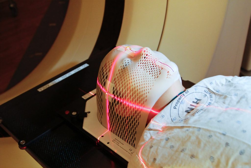
New guidelines for managing and treating brain metastases have been published in the Journal of Clinical Oncology and are set to improve care for cancer patients and extend and improve the quality of their lives.

The new guidelines come from an expert panel assembled by the American Society of Clinical Oncology (ASCO). The panel included a diverse range of top cancer doctors, as well as a patient representative.

The guidelines reflect the enormous advances in care for brain metastases  over the last few decades. In the 1970s, early attempts to develop guidelines largely emphasised steroids and whole-brain radiation therapy, without controlled, randomised studies to guide the use of surgery and chemotherapy.

Far more encompassing and far more evidence-based, the new guidelines will help doctors and patients make the best treatment decisions and achieve the best outcomes.

“When I started in this field 30 years ago, the average survival with brain metastases was 4 months, and most patients died from the brain disease. With improvements in therapies, fewer than one-quarter of patients die from the brain metastases, and some patients live years or are even cured,” said David Schiff, MD, a co-chair of the ASCO panel and the co-director of UVA Cancer Center’s Neuro-Oncology Center. “Equally importantly, the use of advanced localised radiation techniques and new targeted chemotherapies and immunotherapies have improved the quality of survival for most patients suffering from brain metastases.”

Up to 30% of cancer patients will have it spread to the brain, where it can be extremely difficult to treat. In the United States, approximately 200 000 new brain metastases are diagnosed each year.

The likelihood of a solid tumour spreading to the brain varies by cancer type, with approximately 20% of lung cancers spreading to the brain within a year after diagnosis. For patients with breast cancer, renal cell cancer or melanoma, that number is up to 7%. That is in addition to the patients found to have brain metastases at the time of their initial diagnosis.

Bringing together a diverse range of disciplines, the ASCO panel incorporated the results of more than 30 randomised trials published since 2008. The resulting guidelines cover everything from when surgery is appropriate and when and in what form radiation should be used to those circumstances in which medication alone may be employed.

The guidelines emphasise the importance of local therapies (surgery or stereotactic radiosurgery) for symptomatic brain metastases and lay out when these options are feasible. They highlight situations in which local therapy or whole brain radiotherapy can be deferred in place of chemotherapy, targeted therapy or immunotherapy depending on tumour type and molecular features. They also lay out how, in many cases, doctors can avoid the cognitive toxicity of whole brain radiotherapy by using either stereotactic radiosurgery or hippocampal-avoidant whole brain radiotherapy with the drug memantine.

“Patients with brain metastases may initially see a neurosurgeon, radiation or medical oncologist. The rigorous analysis underpinning these guidelines will provide each subspecialist a comprehensive picture of the treatment options appropriate for a given patient,” Dr Schiff said. “The result will allow patients the optimal personalised approach to maximise long-term control of brain metastases with good functional outcome.”

 Additional information is available at the ASCO website.

Source: EurekAlert!