Tag: bone repair

A Revolutionary Coral-inspired Material for Bone Repair

(Left) An image of a 3D-printed material implanted in vivo for 4 weeks. The photo was taken using a scanning electron microscope. Credit: Dr Zhidao Xia.
(Right) A photo of coral. Credit: Jesus Cobaleda.

Researchers at Swansea University have developed a revolutionary bone graft substitute inspired by coral which not only promotes faster healing but dissolves naturally in the body after the repair is complete.

This groundbreaking research, led by Dr Zhidao Xia from Swansea University Medical School in collaboration with colleagues from the Faculty of Science and Engineering and several external partners, has been patented and published in the leading journal Bioactive Materials.

Bone defects caused by conditions like fractures, tumours, and non-healing injuries are one of the leading causes of disability worldwide. Traditionally, doctors use either a patient’s own bone (autograft) or donor bone (allograft) to fill these gaps. However, these methods come with challenges, including a limited supply, the risk of infection and ethical concerns.

By using advanced 3D-printing technology, the team have developed a biomimetic material that mimics the porous structure and chemical composition of coral-converted bone graft substitute, blending perfectly with human bone and offering several incredible benefits:

  • Rapid Healing – It helps new bone grow within just 2–4 weeks.
  • Complete Integration – The material naturally degrades within 6–12 months after enhanced regeneration, leaving behind only healthy bone.
  • Cost-Effective – Unlike natural coral or donor bone, this material is easy to produce in large quantities.

In preclinical in vivo studies, the material showed remarkable results: it fully repaired bone defects within 3–6 months and even triggered the formation of a new layer of strong, healthy cortical bone in 4 weeks.

Most synthetic bone graft substitutes currently on the market can’t match the performance of natural bone. They either take too long to dissolve, don’t integrate well, or cause side effects like inflammation. This new material overcomes these problems by closely mimicking natural bone in both structure and biological behaviour.

Dr Xia explained: “Our invention bridges the gap between synthetic substitutes and donor bone. We’ve shown that it’s possible to create a material that is safe, effective, and scalable to meet global demand. This could end the reliance on donor bone and tackle the ethical and supply issues in bone grafting.”

Innovations like this not only promise to improve patient quality of life but also reduce healthcare costs and provide new opportunities for the biomedical industry.

The Swansea University team is now looking to partner with companies and healthcare organisations to bring this life-changing technology to patients around the world.

Source: Swansea University

Bone Cells Growing on Biomaterials Like Curvatures the Best

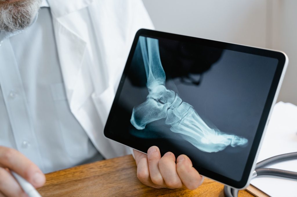
Doctor shows an X-ray of a foot
Photo by Tima Miroshnichenko on Pexels

TU Delft engineers have shown that the curvature of biomaterials inhibits or stimulates bone cells to make new tissue. Their findings are reported in Nature Communications, and their study of geometries could be an important step in research into repairing damaged tissues.

Living cells can perceive and respond to the geometry of their environment. “Cells sense and respond to the geometry of the surfaces they are exposed to. Depending on their curvature, surfaces can either encourage cells to create new tissue or prevent them from doing so,” says Amir Zadpoor, professor of Biomaterials and Tissue Biomechanics, supervisor of the study. “Stimulating curvatures made by a 3D printer are an easy and safe way to promote tissue growth. As compared to drugs, they are also much cheaper.”

The researchers grew bone cells in vitro surrounded by small moulds made from biomaterials with which the researchers have experience. Depending on the curvatures in the moulds, the cells tended to grow, divide, and form tissue to different extents.

Cells like a saddle shape

Although curved shapes seem to exist in endless variations, they always fall roughly into one of these three categories: a ball that has a convex curvature, a saddle that has a concave curvature, and a plate that is flat. One of the authors, assistant professor of Biomaterials Lidy Fratila-Apachitei: “Cells prefer a saddle shape. If they perceive a saddle shape nearby, growth is stimulated. The study also shows that cells prefer valleys over hills.”

Rather aligned than bent

First author Sebastien Callens did the experiments and analysis in the study. “Cells also have a skeleton, which consists of fibres that are under tension to different degrees. How tension builds up in those fibres strongly influences the behaviour of cells. Our study shows that cells collectively align their stress fibres with the curvatures they experience to minimise their need to bend. I could see that cells prefer to align than to bend.”

Budget of saddle curvature

You can’t have only saddle curves around cells. Just as the three angles of a triangle always add up to 180 degrees, the sum of all curvatures must also equal some fundamental numbers. “You always have a limited budget of saddle shapes,” says Zadpoor. “If you use too much negative curvature somewhere, you must use positive curvatures somewhere else to keep the sum constant. You need to use your budget wisely to encourage maximum tissue regeneration.”

New biomaterials

The study provides guidance on the optimal geometry of biomaterials and implants to maximise tissue regeneration. The complex geometric designs required are made using high-precision 3D printing techniques to make the shapes so small that they are perceptible to cells. Callens: “We have now discovered new playing rules by which biomaterials can stimulate tissue growth. In follow-up research, we will try to apply those rules optimally.”

Source: Delft University of Technology

Bone Tissue has Lymphatic Vessels – and They Aid Healing

Doctor shows an X-ray of a foot
Photo by Tima Miroshnichenko on Pexels

To date, it has been assumed that bones lack lymphatic vessels, but new research published in the journal Cell not only mapped them within bone tissue, but demonstrated their role in bone and blood cell regeneration and reveals changes associated with ageing.

The network of vessels that form the lymphatic system plays an important role in draining excess fluid from tissues, clearing waste products and supporting immune responses.

The fine network of lymph vessels extends throughout the body, but a small number of sites such as the brain, eye and bone were previously assumed to lack lymph tissue. The hard tissue of bone in particular has traditionally made studying the distribution and role of blood and lymph more difficult.

Researchers used light-sheet imaging to identify and visualise the lymphatic vessels of bone in high-resolution 3D, revealing an active network of lymph vessels within bone. The researchers further identified some of the key signals happening between lymph vessels, blood stem cells and bone stem cells.

Dr Lincoln Biswas, co-first author of this study, said: ‘Interestingly after injury, lymphatic vessels in bone show dynamic crosstalk with blood stem cells and with specialised perivascular cells in order to accelerate bone healing. Such interactions between lymphatics and bone stem cells can harnessed to promote bone healing such as in fracture repair.’

The researchers found that lymphatic vessels in bone increase during injury via a signalling molecule called IL6, and trigger expansion of bone progenitor cells by secreting a different signal, called CXCL12. Dr Junyu Chen, a co-first author of the study now based at Sichuan University said: “Ageing is associated with diminished capacity for bone repair, and our findings show that lymphatic signalling is impaired in aged bones. Remarkably, the administration of young lymphatic endothelial cells restores healing of aged bones, thus providing a future direction to promote bone healing in elderly.”

Dr Anjali Kusumbe, who led the research said: “I am very excited as these findings not only demonstrate that lymphatic vessels do exist in bone but also reveal their critical interactions with blood stem cells and perivascular bone stem cells after injury to promote healing, thereby presenting lymphatics as a therapeutic avenue to stimulate bone and blood regeneration. Further, these findings are very fundamental, opening doors for understanding the impact of bone lymphatics on the immune system and their role in bone and blood diseases.”

Source: Oxford University

Sound Waves Used to Regrow Bone

Image by Pawel Czerwinski on Unsplash

In a significant advance for the field of tissue engineering, researchers have used sound waves to turn stem cells into bone cells, a technology which may help regrow bone lost by cancer or disease.

Described in the journal Small, the innovative stem cell treatment from researchers at RMIT University offers a smart way forward for overcoming some of the field’s biggest challenges, through the precision power of high-frequency sound waves.

Tissue engineering is an emerging field that aims to rebuild bone and muscle by harnessing the human body’s natural ability to heal itself. A key challenge in regrowing bone is having sufficient amounts of bone cells that can thrive once implanted in the target area.

So far, turning stem cells into bone cells has needed complicated and expensive equipment, making widespread clinical use unrealistic.

The few clinical trials trying to regrow bone mostly used stem cells painfully extracted from a patient’s bone marrow.

In a new study published in the journal Small, the RMIT research team showed stem cells treated with high-frequency sound waves turned into bone cells quickly and efficiently.

Importantly, the treatment was effective on multiple types of cells including fat-derived stem cells, which are far less painful to extract from a patient.

Co-lead researcher Dr Amy Gelmi said the new approach was faster and simpler than other methods.

“The sound waves cut the treatment time usually required to get stem cells to begin to turn into bone cells by several days,” said Dr Gelmi. “This method also doesn’t require any special ‘bone-inducing’ drugs and it’s very easy to apply to the stem cells.

“Our study found this new approach has strong potential to be used for treating the stem cells, before we either coat them onto an implant or inject them directly into the body for tissue engineering.”

The high-frequency sound waves used in the stem cell treatment were generated on a low-cost microchip device developed by RMIT.

Co-lead researcher Distinguished Professor Leslie Yeo and his team have spent over a decade researching the interaction of sound waves at frequencies above 10MHz with different materials.

The sound wave-generating device they developed can be used to precisely manipulate cells, fluids or materials.

“We can use the sound waves to apply just the right amount of pressure in the right places to the stem cells, to trigger the change process,” Prof Yeo said.

“Our device is cheap and simple to use, so could easily be upscaled for treating large numbers of cells simultaneously – vital for effective tissue engineering.”

The next stage in the research is investigating methods to upscale the platform, working towards the development of practical bioreactors to drive efficient stem cell differentiation.

Source: RMIT

Regenerating Bone with Messenger RNA

Photo by Cottonbro on Pexels

Researchers have developed new way to get bone to regenerate with messenger RNA, which promises to be cheaper and less expensive while having fewer side effects than the current treatment.

Although fractures normally heal, bone will not regenerate under several circumstances. When bone does not regenerate, major clinical problems could result, including amputation.

One treatment is recombinant human bone morphogenetic protein-2, or BMP-2. However, it is expensive and only moderately effective. It also produces side effects, which can be severe.

Researchers at Mayo Clinic, along with colleagues in the Netherlands and Germany, may have a viable, less risky alternative: messenger RNA. 

A study conducted on rats and published in Science Advancesshows that messenger RNA can be used at low doses to regenerate bone – and without side effects. The resulting new bone quality and biomechanical properties are also superior to that of BMP-2. Additionally, messenger RNA is a good choice for bone regeneration because it may not need repeat administrations.

Human bone develops in one of two ways: direct formation of bone cells from mesenchymal progenitor cells, or through endochondral ossification, in which cartilage forms first and then converts to bone. The BMP-2 therapy uses the former method, and the messenger RNA approach uses the latter. In general, the researchers say their work proves that this method “can heal large, critical-sized, segmental osseous defects of long bones in a superior fashion to its recombinant protein counterpart.”

Further studies are required in larger animals than rats before any translation can be considered for clinical trials.

Source: Mayo Clinic

New Biomaterial Produced from Frog Skin and Fish Scales

Photo by Robert Zunikoff on Unsplash

Researchers at Nanyang Technological University, Singapore (NTU Singapore) have developed a new biomaterial made entirely from discarded bullfrog skin and fish scales that could help in bone repair.

The porous biomaterial, which contains the same compounds that are predominant in bones, acts as a scaffold for osteoblasts, or bone-forming cells, to adhere to and multiply, leading to new bone formation. Bone-forming cells successfully latched onto the biomaterial and started growing, and it was found to have a low inflammatory risk.

This kind of scaffold could help regenerate bone tissue lost to disease or injury, such as jaw defects from trauma or cancer surgery. It could also assist bone growth around surgical implants such as dental implants.

The current standard practice of using a patient’s own tissues means extra surgery is needed for bone extraction. The biomaterial used, frog skin and fish scales, are a significant waste stream produced by Singapore’s aquaculture industry and using them helps repurpose this waste.

‘Waste-to-resource’

“We took the ‘waste-to-resource’ approach in our study and turned discards into a high-value material with biomedical applications, closing the waste loop in the process,” said Dalton Tay, Assistant Professor, Nanyang Technological University. “Our lab studies showed that the biomaterial we have engineered could be a promising option that helps with bone repair. The potential for this biomaterial is very broad, ranging from repairing bone defects due to injury or ageing, to dental applications for aesthetics. Our research builds on NTU’s body of work in the area of sustainability and is in line with Singapore’s circular economy approach towards a zero-waste nation.”

To make the biomaterial, the team first extracted Type 1 tropocollagen (many molecules of which form collagen fibres) from the discarded skins of the American bullfrog and hydroxyapatite (a calcium-phosphate compound) from the scales of snakehead fish, commonly known as the Toman fish.

Collagen and hydroxyapatite (HA) are two predominant components found in bones, thus conferring on the biomaterial a structure, composition, and ability to promote cell attachment similar to bone, as well as toughness.

The scientists removed all impurities from the bullfrog skin, then blended it to form a thick collagenous paste that is diluted with water, from which collagen was extracted. “Using this approach, we were able to obtain the highest ever reported yield of collagen of approximately 70 per cent from frog skin, thus making this approach commercially viable,” said Asst Prof Tay, who is also from the NTU School of Biological Sciences (SBS).

HA was harvested from discarded fish scales through calcination – a purification process that requires high heat – to remove the organic matter, and then air-dried.

The biomaterial was synthesised by adding HA powder to the extracted collagen, then cast into a mould to make a 3D porous scaffold — a two-week process which the team believes can be shortened.

Testing the biomaterial

To assess the biological performance of the porous biomaterial scaffold for bone repair, the scientists seeded bone-forming cells onto the scaffold.

The cells proliferated, and after a week, the cells were uniformly distributed across the scaffold – an indicator that the scaffold could promote proper cellular activities and eventually lead to tissue formation. The scientists also found that the presence of HA in the biomaterial significantly enhanced bone formation.

The biomaterial was also tested for its tendency to cause an inflammatory response, which is common after a biomaterial is implanted in the body.

Using real-time polymerase chain reaction, the scientists found that the expression of pro-inflammatory genes in human immune cells exposed to the biomaterial stayed “relatively modest” compared to a control exposed to endotoxins, a compound known to stimulate immune response, said Asst Prof Tay.

For instance, the expression of the gene IL6 in the biomaterial group was negligible and at least 50 times lower than that of the endotoxins-exposed immune cells. This suggests that the risk of the NTU-developed biomaterial to trigger an excessive acute inflammatory response is low.

The team is now further evaluating the long-term safety and efficacy of the biomaterial as dental products. Further research would involve studying how the body responds to this biomaterial in the long term, as well its use in other applications such as skin wounds, along with further development of the waste-to-resource pipeline.

A preprint copy of the article is available as a PDF for download.

Source: Nanyang Technical University