Tag: bone mineral density

Radiology’s Role in Monitoring the Silent Disease – Osteoporosis

Images of a hip and lumbar spine, where bone density is typically measured.

Osteoporosis is often called a ‘silent disease,’ because it progresses, without symptoms, until a fracture occurs most commonly in your hips, spine and wrists.  However, a bone density scan can alert doctors to the disease before a patient has experienced any symptoms.

Radiology imaging techniques play a crucial role in the early diagnosis, management and monitoring of low bone density. The rapid evolution of high-quality imaging techniques, using reduced radiation doses, has positioned radiology ideally for this role.

What is osteoporosis

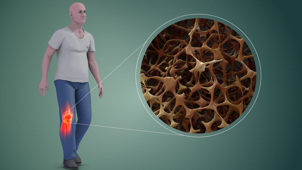
A healthy bone viewed under a microscope, looks like honeycomb. Osteoporosis, put simply, is when the ‘holes and spaces’ in the honeycomb increase in size, causing the bones to lose density or mass and develop abnormal tissue structure. This is caused by the body losing too much bone or making too little bone because of a lack of calcium, vitamin D and not doing any weight-bearing exercises or both. This can lead to a decrease in bone strength which, in turn, can increase the risk of broken or fractured bones.

There are degrees of bone density loss which are determined by radiologists doing a DEXA scan.

‘The standard method of determining your bone density,’ says Dr Hein Els, director at SCP Radiology, ‘is a DEXA scan (dual-energy X-ray absorptiometry). This involves using two X-ray beams, at different energy levels. to measure the bone mineral density. It has a high accuracy for overall bone density and is commonly found in clinics and hospitals.’ 

The scan uses a low radiation exposure making it safer for routine screening and follow-up.

‘The amount of radiation is minimal,’ says Dr Els, ‘it’s equivalent to 1 or 2 days of background radiation at sea level.’ 

Osteoporosis vs osteopenia

Osteoporosis and osteopenia are both conditions measured on a DEXA scan and characterised by decreased bone density. While they are related, they differ in severity and implications for bone health.

The fracture risk is higher in osteoporosis due to more significant bone fragility.

Understanding and managing both conditions are crucial for maintaining bone health and preventing fractures.

Measuring bone density

We measure your bone mass density by comparing it to that of a healthy, young adult. The result will tell us how much lower (or higher) your bone mass score,’ explains Dr Els. ‘Software is also used to calculate a predicted 10-year fracture risk for a major osteoporotic fracture and a hip fracture. The result is a T-score which you will be given by your doctor.’

Who is at greater risk

The vast majority of patients referred for a DEXA scan are women.  However, men over the age of 50 are also at risk, though not to the same degree as women.  The aim is to prevent fractures later in life by maintaining healthy bone mineral density, which means it is beneficial to know your bone mineral density. Fractures in the elderly population are a significant cause of morbidity and mortality.

Apart from diagnosing osteoporosis and osteopenia and assessing fracture risk, DEXA scans are helpful in the following ways:

  • Monitoring bone density changes over time: For individuals diagnosed with osteoporosis or those undergoing treatment for bone loss, DEXA scans are used to monitor changes in bone density. This helps in evaluating the effectiveness of treatment
  • Postmenopausal women: Are at a higher risk of developing osteoporosis due to decreased oestrogen levels. DEXA scans are recommended for postmenopausal women, especially those with additional risk factors
  • Men over 50 can also be at risk of osteoporosis
  • A family history of osteoporosis or fractures can increase an individual’s risk. DEXA scans can help assess bone density in those with a genetic predisposition
  • Individuals with a low body mass index (BMI) are at a higher risk for osteoporosis and may benefit from bone density testing
  • Smokers and heavy alcohol users are risk factors for osteoporosis
  • Patients with fragility fractures: Individuals who have experienced fractures from minor falls or injuries may undergo DEXA scans to determine if osteoporosis is the underlying cause

How do you treat low bone mass density?

This can be done through medication such as bisphosphonates, hormone-related therapy and other bone-building medications or through lifestyle changes. This includes an adequate intake of calcium and vitamin D, regular weight-bearing exercise, quitting smoking and limiting alcohol.

The DEXA scan is the safest, most reliable method of determining your bone loss and whether your bones are normal or if you are osteopenic or osteoporotic – the precursor to osteoporosis or full-blown osteoporosis. Regular medical check-ups and proactive lifestyle changes can significantly mitigate the risks associated with these conditions.

‘There is no need to be harbouring this silent disease,’ says Dr Els, ‘when radiography is available to test for these and can put you on a path to wellness.’  

Vitamin D Supplements don’t Prevent Fractures in Children

Photo by cottonbro studio

A major clinical trial has found that vitamin D supplements do not increase bone strength or prevent bone fractures in children with vitamin D deficiency. The findings, published in Lancet Diabetes & Endocrinology, challenge widely held perceptions relating to the effects of vitamin D on bone health.

Around one-third of children have at least one fracture before the age of 18. This is a major global health issue, as childhood fractures can lead to life years of disability and/or poor quality of life. The potential for vitamin D supplements to improve bone strength has attracted growing interest in recent years, based on vitamin D’s role in promoting bone mineralisation. But there have been no clinical trials to test whether vitamin D supplements can prevent bone fractures in children.

Working with partners in Mongolia, a setting with a particularly high fracture burden and where vitamin D deficiency is highly prevalent, researchers from led by Queen Mary University of London and the Harvard T.H. Chan School of Public Health conducted a clinical trial to determine if vitamin D supplementation would decrease risk of bone fractures or increase bone strength in schoolchildren.

This study is also the largest randomised controlled trial of vitamin D supplementation ever conducted in children. Over the course of three years, 8851 schoolchildren aged 6-13 living in Mongolia received a weekly oral dose of vitamin D supplementation.

Testing revealed that 95.5% of participants had vitamin D deficiency at baseline, and study supplements were highly effective in boosting vitamin D levels into the normal range. No effect was seen on fracture risk or on bone strength, measured in a subset of 1438 participants using quantitative ultrasound.

The trial findings are likely to prompt scientists, doctors and public health specialists to re-consider the effects of vitamin D supplements on bone health.

Dr Ganmaa Davaasambuu, Associate Professor at the Harvard T.H. Chan School of Public Health, said:

“The absence of any effect of sustained, generous vitamin D supplementation on fracture risk or bone strength in vitamin D deficient children is striking. In adults, vitamin D supplementation works best for fracture prevention when calcium is given at the same time – so the fact that we did not offer calcium alongside vitamin D to trial participants may explain the null findings from this study.”

Professor Adrian Martineau, Lead of the Centre for Immunobiology at Queen Mary University of London, added:

“It is also important to note that children who were found to have rickets during screening for the trial were excluded from participation, as it would not have been ethical to offer them placebo (dummy medication). Thus, our findings only have relevance for children with low vitamin D status who have not developed bone complications. The importance of adequate vitamin D intake for prevention of rickets should not be ignored, and UK government guidance recommending a daily intake of 400 IU vitamin D remains important and should still be followed.”

Source: Queen Mary University of London

Repeated Concussions can Result in Skull Thickening

MRI images of the brain
Photo by Anna Shvets on Pexels

Published in the journal Scientific Reports, a study led by Associate Professor Bridgette Semple from Monash University, found that repeated concussions resulted in thicker, denser bones in the skull.

Although bones are considered a mostly structural component of the human body, bones are in fact active living tissues that can respond to applied mechanical forces. For example, martial arts training, with its kicks, punches and throws, has been shown to increase bone mineral density in the arms, legs and spines of practitioners.

At present, it is unclear whether this thickening of the skull is beneficial or detrimental: theoretically, a thicker skull is a stronger skull, suggesting that this may be the bone’s attempt to protect the brain from subsequent impacts.

“This is a bit of a conundrum,” Assoc Prof Semple said. “As we know, repeated concussions can have negative consequences for brain structure and function. Regardless, concussion is never a good thing.”

The team hopes that the microstructural skull alterations caused by concussion are now considered by researchers in the field to better understand how concussions affect the whole body.

A form of mild traumatic brain injury, concussion have been linked to long-term neurological consequences if they happen repetition.

While most studies focus on its effect on the brain and its function, they largely ignore the overlying skull bones.

Study collaborator Professor Melinda Fitzgerald, from Curtin University and the Perron Institute in Western Australia, has previously shown that repeated concussive impacts lead to subtle problems with memory, and evidence of brain damage.

In this new study, high-resolution neuroimaging and tissue staining techniques were used in a pre-clinical animal model, and revealed an increase in bone thickness and density, in close proximity to the site of injury.

“We have been ignoring the potential influence of the skull in how concussive impacts can affect the brain,” Associate Professor Semple said. “These new findings highlight that the skull may be an important factor that affects the consequences of repeated concussions for individuals.”

Future studies are planned, with collaborator and bone expert Professor Natalie Sims from St Vincent’s Institute of Medical Research in Melbourne, to understand if a thickened skull resulting from repeated concussions alters the transmission of impact force through the skull and into the vulnerable brain tissue underneath.

Source: Monash University

PFAS and Phthalates Linked to Reduced Bone Density in Teen Boys

Photo by Gayatri Malhotra on Unsplash

Per- and polyfluoroalkyl substances (PFAS) and phthalates (two types of endocrine-disrupting chemicals) may be associated with lower areal bone mineral density (aBMD) in teenage boys, according to a new study published in the Journal of Clinical Endocrinology and Metabolism.

Endocrine disrupting chemicals (EDCs) and potential EDCs are mostly man-made found in various materials. By interfering with the body’s endocrine system, endocrine disruptors produce adverse developmental, reproductive, neurological, and immune effects in humans, abnormal growth patterns and neurodevelopmental delays in children. These include per- and polyfluoroalkyl substances (PFAS) are used in non-stick pots and pans, clothing and food packaging, and are increasingly being found in US water supplies. Phthalates are used in medical devices, personal care products, food processing and children’s toys.

“Adolescence is an important time when our bodies build up bone. Almost all US children and adolescents are exposed to PFAS and phthalates, but few studies have looked at how these chemicals could be impacting our bone health,” said Abby F. Fleisch, MD, MPH, of the Maine Medical Center Research Institute and Maine Medical Center. “Our research found an association between certain PFAS and phthalates and reduced bone mineral density in adolescent males. Because bone accrual primarily occurs during adolescence, if replicated, this finding may have implications for lifelong bone health.”

The researchers accessed data on urine and blood samples from 453 boys and 395 girls from the National Health and Nutrition Examination Survey (NHANES). Participants were on average 15.1 years old, and found that higher levels of PFAS and phthalates may be associated with lower aBMD in adolescent males. The same effect was not found in girls; rather a slight increase in aBMD was observed for certain PFAS and phthalates.

The researchers noted that bone mineral density tracks across a lifetime, so if the same results are seen in longitudinal cohorts, this finding may have implications for lifelong skeletal health.

Source: The Endocrine Society