Tag: bone marrow

How Breast Cancer Cells Survive in Bone Marrow after Remission

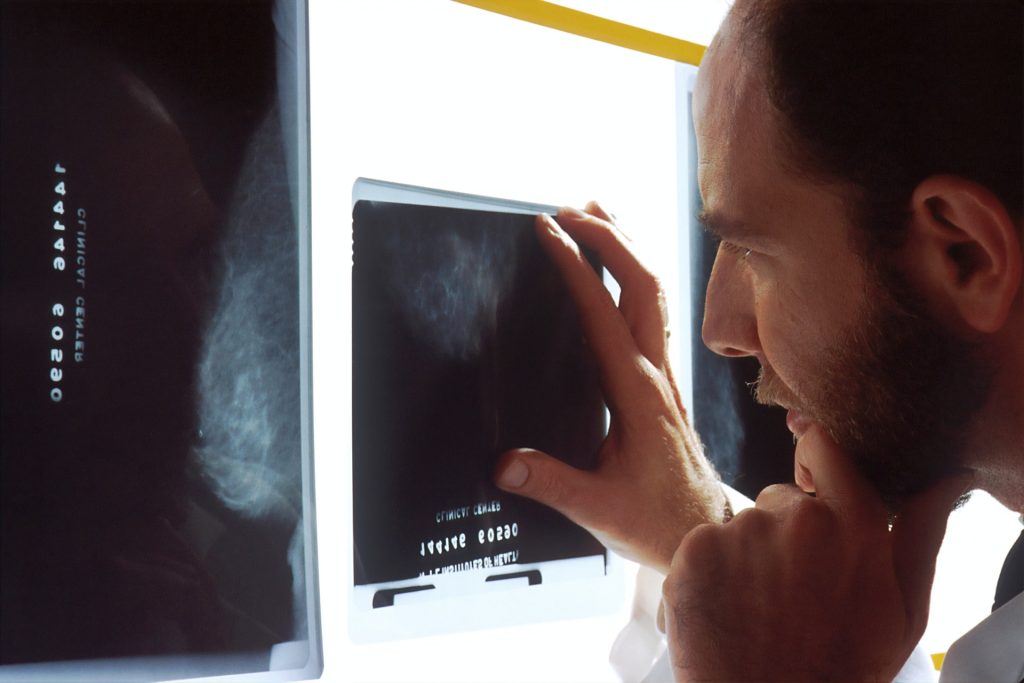
Photo by National Cancer Institute on Unsplash

A new study has shed light on a previously poorly understood aspect of breast cancer recurrence: how cancer cells survive in bone marrow despite targeted therapies. The paper appears in the Journal of Clinical Investigation

Oestrogen receptor positive (OR+) breast cancer is the most common form of the disease, and cancer cells of this kind can live for years in bone marrow after remission. The persistence of these cells in marrow leads to the disease recurring about 40% of patients. This return can take the form of especially aggressive bone cancer with symptoms such as bone fractures and hypercalcaemia. 

The cells can also spread to other organs, causing recurrent disease that is currently incurable. 

To better understand how these cancer cells survive, and why they cause such aggressive returning disease, researchers investigated what happens to these dispersed cells in bone marrow. 

Their key finding was the mechanism by which a normal cell type, mesenchymal stem cells, in the bone marrow supports the cancer cells.

“We discovered that the breast cancer cells require direct contact with mesenchymal stem cells,” said Gary Luker, MD, senior author on the paper.  

“The cancer cells physically borrow molecules – proteins, messenger RNA – directly from the mesenchymal stem cells. Essentially the mesenchymal stem cells act as very generous neighbours in donating things that make the cancer cells more aggressive and drug resistant.”

In laboratory experiments, contact between cancer cells and mesenchymal stem cells induced changes in hundreds of proteins. Further analysis of which proteins allowed for survival of breast cancer cells led researchers to focus on GIV, also known as Girdin. The paper notes that GIV drives “invasiveness, chemoresistance, and acquisition of metastatic potential in multiple cancers.”

GIV makes these cancer cells specifically resistant to oestrogen-targeted therapies, such as the drug Tamoxifen. The researchers hope this understanding of the mechanism of cancer cell survival will one day lead to treatments that prevent OR+ breast cancers from returning.

Sleeper cells can awaken

“Sleeper cells can be reawakened and cause oestrogen receptor positive breast cancers to relapse years –in some cases as long as a decade – after patients were believed to be in remission,” said study author Pradipta Ghosh, M.D., a professor in the Departments of Medicine and Cellular and Molecular Medicine at UC San Diego School of Medicine.

“Since these cancer cells ‘borrow’ essential proteins from stem cells in the bone marrow through cellular tunnels – much like smuggling – approaches for targeting the tunnels or proteins they smuggle could help prevent the relapse and metastasis of oestrogen receptor positive breast cancer.”

Source: University of Michigan

Familial Alzheimer’s Disease Transferred via Bone Marrow Transplant in Mice Experiment

Photo by Mari Lezhava on Unsplash

Familial Alzheimer’s disease can be transferred via bone marrow transplant, researchers show in the journal Stem Cell Reports. When the team transplanted bone marrow stem cells from mice carrying a hereditary version of Alzheimer’s disease into normal lab mice, the recipients developed Alzheimer’s disease – and at an accelerated rate.

The study highlights the role of amyloid that originates outside of the brain in the development of Alzheimer’s disease, which changes the paradigm of Alzheimer’s from being a disease that is exclusively produced in the brain to a more systemic disease. Based on their findings, the researchers say that donors of blood, tissue, organ, and stem cells should be screened for Alzheimer’s disease to prevent its inadvertent transfer during blood product transfusions and cellular therapies.

“This supports the idea that Alzheimer’s is a systemic disease where amyloids that are expressed outside of the brain contribute to central nervous system pathology,” says senior author and immunologist Wilfred Jefferies, of the University of British Columbia. “As we continue to explore this mechanism, Alzheimer’s disease may be the tip of the iceberg and we need to have far better controls and screening of the donors used in blood, organ and tissue transplants as well as in the transfers of human derived stem cells or blood products.”

To test whether a peripheral source of amyloid could contribute to the development of Alzheimer’s in the brain, the researchers transplanted bone marrow containing stem cells from mice carrying a familial version of the disease — a variant of the human amyloid precursor protein (APP) gene, which, when cleaved, misfolded and aggregated, forms the amyloid plaques that are a hallmark of Alzheimer’s disease. They performed transplants into two different strains of recipient mice: APP-knockout mice that lacked an APP gene altogether, and mice that carried a normal APP gene.

In this model of heritable Alzheimer’s disease, mice usually begin developing plaques at 9 to 10 months of age, and behavioural signs of cognitive decline begin to appear at 11 to 12 months of age. Surprisingly, the transplant recipients began showing symptoms of cognitive decline much earlier – at 6 months post-transplant for the APP-knockout mice and at 9 months for the “normal” mice.

“The fact that we could see significant behavioural differences and cognitive decline in the APP-knockouts at 6 months was surprising but also intriguing because it just showed the appearance of the disease that was being accelerated after being transferred,” says first author Chaahat Singh of the University of British Columbia.

In mice, signs of cognitive decline present as an absence of normal fear and a loss of short and long-term memory. Both groups of recipient mice also showed clear molecular and cellular hallmarks of Alzheimer’s disease, including leaky blood-brain barriers and buildup of amyloid in the brain.

Observing the transfer of disease in APP-knockout mice that lacked an APP gene altogether, the team concluded that the mutated gene in the donor cells can cause the disease and observing that recipient animals that carried a normal APP gene are susceptible to the disease suggests that the disease can be transferred to health individuals.

Because the transplanted stem cells were hematopoietic cells, meaning that they could develop into blood and immune cells but not neurons, the researchers’ demonstration of amyloid in the brains of APP knockout mice shows definitively that Alzheimer’s disease can result from amyloid that is produced outside of the central nervous system.

Finally the source of the disease in mice is a human APP gene demonstrating the mutated human gene can transfer the disease in a different species.

In future studies, the researchers plan to test whether transplanting tissues from normal mice to mice with familial Alzheimer’s could mitigate the disease and to test whether the disease is also transferable via other types of transplants or transfusions and to expand the investigation of the transfer of disease between species.

“In this study, we examined bone marrow and stem cells transplantation. However, next it will be important to examine if inadvertent transmission of disease takes place during the application of other forms of cellular therapies, as well as to directly examine the transfer of disease from contaminated sources, independent from cellular mechanisms,” says Jefferies.

Source: Cell Press

More Stem Cell Donors Needed to Increase the Aplastic Anaemia Survival Rate

Photo by National Cancer Institute on Unsplash

Despite being one of the rarest blood disorders, Aplastic Anaemia is one of the deadliest, with about 70% of patients having a life expectancy of only one year if untreated.

Among the treatment options available, stem cell transplants offer hope, boasting a 96% survival rate that increases to 100% in children and adults under 40. Unfortunately, however, there are only 76 019 donors on the South African registry, meaning that the chances of Aplastic Anaemia patients finding a suitable match are slim.

“The chances are even slimmer for Black Aplastic Anaemia patients as only 33% of the registry is comprised of Black donors,” says Palesa Mokomele, Head of Community Engagement and Communications at DKMS Africa, who explains that a patient’s best chance of a match comes from within their own ethnic group.

Currently, the non-profit organisation is trying to find matching donors for at least seven South African Aplastic Anaemia patients between the ages of seven and 36. Some of these patients, like 21-year-old Kholiwe, have been on the waiting list since as far back as 2020.

During her matric year, she started experiencing symptoms and after being rushed to the hospital following a fainting spell, received the shocking diagnosis. Compounding the situation for the aspiring drama student was the withdrawal of support from her family, leaving her all alone to cope emotionally and financially with the disease. As she waits for a matching donor to be found, Kholiwe now has the challenge of finding permanent employment while simultaneously undergoing medical treatments to keep her alive. Despite these hardships, she remains hopeful about finding a matching stem cell donor. “Kholiwe’s future, and those of other Aplastic Anaemia patients, depends on this,” says Mokomele.

Explaining what Aplastic Anaemia is, she shares that, based on information gathered by DKMS Africa in conjunction with BLOODSA, the condition occurs when our bone marrow stops making enough blood cells. “This can lead to anaemia, a weak immune system, and an increased risk of bleeding and bruising.”

As for the cause of Aplastic Anaemia, Mokomele points out that this is due to bone marrow damage. “While some people are born with compromised bone marrow, others develop this as a consequence of pregnancy, genetic disorders, certain medicines or chemicals, an overactive immune system or viral infections such as HIV and Hepatitis.”

In light of Aplastic Anaemia Awareness Day on 4 March, she encourages all South Africans to familiarise themselves with the symptoms. “These include tiredness; feeling weak; pale skin and tongue; bruising and bleeding easily; rapid heartbeat; trouble breathing; frequent infections; headaches and dizziness.”

Similarly, 10-year-old Mesuli’s experience highlights the challenges faced by Aplastic Anaemia patients and their families. His journey began with drastic weight loss and constant fatigue. The once energetic and always bubbly little boy grew weak and started having severe nose bleeds. That’s when his aunt Nonhle, who is caring for him following the passing of his mother, consulted a doctor. With his illness forcing him to leave school, Mesuli’s new reality consists of going to the hospital every Wednesday for a blood transfusion.

“It hurts to see him in pain because it hurts me too. All I want is for him to grow and live out his dreams. I am begging each South African to think of Mesuli, spread the word and get your family and friends registered to help save the life of my boy. His life hangs in the hands of a perfect stranger,” pleads Nonhle.

Mesuli hopes to one day become a doctor and save lives, the same way he hopes his life will be saved.

“Bearing Aplastic Anaemia Awareness Day in mind, we encourage South Africans to pay close attention to their health and that of their loved ones, especially as the rarity of the disease does not diminish its severity. But, more importantly, we need those who are healthy to register as stem cell donors and save the lives of patients with this deadly disease,” concludes Mokomele.

Register today at https://www.dkms-africa.org/register-now

For more information, contact DKMS Africa on 0800 12 10 82.

B Cells Play a Surprising Role in Bone Marrow Output

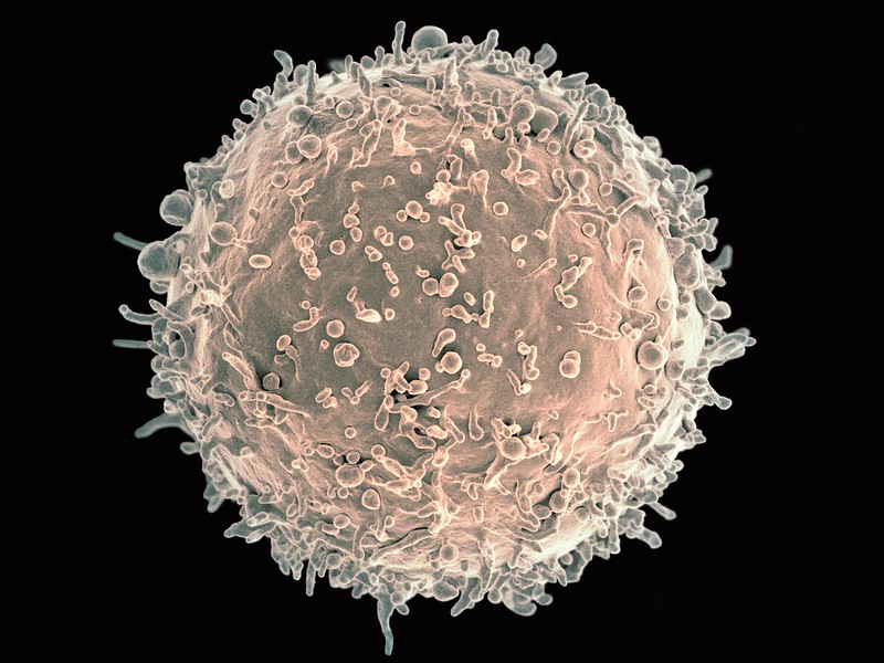
Scanning electron micrograph image of a human B cell. Credit: NIH/NIAID

New research in Nature Immunology has found that B cells play a surprising role in increasing or decreasing the bone marrow’s output of white blood cells. The findings may lead to new treatments for conditions that arise when white blood cell production goes out of balance.

Professor Matthias Nahrendorf, senior author of the study, explained that the nervous system plays a role in controlling blood cell production through neurotransmitters. “This is for instance important in people exposed to stress, where stress hormones — part of the ‘fight-or-flight’ response controlled by the sympathetic nervous system — may increase bone marrow activity and cardiovascular inflammation in response to the neurotransmitter noradrenaline,” he said. The parasympathetic nerves on the other hand, slow down responses and bring about a state of calm to the body, mainly through the neurotransmitter acetylcholine.

Because acetylcholine can have a protective effect against inflammation and heart disease, the researchers studied this neurotransmitter in the bone marrow. “When we looked into how acetylcholine acts on the production of blood cells, we found that it does the expected — it reduces white blood cells, as opposed to noradrenaline, which increases them,” said Prof Nahrendorf. “What was unexpected though was the source of the neurotransmitter acetylcholine.”

In the bone marrow, the typical nerve fibres that are known to release acetylcholine were not found. Instead, it was the antibody-producing B cells supplied the acetylcholine in the bone marrow. “Thus, B cells counter inflammation — even in the heart and the arteries — via dampening white blood cell production in the bone marrow. Surprisingly, they use a neurotransmitter to do so,” said Prof Nahrendorf.

Tapping into this process may help investigators develop strategies to block inflammation in cardiovascular conditions such as atherosclerosis. “Ultimately this may lead to new therapeutics that combat myocardial infarction, stroke, and heart failure,” said Prof Nahrendorf.

Source: Massachusetts General Hospital

A Novel Therapy for Bone Marrow Cancer

Source: NCI on Unsplash

Researchers have found that a novel therapy for the bone marrow cancer myelofibrosis to be safe and well-tolerated, and is associated with modest improvements in patients in an early clinical trial. They shared their findings during an oral presentation at the American Society of Hematology annual meeting in December.

The therapy AVID200 showed improvements in patients’ symptom burden, anaemia, and spleen enlargement. The results from the Phase 1b clinical trial showed that the therapy was safe and displayed some evidence of efficacy (although safety and finding optimal dosage was the main goal) and researchers concluded that the therapy would need to be combined with other drugs to optimise effectiveness in patients.

“This is a real testament to cutting-edge translational research at The Tisch Cancer Institute,” said John Mascarenhas, MD, Director of the Institute’s newly launched Center of Excellence for Blood Cancer and Myeloid Disorders. “Our scientists tested this therapy in the lab, physician-scientists conducted a successful phase 1 trial, and now the optimal combination therapy approach is the subject of ongoing laboratory studies at Mount Sinai. The most interesting finding in this trial was that a subset of patients had a lasting improvement in their platelet counts – including three whose counts were normalised – supporting the preclinical studies conducted.”

Myelofibrosis is a bone marrow cancer type that disrupts normal blood cell production, causing an enlarged spleen, extensive scarring in the bone marrow, and low levels of red blood cells and platelets, increasing bleeding risk. Myelofibrosis patients who have failed the available first-line therapy face a well-documented poor prognosis, so additional therapies are urgently needed to help these patients.

Twenty-one patients enrolled in this multicenter trial were given AVID200, and while this trial’s main purpose was to test safety, some patients had an increase in platelets and there was a decrease in the size of their enlarged spleens. However, in spite of the other clinical benefits seen. patients’ bone marrow scarring did not decline, indicating that AVID200 would need to be combined with other rational therapies in the future.   

Source: EurekAlert!

A ‘Fountain of Youth’ for Bone Marrow Stem Cells

Source: National Cancer Institute on Unsplash

Scientists have shown that reduced bone marrow stem cell function with ageing is due to changes in their epigenome, and they were able to reverse these changes in isolated stem cells by adding acetate. This ‘fountain of youth’ for the epigenome could become important for the treatment of diseases such as osteoporosis.

One responsible mechanism for age-related osteoporosis and fracture risk involves the impaired function of the bone-marrow stem cells, which are required for the maintenance of bone integrity. 

For a long time, researchers have looked at epigenetics as a cause of ageing. Epigenetics looks at changes that affect the activity of genes. One of these is changes in proteins called histones, which package and thus control access to DNA. In this study, the researchers investigated the epigenome of mesenchymal stem cells, which are found in bone marrow and can give rise to different types of cells such as cartilage, bone and fat cells.

“We wanted to know why these stem cells produce less material for the development and maintenance of bones as we age, causing more and more fat to accumulate in the bone marrow. To do this, we compared the epigenome of stem cells from young and old mice,” explained Andromachi Pouikli, first author of the study. “We could see that the epigenome changes significantly with age. Genes that are important for bone production are particularly affected.”

The researchers then sought to find out if it was possible to rejuvenate the epigenome of stem cells. To do this, they treated isolated stem cells from mouse bone marrow with a nutrient solution which contained sodium acetate. The cell converts the acetate into a building block that enzymes can attach to histones to increase access to genes, thereby boosting their activity. “This treatment impressively caused the epigenome to rejuvenate, improving stem cell activity and leading to higher production of bone cells,” Pouikli said.

To see if this change could also be responsible for increased fracture risk and osteoporosis with age, the researchers studied human mesenchymal stem cells from hip surgery patients. In elderly patients with osteoporosis, the same epigenetic changes seen with mice were also seen in these human cells.

“Sodium acetate is also available as a food additive, however, it is not advisable to use it in this form against osteoporosis, as our observed effect is very specific to certain cells,” cautioned study leader Peter Tessarz. “However, there are already first experiences with stem cell therapies for osteoporosis. Such a treatment with acetate could also work in such a case. However, we still need to investigate in more detail the effects on the whole organism in order to exclude possible risks and side effects.”

The results were published in the journal Nature Aging.

Source: Max Planck Society

Bone Marrow Cell Mutations That Protect Against Cancers

Source: NIH

People with shortened telomeres caused by rare disorders may be more likely to have blood cancers such as leukaemia or myelodyplastic syndrome (MS). Now researchers have discovered several “self-correcting” genetic mutations in bone marrow that may protect such patients from these cancers.

In a study published in the Journal of Clinical Investigation, these mutations can serve as biomarkers to indicate if patients with short telomere syndromes (STS) are likely to develop blood cancers.

“These are the most common cancers we see in patients with short telomere syndromes,” said Mary Armanios, MD, director of the Telomere Center and professor of oncology at the Sidney Kimmel Comprehensive Cancer Center at Johns Hopkins. “We know that at a certain point, the cells of patients with shortened telomeres either become cancerous or stay healthy.”

Dr Armanios and her team suspected that a self-correcting mechanism in areas of the body with high cell turnover, such as bone marrow, was allowing normal cells to turn malignant. Instead, it appears this mechanism protects against cells from becoming cancerous.

As over 300 billion blood cells are produced in the bone marrow daily, the researchers suspected they could find evidence of cellular self-correction in this area of the body, especially amid the spongey interior of bones, where quick adaptation is crucial for high-volume cell production.
The researchers tested the bone marrow and blood cells of 84 study participants divided into three groups: Those with STS and MS or leukaemia; those with short telomere syndromes and no MS or leukemia; and those in the control group without short telomere syndromes or any cancers.

Using ultra-deep genetic sequencing which picks up hard-to-detect mutations, Armanios and her team observed genetic mutations and self-correction in several telomere-associated genes. Nearly a quarter of patients with STS had these mutations, some even showing multiple mutations.

One such mutation in a gene called TERT enables the production of crucial parts of telomerase, which stabilises telomeres. By boosting telomerase production and overwriting faulty copies of the TERT gene, the researchers found that bone marrow cells seemed to self-correct to avoid becoming cancerous.

“Our findings speak to the versatility of the bone marrow and other areas with high cell turnover in the body,” says Armanios. “Such advantageous mutations provide the body with a better chance to protect itself. These findings may be important in the screening process of shortened telomere patients so that we can predict who may be protected from cancer.”

Source: John Hopkins Medicine