Tag: blood-brain barrier

A New Era of Treating Neurological Diseases at the Blood-brain-immune Interface

This is a pseudo-colored image of high-resolution gradient-echo MRI scan of a fixed cerebral hemisphere from a person with multiple sclerosis. Credit: Govind Bhagavatheeshwaran, Daniel Reich, National Institute of Neurological Disorders and Stroke, National Institutes of Health

The question of what causes complex neurological diseases such as Alzheimer’s or multiple sclerosis continues to confound scientists and doctors, with the unknowns standing in the way of early diagnoses and effective treatments.

Even among identical twins who share the same genetic risk factors, one may develop a particular neurological disease while the other does not.

That’s because unlike diseases such as cystic fibrosis or sickle-cell anaemia, which are caused by a single gene, most neurological disorders are associated with many, sometimes hundreds, of rare genetic variants. And on their own, these variants can’t predict who will develop disease, as neurological conditions are also strongly influenced by environmental factors and vascular risks such as high blood pressure, aging, heart disease, or obesity.

But there’s one often-overlooked thread that connects most neurological diseases, says Katerina Akassoglou, PhD, a senior investigator at Gladstone Institutes: They’re marked by a toxic immune reaction caused by blood that leaks into the brain through damaged blood vessels.

“Interactions between the brain, blood vessels, and the immune system are a common thread in the development and progression of many neurological diseases that have been traditionally viewed as very different conditions,” says Akassoglou. “Knowing that leaked blood is a key driver of brain inflammation, we can now approach these diseases from a different angle.”

She and her collaborators share their insights on this topic in a commentary article published in Cell’s 50th anniversary “Focus on Neuroscience” issue. 

Neutralising the Culprit

Akassoglou and her lab have long investigated how blood that leaks into the brain triggers neurologic diseases, essentially by hijacking the brain’s immune system and setting off a cascade of harmful often-irreversible effects that result in damaged neurons.

One blood protein in particular, fibrin, normally involved in blood coagulation, is responsible for setting off this detrimental cascade. The process has been observed in conditions as diverse as Alzheimer’s, traumatic brain injury, multiple sclerosis, premature birth, and even COVID-19. However, Akassoglou and her team found that the process could be prevented or interrupted by “neutralising” fibrin to deactivate its toxic properties – an approach that appears to protect against many neurological diseases when tested in animal models.

“As a first step, we know that neutralizing fibrin reduces the burden posed by vascular dysfunction,” Akassoglou says. Regardless of what initially caused the blood leaks, be it a head injury, autoimmunity, genetic mutations, brain amyloid or infection, neutralizing fibrin appears to be protective in multiple animal models of disease.

The scientists previously developed a drug, a therapeutic monoclonal antibody, that specifically targets fibrin’s inflammatory properties without affecting its essential role in blood coagulation. This fibrin-targeting immunotherapy has shown, in mice, to protect from multiple sclerosis and Alzheimer’s, and to treat neurological effects of COVID-19. A humanized version of this first-in-class fibrin immunotherapy is already in Phase 1 safety clinical trials by Therini Bio, a biotech company launched to advance discoveries from Akassoglou’s lab.

A New Era of Brain Research

In the Cell commentary, Akassoglou and her colleagues make the case that seemingly disparate neurological diseases must be viewed differently in light of new research on the blood-brain-immune interface.

They say that in the coming decade, scientific breakthroughs will emerge from collaborative networks of immunologists, neuroscientists, haematologists, geneticists, computer scientists, physicists, bioengineers, drug developers, and clinical researchers. These partnerships, forged across academia, industry, and foundations, will catalyse innovation in drug discovery and transform medical practice for neurological diseases.

“This is a new opportunity for drug discovery that goes beyond addressing genes alone or environmental factors alone,” Akassoglou says. “To usher in this new era, we must leverage new technologies and embrace an interdisciplinary approach that accounts for the important roles of immune and vascular systems in neurodegeneration.”

Source: Gladstone Institutes

How the Brain Protects Itself Against Herpes Simplex Virus

Source: CC0

More than half of us are carriers of chronic herpesvirus infections. But even though the herpes simplex virus can infect nerve cells, it rarely causes serious infection of the brain. Researchers from Aarhus University have now discovered a key element of the explanation.

The researchers have discovered a previously unknown defence mechanism in the body that is the reason why herpes infection causes a serious and potentially fatal brain inflammation in only one out of 250 000 cases. The study has recently been published in the scientific journal Nature.

“The study has exciting perspectives because it gives us a better understanding of how the brain defends itself against viral infections,” says Professor Søren Riis Paludan from the Department of Biomedicine at Aarhus University. He is the article’s last author, a Lundbeck Foundation Professor and centre director of the Excellence Centre CiViA.

“We’ve discovered how our body prevents herpesvirus from entering into the brain, even though 50–80% of us are chronically infected with this particular virus. The idea behind CiViA is that we want to understand how the body fights infections without harming itself at the same time. The mechanism we’ve found doesn’t cause inflammatory reactions,” he says.

The answer lies in the protective TMEFF1 gene.

The brain uses a novel mechanism to keep the virus out

Many years of experimenting with the genome-wide CRISPR screening technology and development of mice that lacked the critical gene have finally convinced the researchers that TMEFF1 produces a protein that prevents herpesvirus from entering into nerve cells.   

The study in Nature is accompanied by another article describing two patients with brain inflammation caused by herpesvirus infection, called herpes encephalitic. In a collaborative study led by researchers in New York, the research group in Aarhus discovered that two children who developed herpes encephalitis were carrying a genetic defect that disabled the protective TMEFF1 gene.

“The new study is groundbreaking because it updates the basic understanding of immunity against viral infections,” explains Søren Riis Paludan.

 “This is interesting for immunologists because it illustrates that there are still many immunological mechanisms in the brain that we don’t know about. “The study is also relevant for neuroscience because it sheds light on how the brain, so to say, prevents unwanted visitors from intruding without causing harm to the brain itself, i.e. the neuronal cells,” he says.

May provide a better understanding of Alzheimer’s

Søren Riis Paludan hopes that the study is the first step towards revealing a completely new range of brain defence mechanisms. One of the tracks that the researchers will now investigate is what the discovery may mean for the development of dementia.

Research has already demonstrated a correlation between infection with herpesviruses and later development of Alzheimer’s disease.

“Perhaps our discovery of a new antiviral mechanism in the brain can help to clarify whether individual differences in this particular mechanism or similar mechanisms can give the virus access to the brain and accelerate neurodegenerative processes,” says Søren Riis Paludan.

Source: Aarhus University

New Drug Shows Promise in Clearing HIV from the Brain

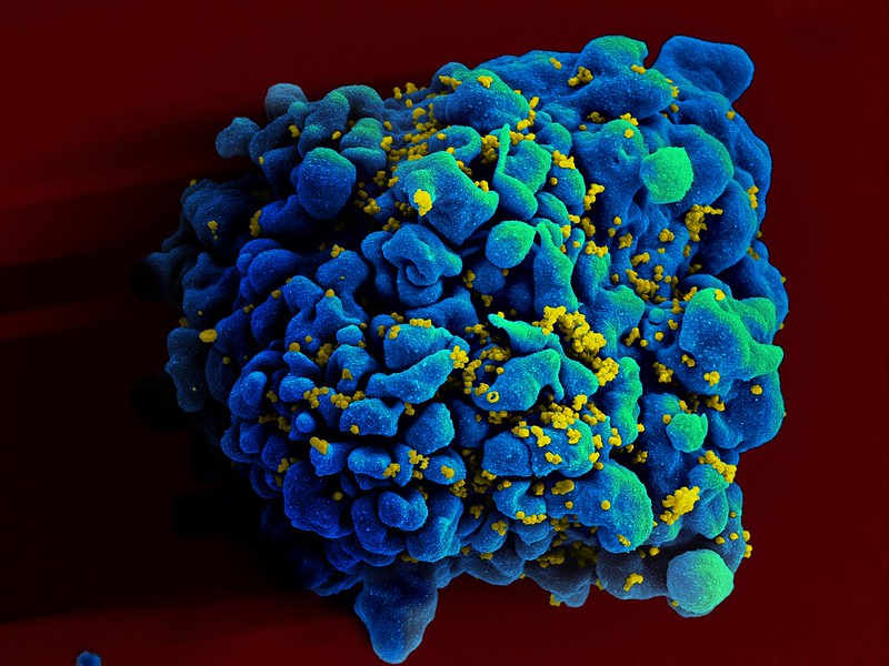
Colourised scanning electron micrograph of HIV (yellow) infecting a human T9 cell (blue). Credit: NIH

An experimental drug originally developed to treat cancer may help clear HIV from infected cells in the brain, according to a new study published in the journal Brain. For the first time, researchers at Tulane University found that a cancer drug significantly reduced levels of SIV, the nonhuman primate equivalent of HIV, in the brain by targeting and depleting certain immune cells that harbour the virus.

This discovery marks a significant step toward eliminating HIV from hard-to-reach reservoirs where the virus evades otherwise effective treatment.

“This research is an important step in tackling brain-related issues caused by HIV, which still affect people even when they are on effective HIV medication,” said lead study author Woong-Ki Kim, PhD, associate director for research at Tulane National Primate Research Center. “By specifically targeting the infected cells in the brain, we may be able to clear the virus from these hidden areas, which has been a major challenge in HIV treatment.”

Antiretroviral therapy (ART) is an essential component of successful HIV treatment, but the virus persists in “viral reservoirs” in the brain, liver, and lymph nodes, where it remains out of reach of ART.

The brain has been a particularly challenging area for treatment due to the blood-brain barrier preventing treatments from reaching the virus. In addition, macrophages are extremely long-lived, making them difficult to eradicate once they become infected.

Infection of macrophages is thought to contribute to neurocognitive dysfunction, experienced by nearly half of those living with HIV. Eradicating the virus from the brain is critical for comprehensive HIV treatment and could significantly improve the quality of life for those with HIV-related neurocognitive problems.

Researchers focused on macrophages, a type of white blood cell that harbours HIV in the brain. By using a small molecule inhibitor to block a receptor that increases in HIV-infected macrophages, the team successfully reduced the viral load in the brain. This approach essentially cleared the virus from brain tissue, providing a potential new treatment avenue for HIV.

The small molecule inhibitor used, BLZ945, has previously been studied for therapeutic use in amyotrophic lateral sclerosis (ALS) and brain cancer, but never before in the context of clearing HIV from the brain.

The study, which took place at the Tulane National Primate Research Center, utilised three groups to model human HIV infection and treatment: an untreated control group, and two groups treated with either a low or high dose of the small molecule inhibitor for 30 days. The high-dose treatment lead to a notable reduction in cells expressing HIV receptor sites, as well as a 95-99% decrease in viral DNA loads in the brain .

In addition to reducing viral loads, the treatment did not significantly impact microglia, the brain’s resident immune cells, which are essential for maintaining a healthy neuroimmune environment. It also did not show signs of liver toxicity at the doses tested.

The next step for the research team is to test this therapy in conjunction with ART to assess its efficacy in a combined treatment approach. This could pave the way for more comprehensive strategies to eradicate HIV from the body entirely.

Source: Tulane University

Brain Fluid Dynamics is Key to the Mysteries of Migraine

Credit: University of Rochester Medical Center

New research describes how a spreading wave of disruption and the flow of fluid in the brain triggers headaches, detailing the connection between the neurological symptoms associated with aura and the migraine that follows. The study, which appears in Science, also identifies new proteins that could be responsible for headaches and may serve as foundation for new migraine drugs.

“In this study, we describe the interaction between the central and peripheral nervous system brought about by increased concentrations of proteins released in the brain during an episode of spreading depolarization, a phenomenon responsible for the aura associated with migraines,” said lead author Maiken Nedergaard, MD, DMSc, co-director of the University of Rochester Center for Translational Neuromedicine. “These findings provide us with a host of new targets to suppress sensory nerve activation to prevent and treat migraines and strengthen existing therapies.”

It is estimated that one out of 10 people experience migraines and in about a quarter of these cases the headache is preceded by an aura, a sensory disturbance that can includes light flashes, blind spots, double vision, and tingling sensations or limb numbness. These symptoms typically appear five to 60 minutes prior to the headache.

The cause of the aura is a phenomenon called cortical spreading depression, a temporary depolarization of neurons and other cells caused by diffusion of glutamate and potassium that radiates like a wave across the brain, reducing oxygen levels and impairing blood flow. Most frequently, the depolarization event is located in the visual processing centre of the brain cortex, hence the visual symptoms that first herald a coming headache.

While migraines auras arise in the brain, the organ itself cannot sense pain. These signals must instead be transmitted from the central nervous system to the peripheral nervous system. The process of communication between the brain and peripheral sensory nerves in migraines has largely remained a mystery.

Fluid dynamics models shed light on migraine pain origins

Nedergaard and her colleagues at the University of Rochester and the University of Copenhagen are pioneers in understanding the flow of fluids in the brain. In 2012, her lab was the first to describe the glymphatic system, which uses cerebrospinal fluid (CSF) to wash away toxic proteins in the brain. In partnership with experts in fluid dynamics, the team has built detailed models of how the CSF moves in the brain and its role in transporting proteins, neurotransmitters, and other chemicals.

The most widely accepted theory is that nerve endings resting on the outer surface of the membranes that enclose the brain are responsible for the headaches that follow an aura. The new study, which was conducted in mice, describes a different route and identifies proteins, many of which are potential new drug targets, that may be responsible for activating the nerves and causing pain.

As the depolarization wave spreads, neurons release a host of inflammatory and other proteins into CSF. In a series of experiments in mice, the researchers showed how CSF transports these proteins to the trigeminal ganglion, a large bundle of nerves that rests at the base of the skull and supplies sensory information to the head and face.

It was assumed that the trigeminal ganglion, like the rest of the peripheral nervous system, rested outside the blood-brain-barrier, which tightly controls what molecules enter and leave the brain. However, the researchers identified a previously unknown gap in the barrier that allowed CSF to flow directly into the trigeminal ganglion, exposing sensory nerves to the cocktail of proteins released by the brain.

Migraine-associated proteins double during brain wave activity

Analysing the molecules, the researchers identified twelve proteins called ligands that bind with receptors on sensory nerves found in the trigeminal ganglion, potentially causing these cells to activate. The concentrations of several of these proteins found in CSF more than doubled following a cortical spreading depression. One of the proteins, calcitonin gene-related peptide (CGRP), is already the target of a new class of drugs to treat and prevent migraines called CGRP inhibitors. Other identified proteins are known to play a role in other pain conditions, such as neuropathic pain, and are likely important in migraine headaches as well.

“We have identified a new signaling pathway and several molecules that activate sensory nerves in the peripheral nervous system. Among the identified molecules are those already associated with migraines, but we didn’t know exactly how and where the migraine inducing action occurred,” said Martin Kaag Rasmussen, PhD, a postdoctoral fellow at the University of Copenhagen and first author of the study. “Defining the role of these newly identified ligand-receptor pairs may enable the discovery of new pharmacological targets, which could benefit the large portion of patients not responding to available therapies.”

The researchers also observed that the transport of proteins released in one side of the brain reaches mostly the nerves in the trigeminal ganglion on the same side, potentially explaining why pain occurs on one side of the head during most migraines.

Source: University of Rochester Medical Center

Nutrient’s Pathway into the Brain could be Used to Treat Neurological Disorders

Source: CC0

A University of Queensland researcher has found molecular doorways that could be used to help deliver drugs into the brain to treat neurological disorders. Dr Rosemary Cater from UQ’s Institute for Molecular Bioscience led a team which discovered that an essential nutrient called choline is transported into the brain by a protein called FLVCR2.

“Choline is a vitamin-like nutrient that is essential for many important functions in the body, particularly for brain development,” Dr Cater said.

“We need to consume 400-500mg of choline per day to support cell regeneration, gene expression regulation, and for sending signals between neurons.”

Dr Cater said that until now, little was known about how dietary choline travels past the layer of specialised cells that separates the blood from the brain.

“This blood-brain barrier prevents molecules in the blood that are toxic to the brain from entering,” she explained. “The brain still needs to absorb nutrients from the blood, so the barrier contains specialised cellular machines – called transporters – that allow specific nutrients such as glucose, omega-3 fatty acids and choline to enter. While this barrier is an important line of defence, it presents a challenge for designing drugs to treat neurological disorders.”

Dr Cater was able to show that choline sits in a cavity of FLVCR2 as it travels across the blood-brain barrier and is kept in place by a cage of protein residues.

“We used high-powered cryo-electron microscopes to see exactly how choline binds to FLVCR2,” she said. “This is critical information for understanding how to design drugs that mimic choline so that they can be transported by FLVCR2 to reach their site of action within the brain. These findings will inform the future design of drugs for diseases such as Alzheimer’s and stroke.”

The research also highlights the importance of eating choline-rich foods – such as eggs, vegetables, meat, nuts and beans.

The research is published in Nature and funded by the National Institutes of Health.

Source: University of Queensland

Crafting a ‘Key’ to Cross the Blood-brain Boundary

Source: Pixabay CC0

Researchers led by Michael Mitchell of the University of Pennsylvania are close to gaining access through the blood-brain barrier, a long-standing boundary in biology, by granting molecules a special ‘key’ to gain access.

Their findings, published in the journal Nano Letters, present a model that uses lipid nanoparticles (LNPs) to deliver mRNA, offering new hope for treating conditions like Alzheimer’s disease and seizures.

“Our model performed better at crossing the blood-brain barrier than others and helped us identify organ-specific particles that we later validated in future models,” says Mitchell, associate professor of bioengineering at Penn’s School of Engineering and Applied Science, and senior author on the study.

“It’s an exciting proof of concept that will no doubt inform novel approaches to treating conditions like traumatic brain injury, stroke, and Alzheimer’s.”

Search for the key

To develop the model, Emily Han, a PhD candidate and NSF Graduate Research Fellow in the Mitchell Lab and first author of the paper, explains that it started with a search for the right in vitro screening platform, saying, “I was combing through the literature, most of the platforms I found were limited to a regular 96-well plate, a two-dimensional array that can’t represent both the upper and lower parts of the blood-brain barrier, which correspond to the blood and brain, respectively.”

Han then explored high-throughput transwell systems with both compartments but found they didn’t account for mRNA transfection of the cells, revealing a gap in the development process.

This led her to create a platform capable of measuring mRNA transport from the blood compartment to the brain, as well as transfection of various brain cell types including endothelial cells and neurons.

“I spent months figuring out the optimal conditions for this new in vitro system, including which cell growth conditions and fluorescent reporters to use,” Han explains.

“Once robust, we screened our library of LNPs and tested them on animal models. Seeing the brains express protein as a result of the mRNA we delivered was thrilling and confirmed we were on the right track.”

The team’s platform is poised to significantly advance treatments for neurological disorders.

It’s currently tailored for testing a range of LNPs with brain-targeted peptides, antibodies, and various lipid compositions.

However, it could also deliver other therapeutic agents like siRNA, DNA, proteins, or small molecule drugs directly to the brain after intravenous administration.

What’s more, this approach isn’t limited to the blood-brain barrier as it shows promise for exploring treatments for pregnancy-related diseases by targeting the blood-placental barrier, and for retinal diseases focusing on the blood-retinal barrier.

Next Steps

The team is eager to use this platform to screen new designs and test their effectiveness in different animal models.

They are particularly interested in working with collaborators with advanced animal models of neurological disorders.

“We’re collaborating with researchers at Penn to establish brain disease models,” Han says.

“We’re examining how these LNPs impact mice with various brain conditions, ranging from glioblastoma to traumatic brain injuries. We hope to make inroads towards repairing the blood-brain barrier or target neurons damaged post-injury.”

Source: University of Pennsylvania

Microplastics Rapidly Bioaccumulate Everywhere in the Body

Photo by FLY:D on Unsplash

The prevalence of microplastics in the environment is well known, along with their harm to marine organisms, but few studies have examined the potential health impacts on mammals. Now, a new study published in the International Journal of Molecular Sciences has found that in mice, the infiltration of microplastics was as widespread in the body as it is in the environment, leading to behavioural changes, especially in older test subjects.

Study leader University of Rhode Island Professor Jaime Ross and her team focused on neurobehavioural effects and inflammatory response to exposure to microplastics, as well as the accumulation of microplastics in tissues, including the brain.

“Current research suggests that these microplastics are transported throughout the environment and can accumulate in human tissues; however, research on the health effects of microplastics, especially in mammals, is still very limited,” said Ross, an assistant professor of biomedical and pharmaceutical sciences at the Ryan Institute for Neuroscience and the College of Pharmacy. “This has led our group to explore the biological and cognitive consequences of exposure to microplastics.”

Behavioural changes detected

Ross’ team exposed young and old mice to varying levels of microplastics in drinking water over the course of three weeks. They found that microplastic exposure induces both behavioural changes and alterations in immune markers in liver and brain tissues. The study mice began to exhibit behaviours akin to dementia in humans. The results were even more profound in older animals.

“To us, this was striking. These were not high doses of microplastics, but in only a short period of time, we saw these changes,” Ross said. “Nobody really understands the life cycle of these microplastics in the body, so part of what we want to address is the question of what happens as you get older. Are you more susceptible to systemic inflammation from these microplastics as you age? Can your body get rid of them as easily? Do your cells respond differently to these toxins?”

To understand the physiological systems that may be contributing to these changes in behaviour, Ross’ team investigated how widespread the microplastic exposure was in the body, dissecting several major tissues including the brain, liver, kidney, gastrointestinal tract, heart, spleen and lungs. The researchers found that the particles had begun to bioaccumulate in every organ, including the brain, as well as in bodily waste.

“Given that in this study the microplastics were delivered orally via drinking water, detection in tissues such as the gastrointestinal tract, which is a major part of the digestive system, or in the liver and kidneys was always probable,” Ross said. “The detection of microplastics in tissues such as the heart and lungs, however, suggests that the microplastics are going beyond the digestive system and likely undergoing systemic circulation. The brain blood barrier is supposed to be very difficult to permeate. It is a protective mechanism against viruses and bacteria, yet these particles were able to get in there. It was actually deep in the brain tissue.”

Possible mechanism

That brain infiltration also may cause a decrease in glial fibrillary acidic protein (called “GFAP”), a protein that supports many cell processes in the brain, results have shown. “A decrease in GFAP has been associated with early stages of some neurodegenerative diseases, including mouse models of Alzheimer’s disease, as well as depression,” Ross said. “We were very surprised to see that the microplastics could induce altered GFAP signalling.”

She intends to investigate this finding further in future work. “We want to understand how plastics may change the ability for the brain to maintain its homeostasis or how exposure may lead to neurological disorders and diseases, such as Alzheimer’s disease,” she said.

Source: University of Rhode Island

Restoring the Integrity of the Blood–Brain Barrier

Source: CC0

A new paper published in Nature Communications describes a treatment that restores intercellular signalling and which could be instrumental in restoring the barrier’s normal function. Key to the process is ‘frizzled’, a key protein receptor implicated in blood-brain abnormalities.

When the blood-brain barrier isn’t working properly, a variety of conditions can crop up. Barrier-invading cancer cells can develop into tumours, and multiple sclerosis can occur when too many white blood cells slip pass the barrier, leading to an autoimmune attack on the protective layer of brain nerves, hindering their communication with the rest of the body.

“A leaky blood-brain barrier is a common pathway for a lot of brain diseases, so to be able to seal off the barrier has been a long sought-after goal in medicine,” said senior author Calvin Kuo, MD, PhD, professor of hematology.

Methods of repairing the blood-brain barrier remain understudied, according to Kuo. But the recent paper he and colleagues led describes a possible treatment.

“We have evaluated a new therapeutic class of molecules that can be used to treat a leaky blood-brain barrier; previously, there were no treatments directed at the blood-brain barrier specifically,” Kuo said.

The researchers started their quest by looking at WNT signalling, a communication pathway used by cells to promote tissue regeneration and wound healing. WNT signalling helps maintain the blood-brain barrier by promoting cell-to-cell communication that lines brain blood vessels.

“There’s a lot of historical data that indicated that the WNT signalling pathway would be important for maintaining the blood-brain barrier,” Kuo said. “The opportunity arose to test a novel WNT signalling pathway that would turn on signalling in the blood-brain barrier by binding very selectively to a receptor called frizzled.”

Scientists have been focusing on ‘frizzled’, a protein receptor that initiates the WNT pathway, for blood-brain barrier therapies since mouse mutations in the frizzled gene cause blood-brain barrier abnormalities.

How it’s made

Many different molecules bind to frizzled protein receptors, so to narrow their search for a potential therapeutic molecule, the researchers selected only those that specifically target cells that line the brain’s blood vessels.

Chris Garcia, PhD, a professor of molecular and cellular physiology as well as the Younger Family Professor, developed prototype therapeutic WNT pathway molecules in the lab, including a molecule that activates the frizzled receptor FZD4. Building off of the work of Garcia and Kuo, collaborators at a research company created L6-F4-2, a FZD4 binding molecule that activates WNT signalling 100 times more efficiently than other FZD4 binders.

When the team, including Jie Ding, a research scientist and the lead author of the paper, activated WNT signaling at a higher rate, they saw an increase in blood-brain barrier strength.

Keeping the barrier up

The researchers wanted to study what happens when the natural molecular key for frizzled is missing, and whether it can be replaced successfully with L6-F4-2. So they turned to Norrie disease, a genetic abnormality that results in a leaky blood-retinal barrier.

The blood-retinal barrier performs the same function for the eye as the blood-brain barrier does for the brain. In Norrie disease, the development of blood vessels of the retina is hindered, resulting in leaky blood vessel connections, improper development and blindness.

Norrie disease results from mutations in the NDP gene, which provides instructions for making a protein called Norrin, which is the key that fits the lock of the FZDreceptor and turns it on. In the study’s mice, the gene is inactivated, and the key is missing causing a leaky barrier and blindness. The scientists replaced the missing Norrin protein with L6-F4-2, which they call a surrogate.

When L6-F4-2 replaced the missing Norrin protein, the blood-retinal layer was restored in the mice. Researchers knew this because they imaged the blood vessels and found them to be denser, and less leaky, than before treatment. Scientists also showed that, for the blood-brain barrier surrounding the mice cerebellum L6-F4-2 replaced Norrin and activated WNT signalling.

Next, the researchers wanted to study a more common human condition — ischemic stroke (in which blood vessels and the blood-brain barrier are damaged, and fluid, blood and inflammatory proteins involved in cellular communication can leak into the brain. They found that L6-F4-2 reduced the severity of stroke and improved survival of mice compared with mice that had untreated strokes. Importantly, L6-F4-2 reversed the leakiness of brain blood vessels after stroke. Mice treated with L6-F4-2 had increased stroke survival, compared to those that were not treated.

The finding shows that, in mice, the blood-brain barrier could be restored by drugs that activate FZD receptors and the WNT signalling pathway.

Because a variety of disorders have their origin in blood-brain barrier dysfunction, Kuo is excited about the treatment potential for a variety of other neurological diseases, such as Alzheimer’s, multiple sclerosis and brain tumours.

“We hope this will be a first step toward developing a new generation of drugs that can repair the blood-brain barrier, using a very different strategy and molecular target than current medications,” Kuo said.

Source: Stanford Medicine

Antibiotic Regimen may be Ineffective in TB Meningitis

Tuberculosis bacteria
Tuberculosis bacteria. Credit: CDC

Research in animal models published in Nature Communications shows that an approved antibiotic regimen for multidrug-resistant (MDR) tuberculosis (TB) may not work for TB meningitis. Limited human studies also provide evidence that a new combination of drugs is needed to develop effective treatments for TB meningitis due to MDR strains.

In the study from Johns Hopkins Children’s Center, the investigators showed that the Food and Drug Administration (FDA)-approved regimen of three antibiotics – bedaquiline, pretomanid and linezolid (BPaL) – used for treating TB of the lungs due to MDR strains, is not effective in treating TB meningitis because bedaquiline and linezolid struggle to cross the blood-brain barrier.

Tuberculosis, caused by the bacteria Mycobacterium tuberculosis, is a global public health threat. About 1%–2% of TB cases progress into TB meningitis, the worst form of TB, which leads to an infection in the brain that causes increased fluid and inflammation.

“Most treatments for TB meningitis are based on studies of treatments for pulmonary TB, so we don’t have good treatment options for TB meningitis,” explains Sanjay Jain, M.D., senior author of the study and director of the Johns Hopkins Medicine Center for Infection and Inflammation Imaging Research.

In 2019, the FDA approved the BPaL regimen to treat MDR strains of TB, specifically those that lead to pulmonary TB. However, there are limited data on how well these antibiotics cross the blood-brain barrier.

In an effort to learn more, the research team synthesised a chemically identical and imageable version of the antibiotic pretomanid. They conducted experiments in mouse and rabbit models of TB meningitis using positron emission tomography (PET) imaging to noninvasively measure pretomanid penetration into the central nervous system as well as using direct drug measurements in mouse brains. In both models, researchers say PET imaging demonstrated excellent penetration of pretomanid into the brain or the central nervous system. However, the pretomanid levels in the cerebrospinal fluid (CSF) that bathes the brain were many times lower than in the brains of mice.

“When we have measured drug concentrations in the spinal fluid, we have found that many times they have no relation to what’s happening in the brain,” says Elizabeth Tucker, MD, a study first author and an assistant professor of anaesthesiology and critical care medicine. “This finding will change how we interpret data from clinical trials and, ultimately, treat infections in the brain.”

Next, researchers measured the efficacy of the BPaL regimen compared with the standard TB treatment for drug-susceptible strains, a combination of the antibiotics rifampin, isoniazid and pyrazinamide. Results showed that the antibacterial effect in the brain using the BPaL regimen in the mouse model was about 50 times lower than the standard TB regimen after six weeks of treatment, likely due to restricted penetration of bedaquiline and linezolid into the brain. The bottom line, says Jain, is that the “regimen that we think works really well for MDR-TB in the lung does not work in the brain.”

In another experiment involving healthy participants, three male and three female aged 20–53 years, first-in-human PET imaging was used to show pretomanid distribution to major organs, according to researchers.

Similar to the work with mice, this study revealed high penetration of pretomanid into the brain or central nervous system with CSF levels lower than those seen in the brain. “Our findings suggest pretomanid-based regimens, in combination with other antibiotics active against MDR strains with high brain penetration, should be tested for treating MDR-TB meningitis,” says study author Xueyi Chen, MD, a paediatric infectious diseases fellow, who is now studying combinations of such therapies.

Limitations included the small quantities of the imageable version of pretomanid per subject (micrograms) used. However, current evidence suggests that studies with small quantities of a drug are a reliable predictor of the drug biodistribution.

Source: Johns Hopkins Medicine

Early Sensing of Malaria in the Brain Leads to Cerebral Malaria

Colourised scanning electron micrograph of red blood cell infected with malaria parasites, which are colourised in blue. The infected cell is in the centre of the image area. To the left are uninfected cells with a smooth red surface. Credit: National Institute of Allergy and Infectious Diseases, NIH

A recent study published in PNAS revealed that endothelial cells in the brain are able to sense the infection by the malaria parasite at an early phase, triggering the inflammation underlying cerebral malaria. This discovery identified new targets for adjuvant therapies that could restrain brain damage in initial phases of the disease and avoid neurological sequelae.

Cerebral malaria is a severe complication of infection with Plasmodium falciparum, the most lethal of the parasites causing malaria. This form of the disease manifests through impaired consciousness and coma and affects mainly children under 5, being one of the main causes of death in this age group in countries of Sub-Saharan Africa. Survivors are frequently affected by debilitating neurological sequelae, such as motor deficits, paralysis, and speech, hearing, and visual impairment.

To prevent certain molecules and cells from reaching the brain, which would disturb its normal functioning, endothelial cells forming a tight barrier between the blood and this organ. Cerebral malaria results from an unrestrained inflammatory response to infection which leads to significant alterations in this barrier and, consequently, neurological complications.

Over the last years, specialists in this field have turned their attention to a molecule, named interferon-β, which seems to be associated with this pathological process. So called for interfering with viral replication, this highly inflammatory molecule has two sides: it can either be protecting or cause tissue destruction. It is known, for example, that despite its antiviral role in COVID-19, at a given concentration and phase of infection, it can cause lung damage. A similar dynamic is thought to occur in cerebral malaria. However, we still don’t know what leads to the secretion of interferon-β, nor the main cells involved.

The present study revealed that endothelial cells in the brain play a crucial role, being able to sense the infection by the malaria parasite at an early phase. These detect the infection through an internal sensor which triggers a cascade of events, starting with the production of interferon-β. Next, they release a signalling molecule that attracts cells of the immune system to the brain, initiating the inflammatory process.

To reach these conclusions, researchers used mice that mimic several symptoms described in human malaria and a genetic manipulation system that allowed them to delete this sensor in several types of cells. When they deleted this sensor in brain endothelial cells, the animals’ symptoms were not as severe with lower mortality. They then realised these brain cells contributed greatly to the pathology of cerebral malaria. “We thought brain endothelial cells acted in a later phase, but we ended up realising that they are participants from the very beginning”, explained Teresa Pais, a post-doctoral researcher at the IGC and first author of the study. “Normally we associate this initial phase of the response to infection with cells of the immune system. These are already known to respond, but cells of the brain, and maybe other organs, also have this ability to sense the infection because they have the same sensors.”

But what really surprised the researchers was the factor activating the sensor and triggering this cell response. This factor is nothing more nothing less than a by-product of the activity of the parasite. Once in the blood, the parasite invades the host’s red blood cells, where it multiplies. Here, it digests haemoglobin, a protein that transports oxygen, to get nutrients. During this process, a molecule named haeme is formed and it can be transported in tiny particles in the blood that are internalised by endothelial cells. When this happens, haeme acts as an alarm for the immune system. “We weren’t expecting that haeme could enter cells this way and activate this response involving interferon-β in endothelial cells”, the researcher confessed.

This six-year project allowed the researchers to identify a molecular mechanism that is critical for the destruction of brain tissue during infection with the malaria parasite and, with that, new therapeutic targets. “The next step will be to try to inhibit the activity of this sensor inside the endothelial cells and understand if we can act on the host’s response and stop brain pathology in an initial phase,” explained principal investigator Carlos Penha Gonçalves. “If we could use inhibitors of the sensor in parallel with antiparasitic drugs maybe we could stop the loss of neuronal function and avoid sequelae which are a major problem for children surviving cerebral malaria.”

Source: Instituto Gulbenkian de Ciência (IGC)