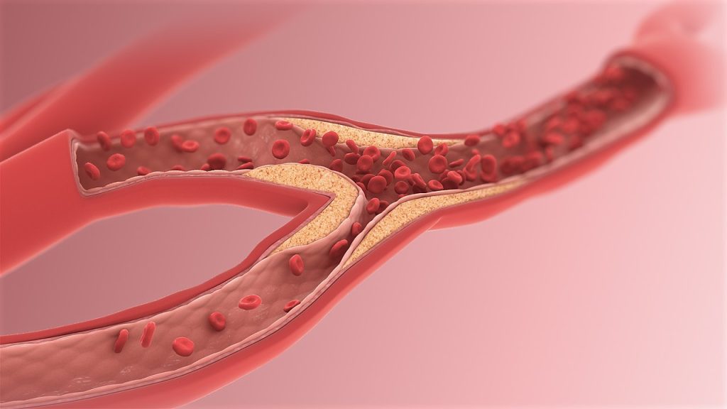
Tag: atherosclerosis

Inflammation may Link Depression and Cardiomyopathy

Photo by Alex Green on Pexels

Coronary artery disease and major depression may be genetically linked via inflammatory pathways to an increased risk for cardiomyopathy, a degenerative heart muscle disease, researchers at Vanderbilt University Medical Center and Massachusetts General Hospital have found.

Their report, published in Nature Mental Health, suggests that drugs prescribed for coronary artery disease and depression, when used in combination, potentially may reduce inflammation and prevent the development of cardiomyopathy.

“This work suggests that chronic low-level inflammation may be a significant contributor to both depression and cardiovascular disease,” said the paper’s corresponding author, Lea Davis, PhD, associate professor of Medicine in the Division of Genetic Medicine and Vanderbilt Genetics Institute.

The connection between depression and other serious health conditions is well known. As many as 44% of patients with coronary artery disease (CAD), the most common form of cardiovascular disease, also have a diagnosis of major depression. Yet the biological relationship between the two conditions remains poorly understood.

A possible connection is inflammation. Changes in the levels of inflammatory markers have been observed in both conditions, suggesting that there may be a common biological pathway linking neuroinflammation in depression with atherosclerotic inflammation in CAD.

In the current study, the researchers used a technique called transcriptome-wide association scans to map single nucleotide polymorphisms (genetic variations) involved in regulating the expression of genes associated with both CAD and depression.

The technique identified 185 genes that were significantly associated with both depression and CAD, and which were “enriched” for biological roles in inflammation and cardiomyopathy.

This suggests that predisposition to both depression and CAD, which the researchers called (major) depressive CAD, or (m)dCAD, may further predispose individuals to cardiomyopathy.

However, when the researchers scanned large electronic health record databases at VUMC, Mass General, and the National Institutes of Health’s All of Us Research Program, they found the actual incidence of cardiomyopathy in patients with the enriched genes for (m)dCAD was lower than in patients with CAD alone.

One possible explanation is that medications prescribed for CAD and depression, such as statins and antidepressants, may prevent development of cardiomyopathy by reducing inflammation, the researchers concluded.

“More research is needed to investigate optimal treatment mechanisms,” Davis added, “but at a minimum this work suggests that patient heart and brain health should be considered together when developing management plans to treat depression or cardiovascular disease.”

Source: Vanderbilt University Medical Center

Hot Days may Drive Inflammation and Accelerate Cardiovascular Disease

Photo by Fandy Much: https://www.pexels.com/photo/toshiba-outdoor-air-conditioner-unit-on-yellow-wall-14086132/

Short-term exposure to higher heat may increase inflammation and interfere with normal immune system functions in the body, which may, in turn, increase susceptibility to infections and accelerate the progression of cardiovascular disease, according to preliminary research be presented at the American Heart Association’s Epidemiology and Prevention – Lifestyle and Cardiometabolic Scientific Sessions 2024, March 18-21.

An inflammatory response that is longstanding (lasting weeks to months) or that occurs in healthy tissues is damaging and plays a key role in the build-up of plaque in the arteries. This may lead to atherosclerosis. Heat waves are known to promote inflammation, however, studies examining air temperature and biomarkers of inflammation have had mixed results.

“Most research only considers temperature as the exposure of interest, which may not be adequate to capture a person’s response to heat,” said lead study author Daniel W. Riggs, PhD, an assistant professor of medicine at the University of Louisville. “In our study, we used alternative measurements of heat in relation to multiple markers of inflammation and immune response in the body to investigate the short-term effects of heat exposure and produce a more complete picture of its health impact.”

Participants visited study sites in Louisville, during the summer months for a blood test, and researchers analysed the blood for multiple markers of immune system function. The researchers then examined associations between the markers of immune system function and heat levels, including temperature, net effective temperature (which factors in relative humidity, air temperature and windspeed) and the Universal Thermal Climate Index (UTCI) on that day. UTCI is a thermo-physiological model developed by the International Society of Biometeorology Commission that factors in temperature, humidity, wind speed and ultraviolet radiation levels, which was used to evaluate participant’s physical comfort.

The analysis found:

  • For every 5°F (2.8°C) increase in UTCI (in this study, the equivalent of going from a day with no thermal stress to a day with moderate thermal stress, Riggs said), there was an increase in blood levels of key inflammatory markers: monocytes (4.2%), eosinophils (9.5%), natural killer T-cells (9.9%) and tumour necrosis factor-alpha (7.0%). These immune molecules indicate activation of the body’s innate immune system, which spurs a fast and non-specific inflammatory response throughout the body to protect against pathogens and injury.
  • A decrease in B-cells (-6.8%), indicating the body’s adaptive immune system that remembers specific viruses and germs and creates antibodies to fight them, was lowered.
  • A lesser impact on the immune system was found when heat was measured by average 24-hour temperature or by net effective temperature, which incorporates humidity and wind but not sunshine.

“Our study participants only had minor exposure to high temperatures on the day of their blood test, however, even minor exposure may contribute to changes in immune markers,” Riggs said. “With rising global temperatures, the association between heat exposure and a temporarily weakened response from the immune system is a concern because temperature and humidity are known to be important environmental drivers of infectious, airborne disease transmission. Thus, during the hottest days of summer people may be at higher risk of heat exposure, they may also be more vulnerable to disease or inflammation.”

Adults over 60 years and adults with existing cardiovascular disease are particularly at risk for heat-related cardiovascular events and deaths, Riggs explained.

“It’s important for physicians to communicate with patients about the risk of adverse health effects from heat exposure. For example, cardiologists could conduct customised consultations and assessments to increase patient awareness about their susceptibility to the effects of high temperatures. Also, changes to treatment regimens may be important to consider to address other risks. For example, some medications could make people more susceptible to heat-related illness or some may not be as effective when the body is exposed to high temperatures,” Riggs said.

Source: American Heart Association

A Better View of Atherosclerotic Plaques with New Imaging Technique

Source: Wikimedia CC0

Researchers have developed a new catheter-based device that combines two powerful optical techniques to image atherosclerotic plaques that can build up inside the heart’s coronary arteries. By providing new details about plaque, the device could help clinicians and researchers improve treatments for preventing heart attacks and strokes.

“Atherosclerosis, leading to heart attacks and strokes, is the number one cause of death in Western societies – exceeding all combined cancer types – and, therefore, a major public health issue,” said research team member leader Laura Marcu from University of California, Davis. “Better clinical management made possible by advanced intravascular imaging tools will benefit patients by providing more accurate information to help cardiologists tailor treatment or by supporting the development of new therapies.”

In the Optica Publishing Group journal Biomedical Optics Express, researchers describe their new flexible device, which combines fluorescence lifetime imaging (FLIM) and polarisation-sensitive optical coherence tomography (PSOCT) to capture rich information about the composition, morphology and microstructure of atherosclerotic plaques. The work was a collaborative project with Brett Bouma and Martin Villiger, experts in OCT from the Wellman Center for Photomedicine at Massachusetts General Hospital.

“With further testing and development, our device could be used for longitudinal studies where intravascular imaging is obtained from the same patients at different timepoints, providing a picture of plaque evolution or response to therapeutic interventions,” said Julien Bec, first author of the paper. “This will be very valuable to better understand disease evolution, evaluate the efficacy of new drugs and treatments and guide stenting procedures used to restore normal blood flow.”

Gaining an unprecedented view

Most of what scientists know about how atherosclerosis forms and develops over time comes from histopathology studies of postmortem coronary specimens. Although the development of imaging systems such as intravascular ultrasound and intravascular OCT has made it possible to study plaques in living patients, there is still a need for improved methods and tools to investigate and characterise atherosclerosis.

To address this need, the researchers embarked on a multi-year research project to develop and validate multispectral FLIM as an intravascular imaging modality. FLIM can provide insights into features such as the composition of the extracellular matrix, the presence of inflammation and the degree of calcification inside an artery. In earlier work, they combined FLIM with intravascular ultrasound, and in this new work they combined it with PSOCT. PSOCT provides high-resolution morphological information along with birefringence and depolarisation measurements. When used together, FLIM and PSOCT provide an unprecedented amount of information on plaque morphology, microstructure and biochemical composition.

“Birefringence provides information about the plaque collagen, a key structural protein that helps with lesion stabilization, and depolarisation is related to lipid content that contributes to plaque destabilization,” said Bec. “Holistically, this hybrid approach can provide the most detailed picture of plaque characteristics of all intravascular imaging modalities reported to date.”

Getting two imaging modalities into one device

The development of multimodal intravascular imaging systems compatible with coronary catheterisation is technologically challenging. It requires flexible catheters < 1mm diameter that can operate in vessels with sharp twists and turns. A high imaging speed of around 100 frames/second is also necessary to limit cardiac motion artefacts and ensure proper imaging inside an artery.

To integrate FLIM and PSOCT into a single device without compromising the performance of either imaging modality, the researchers used optical components previously developed by Marcu’s lab and other research groups. Key to achieving high PSOCT performance was a newly designed rotary collimator with high light throughput and a high return loss, ie the ratio of power reflected back toward the light source compared to the power incident on the device. The catheter system they developed has similar dimensions and flexibility as the intravascular imaging devices that are currently in clinical use.

After testing the new system with artificial tissue to demonstrate basic functionality on well characterized samples, the researchers also showed that it could be used to measure properties of a healthy coronary artery removed from a pig. Finally, in vivo testing in swine hearts demonstrated that the hybrid catheter system’s performance was sufficient to support work toward clinical validation. These tests all showed that the FLIM-PSOCT catheter system could simultaneously acquire co-registered FLIM data over four distinct spectral bands and PSOCT backscattered intensity, birefringence and depolarization information.

Next, the researchers plan to use the intravascular imaging system to image plaques in ex vivo human coronary arteries. By comparing the optical signals acquired using the system with plaque characteristics identified by expert pathologists, they can better understand which features can be identified by FLIM-PSOCT and use this to develop prediction models. They also plan to move forward with testing in support of clinical validation of the system in patients.

Source: Optica

Heart Disease Research Challenges ‘One Size Fits All’ Aspirin Guidelines

Analysis of results from international trials question whether current aspirin recommendations apply to all patients

Photo by cottonbro studio

Heart disease researchers have identified a group of patients in whom international guidelines on aspirin use for heart health may not apply. In a study published in the medical journal Circulation, the findings of a review of data from three clinical trials challenge current best practice for use of the drug for primary prevention of heart disease or stroke – otherwise known as atherosclerotic cardiovascular disease.

The research examined the results from clinical trials involving more than 47 000 patients in 10 countries, including the US, the UK and Australia, which were published in 2018.

The analysis focused on findings for a subgroup of 7222 patients who were already taking aspirin before the three trials commenced. Those studied were at increased risk for cardiovascular disease and were taking aspirin to prevent the first occurrence of a heart attack or stroke.

The data showed a higher risk of heart disease or stroke – 12.5% versus 10.4% – for patients who were on aspirin before the trials and who then stopped, compared to those who stayed on the drug.

Analyses also found no significant statistical difference in the risk for major bleeding between the two groups of patients.

The research was led by Professor J. William McEvoy, Established Professor of Preventive Cardiology at University of Galway and Consultant Cardiologist at Saolta University Health Care Group, in collaboration with researchers in University of Tasmania and Monash University, Melbourne.

Professor McEvoy said: “We challenged the notion that aspirin discontinuation is a one-size-fits-all approach.”

The research team noted results from observational studies which suggest a 28% higher risk of heart disease or stroke among adults who were prescribed aspirin to reduce the risk for a first heart attack or stroke, but who subsequently chose to stop taking the aspirin without being told to do so by their doctor.

Based in large part on three major clinical trials published in 2018, international guidelines no longer recommend the routine use of aspirin to prevent the first occurrence of heart attack or stroke.

Importantly, aspirin remains recommended for high-risk adults who have already had a heart disease or stroke event, to reduce the risk of a second event.

The move away from primary prevention aspirin in recent guidelines is motivated by the increased risk of major bleeding seen with this common medication in the three trials, albeit major bleeding is relatively uncommon on aspirin and was most obvious only among trial participants who were started on aspirin during the trial, rather than those who were previously taking aspirin safely.

These trials primarily tested the effect of starting aspirin among adults who have not previously been treated with the drug to reduce the risk of atherosclerotic cardiovascular disease. Less is known about what to do in the common scenario of adults who are already safely taking aspirin for primary prevention.

Professor McEvoy said: “Our findings of the benefit of aspirin in reducing heart disease or stroke without an excess risk of bleeding in some patients could be due to the fact that adults already taking aspirin without a prior bleeding problem are inherently lower risk for a future bleeding problem from the medication. Therefore, they seem to get more of the benefits of aspirin with less of the risks.

“These results are hypothesis-generating, but at present are the best available data. Until further evidence becomes available, it seems reasonable that persons already safely treated with low-dose aspirin for primary prevention may continue to do so, unless new risk factors for aspirin-related bleeding develop.”

Source: University of Galway

Excessive Protein Consumption Increases Atherosclerosis Risk

Cardiovascular pitfalls to increasing protein intake discovered

Image by Scientific Animations, CC4.0

University of Pittsburgh School of Medicine researchers discovered a molecular mechanism by which excessive dietary protein could increase atherosclerosis risk. The study, published in Nature Metabolism, combined small human trials with in vitro human and mouse cell experiments.

It showed that consuming over 22% of dietary calories from protein can lead to increased activation of immune cells that play a role in atherosclerotic plaque formation, driving the disease risk.

Furthermore, the scientists showed that one amino acid, leucine, seems to have a disproportionate role in driving the pathological pathways linked to atherosclerosis, or stiff, hardened arteries.

“Our study shows that dialling up your protein intake in pursuit of better metabolic health is not a panacea. You could be doing real damage to your arteries,” said senior and co-corresponding author Babak Razani, MD, PhD, professor of cardiology at Pitt.

“Our hope is that this research starts a conversation about ways of modifying diets in a precise manner that can influence body function at a molecular level and dampen disease risks.”

According to a survey of an average American diet over the last decade, Americans generally consume a lot of protein, mostly from animal sources.

Further, nearly a quarter of the population receives over 22% of all daily calories from protein alone.

That trend is likely driven by the popular idea that dietary protein is essential to healthy living, says Razani.

But his and other groups have shown that overreliance on protein may not be such a good thing for long-term health.

Following their 2020 research, in which Razani’s laboratory first showed that excess dietary protein increases atherosclerosis risk in mice, his next study in collaboration with Bettina Mittendorfer, PhD, a metabolism expert at the University of Missouri, Columbia, delved deeper into the potential mechanism and its relevance to the human body.

To arrive at the answer, Razani’s laboratory, led by first-authors Xiangyu Zhang, Ph.D., and Divya Kapoor, M.D., teamed up with Mittendorfer’s group to combine their expertise in cellular biology and metabolism and perform a series of experiments across various models, from cells to mice to humans.

“We have shown in our mechanistic studies that amino acids, which are really the building blocks of the protein, can trigger disease through specific signaling mechanisms and then also alter the metabolism of these cells,” Mittendorfer said.

“For instance, small immune cells in the vasculature called macrophages can trigger the development of atherosclerosis.”

Based on initial experiments in healthy human subjects to determine the timeline of immune cell activation following ingestion of protein-enriched meals, the researchers simulated similar conditions in mice and in human macrophages, immune cells that are shown to be particularly sensitive to amino acids derived from protein.

Their work showed that consuming more than 22% of daily dietary calories through protein can negatively affect macrophages that are responsible for clearing out cellular debris, leading to the accumulation of a “graveyard” of those cells inside the vessel walls and worsening of atherosclerotic plaques overtime.

Interestingly, the analysis of circulating amino acids showed that leucine, an amino acid enriched in animal-derived foods like beef, eggs and milk, is primarily responsible for abnormal macrophage activation and atherosclerosis risk, suggesting a potential avenue for further research on personalized diet modification, or “precision nutrition.”

Razani is careful to note that many questions remain to be answered, mainly: What happens when a person consumes between 15% of daily calories from protein as recommended by the USDA and 22% of daily calories from protein, and if there is a ‘sweet spot’ for maximising the benefits of protein (such as muscle gain) while avoiding kick-starting a molecular cascade of damaging events leading to cardiovascular disease.

The findings are particularly relevant in hospital settings, where nutritionists often recommend protein-rich foods for the sickest patients to preserve muscle mass and strength.

“Perhaps blindly increasing protein load is wrong,” Razani said.

“Instead, it’s important to look at the diet as a whole and suggest balanced meals that won’t inadvertently exacerbate cardiovascular conditions, especially in people at risk of heart disease and vessel disorders.”

Razani also notes that these findings suggest differences in leucine levels between diets enriched in plant and animal protein might explain the differences in their effect on cardiovascular and metabolic health.

“The potential for this type of mechanistic research to inform future dietary guidelines is quite exciting,” he said.

Source: University of Pittsburgh

Potential New Treatment to Reverse Inflammation and Atherosclerosis in Rheumatoid Arthritis

Image by Scientific Animations, CC4.0

Researchers from Queen Mary University of London have found that the molecule RvT4 enhances the body’s natural defences against atherosclerosis in patients with rheumatoid arthritis.

The mouse-based study, published in Nature Communications, shows that increasing levels of the RvT4 molecule in the body improves the ability of the body’s own defence mechanisms [macrophages] to reduce local inflammation and remove blockages in blood vessels.

This breakthrough in understanding the processes involved could lead to better treatments for people who have rheumatoid arthritis (RA), and who are at higher risk of developing cardiovascular disease.

Alongside the more widely-known symptoms of joint inflammation, people with the condition are also twice as likely as others to develop blood vessel disease.

One type of blood vessel disease seen in people with RA is atherosclerosis, which is caused by a build-up of ‘plaque’ along the artery walls, which can break free and cause heart attacks and strokes.

Understanding the reasons why RA patients are at increased risk of these cardiovascular problems is critical in developing better treatments for this group and others.

To gain a better understanding of the causes of blood vessel disease in patients with RA, researchers explored the role of a group of molecules called 13-series resolvins (RvTs). In experimental arthritis the levels of one of these molecules, RvT4, are markedly reduced, a phenomenon that associates with a higher degree of blood vessel disease.

This study was designed to explore why this might be the case.

The findings

The study found that treating arthritic mice with RvT4 reduced blood vessel inflammation by re-programming macrophages, which accumulate in the diseased vessels, to release stored lipids.

Researchers observed that these lipids were preventing the macrophage from carrying out their usual work of clearing dead cells and reducing localised inflammation in blood vessels.

Once freed of their lipid burden, the macrophages were able to move and work much more effectively to reduce the causes of atherosclerosis.

The observation that RvT4 restores protective macrophage biological activities is an exciting finding.

RA patients also often present with metabolic dysfunction and this is thought to exacerbate vascular disease.

The study found that administration of RvT4 to mice engineered to develop characteristics of metabolic dysfunction, advanced atherosclerosis, and arthritis led to an overall decrease in lipoprotein-associated cholesterol in plasma and an increase in the ratio of HDL-associated cholesterol to total cholesterol.

Jesmond Dalli, Professor in Molecular Pharmacology and Lipid Mediator Unit Director at the William Harvey Institute, Queen Mary University of London, said: “The study is important because it identifies for the first time the loss of RvT4 production as a potential new cause of blood vessel inflammation in the context of arthritis, offering a mechanistic explanation on the cause of this important disease in RA patients. It also showed that RvT4 restores the biological activities of lipid loaded macrophages by promoting lipid breakdown and efflux from the cells, an observation that can guide the development of new treatments to limit the incidence and/or severity of cardiovascular disease in patients with RA.”

Source: Queen Mary University of London

Night Owls have Nearly Double the Incidence of Atherosclerosis

Image by Scientific Animations, CC4.0

Atherosclerosis is almost twice as common in night owls compared to early birds, according to a study from the University of Gothenburg, Sweden. Circadian function appears to be particularly important during the early stages of cardiovascular disease.

Atherosclerosis involves fatty deposits gradually accumulating on the inside of the arteries, making it harder for blood to pass through. The disease is usually not noticed until it leads to blood clots causing angina, heart attack, or stroke.

Previous research has shown that people with late-night habits have an increased risk of cardiovascular disease, but this is the first study to show how circadian rhythms specifically affect calcification of the arteries.

Coronary artery calcification

The study, which has been published in the journal Sleep Medicine, involved 771 men and women aged between 50 and 64, all of whom are part of the larger population study SCAPIS.

The degree of artery calcification in the heart’s coronary arteries was examined using computer tomography.

Participants themselves indicated their so called chronotype on a five-point scale: extreme morning type, moderate morning type, intermediate type, moderate evening type, or extreme evening type.

Of the 771 participants, 144 identified as extreme morning types, and 128 as extreme evening types.

Among the group who were most alert in the morning, 22.2% had pronounced artery calcification — the lowest proportion of all five chronotypes.

The extreme evening type group had the highest prevalence of severe coronary artery calcification, at 40.6%.

The first author of the study is Mio Kobayashi Frisk, a doctoral student at Sahlgrenska Academy, University of Gothenburg:

“Our results indicate that extreme evening chronotype may be linked not only to poorer cardiovascular health in general, but also more specifically to calcification in the coronary arteries calcification and atherosclerosis,” Mio Kobayashi Frisk says.

Preventive treatment

The statistical analysis considered a range of other factors that can affect the risk of atherosclerosis, including blood pressure, blood lipids, weight, physical activity, stress level, sleep, and smoking.

The last author of the study is Ding Zou, a researcher at Sahlgrenska Academy, University of Gothenburg:

“As well as the previously known factors, the individual circadian rhythm also appears to be an important risk factor for atherosclerosis. We interpret our results as indicating that circadian rhythms are more significant early in the disease process. It should therefore particularly be considered in the preventive treatment of cardiovascular diseases,” says Ding Zou.

Self-reported chronotype

Those who had experienced a heart attack were excluded from the study, meaning that the study participants were healthier than the general population.

Another weakness identified by the researchers is that participants themselves provided their chronotype.

Each chronotype can be said to have an average time when half of the night’s sleep has passed.

In a previous study on the same population, though not necessarily the same individuals, this time occurred at 02:55 AM for the extreme morning type group and at 04:25 AM for the extreme evening type group.

With the remaining chronotype groups’ mid-sleep times were somewhere in between these extremes.

Source: University of Gothenburg

Global Trends for Atherosclerosis Still on the Rise

Source: Wikimedia CC0

Atherosclerosis, caused by arterial wall plaque build-up, is a leading cause of death globally, particularly in the developed countries. Although the mortality rates for this condition fell dramatically during the 20th century, the incidence is now ever increasing. Unfortunately, despite the widespread impact of atherosclerosis – and efforts to curb it – data on the global and national trends of the disease is quite limited.

In a recent effort to address this knowledge gap, a research team from China led by Professor Rongchong Huang decided to conduct a detailed statistical analysis on the impact of atherosclerosis at the global and national levels by using GBD data. The study was published in the Chinese Medical Journal.

“It is unknown how global changes in pertinent controllable variables in recent years have affected the burden of atherosclerosis,” remarks Prof Huang.

The researchers analysed publicly available data from the Global Burden of Disease Study 2019 related to the three main clinical presentations of atherosclerosis, ischaemic heart disease (IHD), ischaemic stroke (IS), and peripheral arterial disease (PAD). The data collected, which spanned the period from 1990 to 2019, included participants of all ages with similar epidemiologic characteristics and in relative proximity, from 21 countries.

Overall, the study had four main goals. The first was to determine the global trends in terms of prevalence, mortality, and disability of the three conditions. The second goal was to identify the years that had the biggest shift in the trends of these indicators. Finally, the third and fourth goals were to analyze global trends based on age, gender, and socioeconomic factors and report global and national patterns, respectively. 

According to the results, there was an overall increasing trend in the global incidence of the three clinical manifestations of atherosclerosis from 1990 to 2019. Notably, the main drivers for this rising incidence were adults aged 20–54. The researchers found this very concerning, given that atherosclerosis with such an early onset is usually caused by preventable factors, such as lack of exercise, dietary habits, and environmental pollution. However, the mortality rates and disability-adjusted life years for IHD and IS declined during this period across all age groups. This could indicate greater awareness regarding these conditions and their early symptoms, as well as advancements in clinical management. 

Nonetheless, the global rise in the incidence of atherosclerosis over the past three decades is a serious problem that warrants special attention towards its root causes. In this regard, Prof Huang explains: “This rise can be attributed to a variety of factors. Firstly, global aging trends have led to a higher prevalence of the disease, given that age is a significant risk factor for atherosclerosis. Secondly, modern lifestyle habits, including high-fat diets, lack of exercise, smoking, and excessive alcohol consumption, have increased atherosclerosis risk. Lastly, there has been a rise in chronic diseases such as diabetes and hypertension, which are significant risk factors for atherosclerosis.”

The researchers also pointed out that the burden posed by atherosclerosis is increasing significantly in low- or middle-income countries, summarising key social and economic development indicators. China, which has the world’s greatest number of deaths due to cardiovascular diseases, is a prime example of this issue.

Taken together, the study paints a grim picture of global cardiovascular health, which is very concerning. “Overall, the burden of atherosclerosis-related disease is still not significantly decreasing and is even trending upward, especially in low- and middle-income countries and in younger populations,” says Prof Huang, “There is an urgent need for more targeted treatment and management in younger populations and in low-middle and middle-income countries.”

Hopefully, the results of these analyses will prompt decision makers, scientists, and medical professionals alike to increase their efforts towards fighting against atherosclerosis and its devastating consequences.

Source: EurekAlert!

Study Explains a Link between COVID and Increased Cardiovascular Risk

Source: Wikimedia CC0

A study published in the journal Nature Cardiovascular Research shows that SARS-CoV-2 can directly infect the arteries of the heart and cause the fatty plaque inside arteries to become highly inflamed, increasing the risk of heart attack and stroke. The findings may help explain why certain people who get COVID have a greater chance of developing cardiovascular disease, or if they already have it, develop more heart-related complications.

In the National Institutes of Health (NIH)-funded study, researchers focused on older people with atherosclerotic plaque, who died from COVID. However, because the researchers found the virus infects and replicates in the arteries no matter the levels of plaque, the findings could have broader implications for anybody who gets COVID.

“Since the early days of the pandemic, we have known that people who had COVID have an increased risk for cardiovascular disease or stroke up to one year after infection,” said Michelle Olive, PhD, acting associate director of the Basic and Early Translational Research Program at the National Heart, Lung, and Blood Institute (NHLBI), part of NIH. “We believe we have uncovered one of the reasons why.”

Though previous studies have shown that SARS-CoV-2 can directly infect tissues such as the brain and lungs, less was known about its effect on the coronary arteries. Researchers knew that after the virus reaches the cells, the body’s immune system sends in macrophages to help clear the virus. In the arteries, macrophages also help remove cholesterol, and when they become overloaded with cholesterol, they morph into a specialised type of cell called foam cells.

The researchers thought that if SARS-CoV-2 could directly infect arterial cells, the macrophages that normally are turned loose might increase inflammation in the existing plaque, explained Chiara Giannarelli, MD, PhD, associate professor in the departments of medicine and pathology at New York University’s Grossman School of Medicine and senior author on the study. To test their theory, Giannarelli and her team took tissue from the coronary arteries and plaque of people who had died from COVID and confirmed the virus was in those tissues. Then they took arterial and plaque cells – including macrophages and foam cells – from healthy patients and infected them with SARS-CoV-2 in a lab dish. They found that the virus had also infected those cells and tissues.

Additionally, the researchers found that when they compared the infection rates of SARS-CoV-2, they showed that the virus infects macrophages at a higher rate than other arterial cells. Cholesterol-laden foam cells were the most susceptible to infection and unable to readily clear the virus. This suggested that foam cells might act as a reservoir of SARS-CoV-2 in the atherosclerotic plaque. Having more build-up of plaque, and thus a greater number of foam cells, could increase the severity or persistence of COVID.

The researchers then looked at the predicted inflammation in the plaque after infecting it with the virus. They observed the release of inflammatory cytokines, also known to promote the formation of even more plaque. The cytokines were released by infected macrophages and foam cells. The researchers said this may help explain why people who have underlying plaque buildup and then get COVID may have cardiovascular complications long after getting the infection.  

“This study is incredibly important as it adds to the larger body of work to better understand COVID,” said Olive. “This is just one more study that demonstrates how the virus both infects and causes inflammation in many cells and tissues throughout the body. Ultimately, this is information that will inform future research on both acute and Long COVID.”

Though the findings conclusively show that SARS-CoV-2 can infect and replicate in the macrophages of plaques and arterial cells, they are only relevant to the original strains of SARS-CoV-2 that circulated in New York City between May 2020 and May 2021. The study was conducted in a small cohort of older individuals, all of whom had atherosclerosis and other medical conditions; therefore, the results cannot be generalised to younger, healthy individuals.

Source: National Institutes of Health

Why Blood Vessel Linings go Wrong and Contribute to Plaque Growth

Source: Wikimedia CC0

University of Virginia Health researchers probing the causes of coronary artery disease have identified why blood vessel lining, which usually secure plaques to stop them drifting, sometimes instead contribute to plaque buildup. The discovery, published in Circulation: Genomic and Precision Medicine, provides new targets for scientists looking for better ways to treat and prevent the disease.

“Smooth muscle cells that make up the bulk of our blood vessels play important roles in coronary artery disease. They undergo pathological transformations as the disease develops inside our arteries,” said researcher Mete Civelek, of the University of Virginia School of Medicine’s Center for Public Health Genomics and the Department of Biomedical Engineering.

“Our results point to a previously underappreciated role for metabolic pathways during this pathological transformation,” he said.

Civelek and his team wanted to unravel a longstanding mystery about the behaviour of smooth muscle cells during plaque formation. These cells, which line blood vessels, protect the body during plaque formation by building stabilising caps over the plaque that prevent the lesions from breaking loose and causing strokes.

But sometimes smooth muscle cells begin to accelerate the plaque development and spur the progression of the disease, scientists believe.

Civelek’s new discovery helps explain why. Noah Perry, a doctoral student on Civelek’s team, analysed smooth muscle cells collected from 151 heart transplant donors and used a sophisticated approach to identify genes responsible for the smooth muscle cells’ behaviour.

After initially identifying 86 groups of genes, the researchers focused in on 18 groups that could explain the mysterious behaviour. Their analysis suggested that the smooth muscle cells’ shift might stem from problems with how the cells use nitrogen and glycogen.

The researchers identified a particular sugar, mannose, that may be contributing to the problems, potentially even triggering them. But determining that, the scientists say, will require more research.

“The metabolic shift in the cells as they transition to a disease state can point to points of intervention and therapy,” said Perry, of UVA’s Department of Biomedical Engineering, the lead author of the study.

By better understanding what triggers the smooth muscle cells to become harmful, Civelek says, doctors may be able to develop ways to prevent that from happening. That could open the door to new ways to treat and prevent coronary artery disease.

“Coronary artery disease is still the leading cause of death worldwide,” Civelek said. “Although cholesterol-lowering therapies and blood pressure control have been very effective tools to prevent deaths from heart attacks, we still need more targets to reduce the suffering of patients and their families from this devastating disease.”

Source: University of Virginia