How Astrocytes Know to Give the Brain an Energy Boost

A key mechanism by which astrocytes detect when an energy boost is needed for the brain has been elucidated by University College of London researchers using mouse-based and in vitro studies.
The findings, published in Nature, could inform new therapies to maintain brain health and longevity, the researchers say, since other studies have found that brain energy metabolism can become impaired late in life and contribute to cognitive decline and the development of neurodegenerative disease.
Lead author Professor Alexander Gourine (UCL Neuroscience, Physiology & Pharmacology) said: “When our brain is more active, such as when we’re performing a mentally taxing task, our brain needs an immediate boost of energy, but the exact mechanisms that ensure on-demand local supply of metabolic energy to active brain regions are not fully understood.”
First and co-corresponding author Dr Shefeeq Theparambil, who began the study at UCL before moving to Lancaster University, said: “The normal activities of the brain require enormous amounts of energy, comparable to that of a human leg muscle running a marathon. This energy is primarily derived from blood glucose. Neurons in the brain consume more than 75% of this energy.”
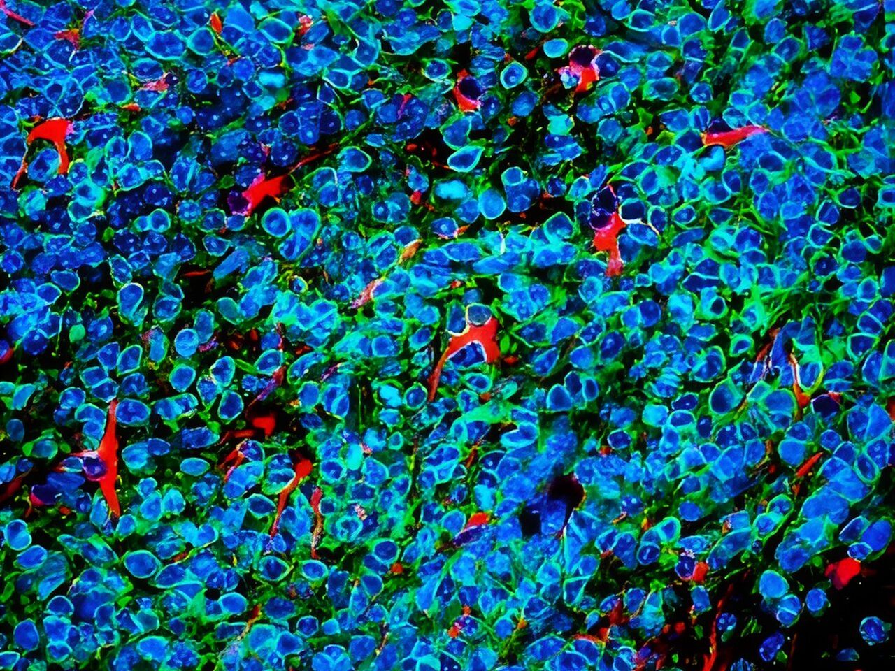
Prior research has shown that numerous brain cells called astrocytes appear to play a role in providing the brain neurons with energy they need. Astrocytes, shaped like stars, are a type of glial cell, which are non-neuronal cells found in the central nervous system. When neighbouring neurons need an increase in energy supply, astrocytes jump into action by rapidly activating their own glucose stores and metabolism, leading to the increased production and release of lactate. Lactate supplements the pool of energy that is readily available for use by neurons in the brain.
Professor Gourine explained: “In our study, we have figured out how exactly astrocytes are able to monitor the energy use by their neighbouring nerve cells, and kick-start this process that delivers additional chemical energy to busy brain regions.”
In a series of experiments using mouse models and cell samples, the researchers identified a set of specific receptors in astrocytes that can detect and monitor neuronal activity, and trigger a signalling pathway involving an essential molecule called adenosine. The researchers found that the metabolic signalling pathway activated by adenosine in astrocytes is exactly the same as the pathway that recruits energy stores in the muscle and the liver, for example when we exercise.
Adenosine activates astrocyte glucose metabolism and supply of energy to neurons to ensure that synaptic function (neurotransmitters passing communication signals between cells) continues apace under conditions of high energy demand or reduced energy supply.
The researchers found that when they deactivated the key astrocyte receptors in mice, the animal’s brain activity was less effective, including significant impairments in global brain metabolism, memory and disruption of sleep, thus demonstrating that the signalling pathway they identified is vital for processes such as learning, memory and sleep.
Dr Theparambil said: “Identification of this mechanism may have broader implications as it could be a way of treating brain diseases where brain energetics are downregulated, such as neurodegeneration and dementia.”
Professor Gourine added: “We know that brain energy homeostasis is progressively impaired in ageing and this process is accelerated during the development of neurodegenerative diseases such as Alzheimer’s disease. Our study identifies an attractive readily druggable target and therapeutic opportunity for brain energy rescue for the purpose of protecting brain function, maintaining cognitive health, and promoting brain longevity.”