Tag: artificial intelligence

Less Invasive Method for Measuring Intracranial Pressure After TBI

Coup and contrecoup brain injury. Credit: Scientific Animations CC4.0

Researchers at Johns Hopkins explored a potential alternative and less-invasive approach to evaluate intracranial pressure (ICP) in patients with serious neurological conditions. This research, using artificial intelligence (AI) to analyse routinely captured ICU data, was published in Computers in Biology and Medicine.

ICP is a physiological variable that can increase abnormally if one has severe traumatic brain injury, stroke or obstruction to the flow of cerebrospinal fluid. Symptoms of elevated ICP may include headaches, blurred vision, vomiting, changes in behaviour and decreased level of consciousness. It can be life-threatening, hence the need for ICP monitoring in selected patients who are at increased risk. But the current standard for ICP monitoring is highly invasive: it requires the placement of an external ventricular drain (EVD) or an intraparenchymal brain monitor (IPM) in the functional tissue in the brain consisting of neurons and glial cells by drilling through the skull.

“ICP is universally accepted as a critical vital sign – there is an imperative need to measure and treat ICP in patients with serious neurological disorders, yet the current standard for ICP measurement is invasive, risky, and resource-intensive. Here we explored a novel approach leveraging Artificial Intelligence which we believed could represent a viable noninvasive alternative ICP assessment method,” says senior author Robert Stevens, MD, MBA, associate professor of anaesthesiology and critical care medicine.

EVD procedures carry a number of risks including catheter misplacement, infection, and haemorrhaging at 15.3 %, 5.8 %, and 12.1 %, respectively, according to recent research. EVD and IPM procedures also require surgical expertise and specialised equipment that is not consistently available in many settings thus underscoring the need for an alternative method in examining and monitoring ICP in patients.

The Johns Hopkins team, a group that included faculty and students from the School of Medicine and Whiting School of Engineering, hypothesised that severe forms of brain injury, and elevations in ICP in particular, are associated with pathological changes in systemic cardiocirculatory function due, for example, to dysregulation of the central autonomic nervous system. This hypothesis suggests that extracranial physiological waveforms can be studied to better understand brain activity and ICP severity.

In this study, the Johns Hopkins team set out to explore the relationship between the ICP waveform and the three physiological waveforms that are routinely captured in the ICU: invasive arterial blood pressure (ABP), photoplethysmography (PPG) and electrocardiography (ECG). ABP, PPG and ECG data were used to train deep learning algorithms, resulting in a level of accuracy in determining ICP that rivals or exceeds other methodologies.

Overall study findings suggest a completely new, noninvasive alternative to monitor ICP in patients.

Stevens says, “with validation, physiology-based AI solutions, such as the one used here, could significantly expand the proportion of patients and health care settings in which ICP monitoring and management can be delivered.” 

Source: John Hopkins Medicine

AI Models that can Identify Patient Demographics in X-rays are Also Unfair

Photo by Anna Shvets

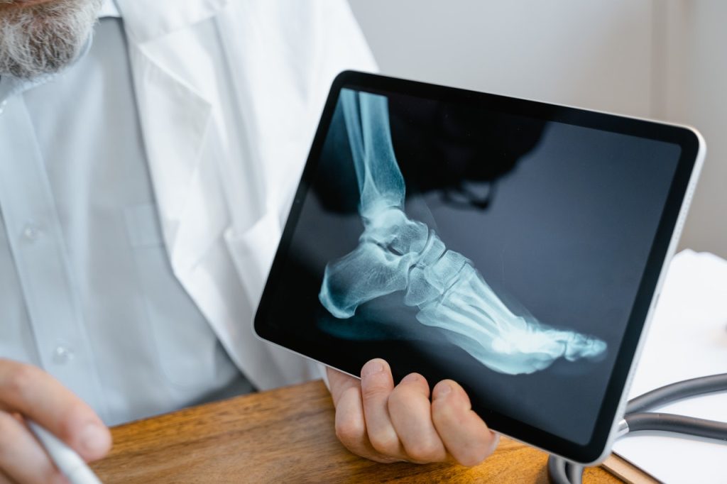
Artificial intelligence models often play a role in medical diagnoses, especially when it comes to analysing images such as X-rays. But these models have been found not perform as well across all demographic groups, usually faring worse on women and people of colour.

These models have also been shown to develop some surprising abilities. In 2022, MIT researchers reported that AI models can make accurate predictions about a patient’s race from their chest X-rays – something that the most skilled radiologists can’t do.

Now, in a new study appearing in Nature, the same research team has found that the models that are most accurate at making demographic predictions also show the biggest “fairness gaps”, ie having reduced accuracy diagnosing images of people of different races or genders. The findings suggest that these models may be using “demographic shortcuts” when making their diagnostic evaluations, which lead to incorrect results for women, Black people, and other groups, the researchers say.

“It’s well-established that high-capacity machine-learning models are good predictors of human demographics such as self-reported race or sex or age. This paper re-demonstrates that capacity, and then links that capacity to the lack of performance across different groups, which has never been done,” says senior author Marzyeh Ghassemi, an MIT associate professor of electrical engineering and computer science.

The researchers also found that they could retrain the models in a way that improves their fairness. However, their approached to “debiasing” worked best when the models were tested on the same types of patients they were trained on, such as patients from the same hospital. When these models were applied to patients from different hospitals, the fairness gaps reappeared.

“I think the main takeaways are, first, you should thoroughly evaluate any external models on your own data because any fairness guarantees that model developers provide on their training data may not transfer to your population. Second, whenever sufficient data is available, you should train models on your own data,” says Haoran Zhang, an MIT graduate student and one of the lead authors of the new paper.

Removing bias

As of May 2024, the FDA has approved 882 AI-enabled medical devices, with 671 of them designed to be used in radiology. Since 2022, when Ghassemi and her colleagues showed that these diagnostic models can accurately predict race, they and other researchers have shown that such models are also very good at predicting gender and age, even though the models are not trained on those tasks.

“Many popular machine learning models have superhuman demographic prediction capacity – radiologists cannot detect self-reported race from a chest X-ray,” Ghassemi says. “These are models that are good at predicting disease, but during training are learning to predict other things that may not be desirable.”

In this study, the researchers set out to explore why these models don’t work as well for certain groups. In particular, they wanted to see if the models were using demographic shortcuts to make predictions that ended up being less accurate for some groups. These shortcuts can arise in AI models when they use demographic attributes to determine whether a medical condition is present, instead of relying on other features of the images.

Using publicly available chest X-ray datasets from Beth Israel Deaconess Medical Center (BIDMC) in Boston, the researchers trained models to predict whether patients had one of three different medical conditions: fluid buildup in the lungs, collapsed lung, or enlargement of the heart. Then, they tested the models on X-rays that were held out from the training data.

Overall, the models performed well, but most of them displayed “fairness gaps” – that is, discrepancies between accuracy rates for men and women, and for white and Black patients.

The models were also able to predict the gender, race, and age of the X-ray subjects. Additionally, there was a significant correlation between each model’s accuracy in making demographic predictions and the size of its fairness gap. This suggests that the models may be using demographic categorisations as a shortcut to make their disease predictions.

The researchers then tried to reduce the fairness gaps using two types of strategies. For one set of models, they trained them to optimise “subgroup robustness,” meaning that the models are rewarded for having better performance on the subgroup for which they have the worst performance, and penalised if their error rate for one group is higher than the others.

In another set of models, the researchers forced them to remove any demographic information from the images, using “group adversarial” approaches. Both strategies worked fairly well, the researchers found.

“For in-distribution data, you can use existing state-of-the-art methods to reduce fairness gaps without making significant trade-offs in overall performance,” Ghassemi says. “Subgroup robustness methods force models to be sensitive to mispredicting a specific group, and group adversarial methods try to remove group information completely.”

Not always fairer

However, those approaches only worked when the models were tested on data from the same types of patients that they were trained on, eg from BIDMC.

When the researchers tested the models that had been “debiased” using the BIDMC data to analyse patients from five other hospital datasets, they found that the models’ overall accuracy remained high, but some of them exhibited large fairness gaps.

“If you debias the model in one set of patients, that fairness does not necessarily hold as you move to a new set of patients from a different hospital in a different location,” Zhang says.

This is worrisome because in many cases, hospitals use models that have been developed on data from other hospitals, especially in cases where an off-the-shelf model is purchased, the researchers say.

“We found that even state-of-the-art models which are optimally performant in data similar to their training sets are not optimal – that is, they do not make the best trade-off between overall and subgroup performance – in novel settings,” Ghassemi says. “Unfortunately, this is actually how a model is likely to be deployed. Most models are trained and validated with data from one hospital, or one source, and then deployed widely.”

The researchers found that the models that were debiased using group adversarial approaches showed slightly more fairness when tested on new patient groups than those debiased with subgroup robustness methods. They now plan to try to develop and test additional methods to see if they can create models that do a better job of making fair predictions on new datasets.

The findings suggest that hospitals that use these types of AI models should evaluate them on their own patient population before beginning to use them, to make sure they aren’t giving inaccurate results for certain groups.

Using AI, Scientists Discover High-risk Form of Endometrial Cancer

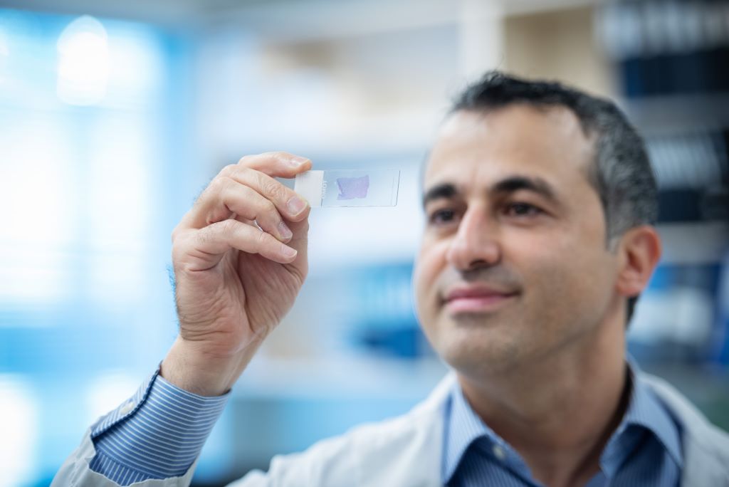
Dr Ali Bashashati observes an endometrial cancer sample on a microscope slide. Credit: University of British Columbia

A discovery by researchers at the University of British Columbia promises to improve care for patients with endometrial cancer, the most common gynaecologic malignancy.  Using artificial intelligence (AI) to spot patterns across thousands of cancer cell images, the researchers have pinpointed a distinct subset of more stubborn endometrial cancer that would otherwise go unrecognised by traditional pathology and molecular diagnostics.

The findings, published in Nature Communications, will help doctors identify patients with high-risk disease who could benefit from more comprehensive treatment.

“Endometrial cancer is a diverse disease, with some patients much more likely to see their cancer return than others,” said Dr Jessica McAlpine, professor at UBC. “It’s so important that patients with high-risk disease are identified so we can intervene and hopefully prevent recurrence. This AI-based approach will help ensure no patient misses an opportunity for potentially lifesaving interventions.”

AI-powered precision medicine

The discovery builds on work by Dr McAlpine and colleagues in the Gynaecologic Cancer Initiative, who in 2013 helped show that endometrial cancer can be classified into four subtypes based on the molecular characteristics of cancerous cells, with each posing a different level of risk to patients.

Dr McAlpine and team then went on to develop an innovative molecular diagnostic tool, called ProMiSE, that can accurately discern between the subtypes. The tool is now used across parts of Canada and internationally to guide treatment decisions.

Yet, challenges remain. The most prevalent molecular subtype, encompassing approximately 50% of all cases, is largely a catch-all category for endometrial cancers lacking discernible molecular features.

“There are patients in this very large category who have extremely good outcomes, and others whose cancer outcomes are highly unfavourable. But until now, we have lacked the tools to identify those at-risk so that we can offer them appropriate treatment,” said Dr McAlpine.

Dr McAlpine turned to long-time collaborator and machine learning expert Dr.Ali Bashashati, an assistant professor of biomedical engineering and pathology and laboratory medicine at UBC, to try and further segment the category using advanced AI methods.

Dr Bashashati and his team developed a deep learning AI model that analyses images of tissue samples collected from patients. The AI was trained to differentiate between different subtypes, and after analysing over 2300 cancer tissue images, pinpointed the new subgroup that exhibited markedly inferior survival rates.

“The power of AI is that it can objectively look at large sets of images and identify patterns that elude human pathologists,” said Dr Bashashati. “It’s finding the needle in the haystack. It tells us this group of cancers with these characteristics are the worst offenders and represent a higher risk for patients.”

Bringing the discovery to patients

The team is now exploring how the AI tool could be integrated into clinical practice alongside traditional molecular and pathology diagnostics.

“The two work hand-in-hand, with AI providing an additional layer on top of the testing we’re already doing,” said Dr McAlpine.

One benefit of the AI-based approach is that it’s cost-efficient and easy to deploy across geographies. The AI analyses images that are routinely gathered by pathologists and healthcare providers, even at smaller hospital sites in rural and remote communities, and shared when seeking second opinions on a diagnosis.

The combined use of molecular and AI-based analysis could allow many patients to remain in their home communities for less intensive surgery, while ensuring those who need treatment at a larger cancer centre can do so.  

“What is really compelling to us is the opportunity for greater equity and access,” said Dr Bashashati. “The AI doesn’t care if you’re in a large urban centre or rural community, it would just be available, so our hope is that this could really transform how we diagnose and treat endometrial cancer for patients everywhere.”

Source: University of British Columbia

AI Analyses Fitbit Data to Predict Spine Surgery Outcomes

Photo by Barbara Olsen on Pexels

Researchers who had been using Fitbit data to help predict surgical outcomes have a new method to more accurately gauge how patients may recover from spine surgery.

Using machine learning techniques developed at the AI for Health Institute at Washington University in St. Louis, Chenyang Lu, the Fullgraf Professor in the university’s McKelvey School of Engineering, collaborated with Jacob Greenberg, MD, assistant professor of neurosurgery at the School of Medicine, to develop a way to predict recovery more accurately from lumbar spine surgery.

The results show that their model outperforms previous models to predict spine surgery outcomes. This is important because in lower back surgery and many other types of orthopaedic operations, the outcomes vary widely depending on the patient’s structural disease but also varying physical and mental health characteristics across patients. The study is published in Proceedings of the ACM on Interactive, Mobile, Wearable and Ubiquitous Technologies.

Surgical recovery is influenced by both preoperative physical and mental health. Some people may have catastrophising, or excessive worry, in the face of pain that can make pain and recovery worse. Others may suffer from physiological problems that cause worse pain. If physicians can get a heads-up on the various pitfalls for each patient, that will allow for better individualized treatment plans.

“By predicting the outcomes before the surgery, we can help establish some expectations and help with early interventions and identify high risk factors,” said Ziqi Xu, a PhD student in Lu’s lab and first author on the paper.

Previous work in predicting surgery outcomes typically used patient questionnaires given once or twice in clinics that capture only one static slice of time.

“It failed to capture the long-term dynamics of physical and psychological patterns of the patients,” Xu said. Prior work training machine learning algorithms focus on just one aspect of surgery outcome “but ignore the inherent multidimensional nature of surgery recovery,” she added.

Researchers have used mobile health data from Fitbit devices to monitor and measure recovery and compare activity levels over time but this research has shown that activity data, plus longitudinal assessment data, is more accurate in predicting how the patient will do after surgery, Greenberg said.

The current work offers a “proof of principle” showing, with the multimodal machine learning, doctors can see a much more accurate “big picture” of all the interrelated factors that affect recovery. Proceeding this work, the team first laid out the statistical methods and protocol to ensure they were feeding the AI the right balanced diet of data.

Prior to the current publication, the team published an initial proof of principle in Neurosurgery showing that patient-reported and objective wearable measurements improve predictions of early recovery compared to traditional patient assessments. In addition to Greenberg and Xu, Madelynn Frumkin, a PhD psychological and brain sciences student in Thomas Rodebaugh’s laboratory in Arts & Sciences, was co-first author on that work. Wilson “Zack” Ray, MD, the Henry G. and Edith R. Schwartz Professor of neurosurgery in the School of Medicine, was co-senior author, along with Rodebaugh and Lu. Rodebaugh is now at the University of North Carolina at Chapel Hill.

In that research, they show that Fitbit data can be correlated with multiple surveys that assess a person’s social and emotional state. They collected that data via “ecological momentary assessments” (EMAs) that employ smart phones to give patients frequent prompts to assess mood, pain levels and behaviour multiple times throughout day.

We combine wearables, EMA -and clinical records to capture a broad range of information about the patients, from physical activities to subjective reports of pain and mental health, and to clinical characteristics,” Lu said.

Greenberg added that state-of-the-art statistical tools that Rodebaugh and Frumkin have helped advance, such as “Dynamic Structural Equation Modeling,” were key in analyzing the complex, longitudinal EMA data.

For the most recent study they then took all those factors and developed a new machine learning technique of “Multi-Modal Multi-Task Learning (M3TL)” to effectively combine these different types of data to predict multiple recovery outcomes.

In this approach, the AI learns to weigh the relatedness among the outcomes while capturing their differences from the multimodal data, Lu adds.

This method takes shared information on interrelated tasks of predicting different outcomes and then leverages the shared information to help the model understand how to make an accurate prediction, according to Xu.

It all comes together in the final package producing a predicted change for each patient’s post-operative pain interference and physical function score.

Greenberg says the study is ongoing as they continue to fine tune their models so they can take these more detailed assessments, predict outcomes and, most notably, “understand what types of factors can potentially be modified to improve longer term outcomes.”

Source: Washington University in St. Louis

AI Helps Clinicians to Assess and Treat Leg Fractures

Photo by Tima Miroshnichenko on Pexels

By using artificial intelligence (AI) techniques to process gait analyses and medical records data of patients with leg fractures, researchers have uncovered insights on patients and aspects of their recovery.

The study, which is published in the Journal of Orthopaedic Research, uncovered a significant association between the rates of hospital readmission after fracture surgery and the presence of underlying medical conditions. Correlations were also found between underlying medical conditions and orthopaedic complications, although these links were not significant.

It was also apparent that gait analyses in the early postinjury phase offer valuable insights into the injury’s impact on locomotion and recovery. For clinical professionals, these patterns were key to optimising rehabilitation strategies.

“Our findings demonstrate the profound impact that integrating machine learning and gait analysis into orthopaedic practice can have, not only in improving the accuracy of post-injury complication predictions but also in tailoring rehabilitation strategies to individual patient needs,” said corresponding author Mostafa Rezapour, PhD, of Wake Forest University School of Medicine. “This approach represents a pivotal shift towards more personalised, predictive, and ultimately more effective orthopaedic care.”

Dr. Rezapour added that the study underscores the critical importance of adopting a holistic view that encompasses not just the mechanical aspects of injury recovery but also the broader spectrum of patient health. “This is a step forward in our quest to optimize rehabilitation strategies, reduce recovery times, and improve overall quality of life for patients with lower extremity fractures,” he said.

Source: Wiley

Admin and Ethics should be the Basis of Your Healthcare AI Stratetgy

Technology continues to play a strong role in shaping healthcare. In 2023, the focus was on how Artificial Intelligence (AI),  became significantly entrenched in patient records, diagnosis and care. Now in 2024 the focus is on the ethical aspects of AI.  Many organisations including practitioner groups, hospitals and medical associations are putting together AI Codes of Conduct, with new legislation planning to be passed in countries such as the USA.

The entire patient journey has benefited from the use of AI, in tangible ways that we can understand. From online bookings, the sharing of information with electronic health records, keyword diagnosis, sharing of visual scans, e-scripts, easy claims, SMS’s and billing, are all examples of how software systems are incorporated into practices to facilitate a streamlined experience for both the patient and doctor. *But although 75% of medical professionals agree on the transformation abilities of AI, only 6% have implemented an AI strategy.

Strategies need to include ethical considerations

CompuGroup Medical South Africa, (CGM SA), a leading international MedTech company that has spent over 20 years designing software solutions for the healthcare industry, has identified one main area that seems to constantly be the topic for ethical consideration.

This is the sharing of patient electronic health records or EHR’s. On one hand the wealth of information provided in each EHR – from a patient’s medical history, demographics, their laboratory test results over time, medicine prescribed, a history of medical procedures, X-rays to any medical allergies – offers endless opportunities for real time patient care. On the other hand, there seems to be a basic mistrust of how these records will be shared and stored, no one wants their personal medical information to end up on the internet.

But there’s also the philosophical view that although you might not want your info to be public record, it still has the ability to benefit the care of thousands of people. If we want a learning AI system that adapts as we do, if we want a decision making support system that is informed by past experiences, then the sharing of data should be viewed as a tool and no longer a privacy barrier.

Admin can cause burnout

Based on their interactions with professionals, CGM has informally noted that healthcare practices spend 73% of their time dealing with administrative tasks. This can be broken down into 38% focusing on EHR documentation and review, 19% related to insurance and billing, 11% on tests, medications and other orders and the final 6% on clinical planning and logistics.

Even during the consultation doctors can spend up to 40% of their time taking clinical notes. Besides the extra burden that this places on health care practices, this also leads to less attention being paid to the patient and still requires 1-2 hours of admin in the evenings. (Admin being the number one cause of burnout in clinicians and too much screen time during interactions being the number one complaint by patients.)

The solution

The ability for medical practitioners to implement valuable and effective advanced technical software, such as Autoscriber, will assist with time saving, data quality and overall job satisfaction. Autoscriber is an AI engine designed to ease the effort required when creating clinical notes by turning the consultation between patient and doctor into a structured summary that includes ICD-10 codes which is the standard method of classification of diseases used by South African medical professionals    

It identifies clinical facts in real time, including medications and symptoms. It then orders and summarises the data in a format ready for import into the EHR, creating a more detailed and standardised report on each patient encounter, allowing for a more holistic patient outcome. In essence, with the introduction of Autoscriber into the South African market, CGM seeks to aid practitioners in swiftly creating precise and efficient clinical records, saving them from extensive after-hours commitments.

Dilip Naran, VP of Product Architecture at CGM SA explains: “It is clear that AI will not replace healthcare professionals, but it will augment their capabilities to provide superior patient care. Ethical considerations are important but should not override patient care or safety. The Autoscriber solution provides full control to the HCP to use, edit or discard the transcribed note ensuring that these notes are comprehensive, attributable and contemporaneous.”

AI-based App can Help Physicians Diagnose Melanomas

3D structure of a melanoma cell derived by ion abrasion scanning electron microscopy. Credit: Sriram Subramaniam/ National Cancer Institute

A mobile app that uses artificial intelligence, AI, to analyse images of suspected skin lesions can diagnose melanoma with very high precision. This is shown in a study led from Linköping University in Sweden where the app has been tested in primary care. The results have been published in the British Journal of Dermatology.

“Our study is the first in the world to test an AI-based mobile app for melanoma in primary care in this way. A great many studies have been done on previously collected images of skin lesions and those studies relatively agree that AI is good at distinguishing dangerous from harmless ones. We were quite surprised by the fact that no one had done a study on primary care patients,” says Magnus Falk, senior associate professor at the Department of Health, Medicine and Caring Sciences at Linköping University, specialist in general practice at Region Östergötland, who led the current study.

Melanoma can be difficult to differentiate from other skin changes, even for experienced physicians. However, it is important to detect melanoma as early as possible, as it is a serious type of skin cancer.

There is currently no established AI-based support for assessing skin lesions in Swedish healthcare.

“Primary care physicians encounter many skin lesions every day and with limited resources need to make decisions about treatment in cases of suspected skin melanoma. This often results in an abundance of referrals to specialists or the removal of skin lesions, which in the majority of cases turn out to be harmless. We wanted to see if the AI support tool in the app could perform better than primary care physicians when it comes to identifying pigmented skin lesions as dangerous or not, in comparison with the final diagnosis,” says Panos Papachristou, researcher affiliated with Karolinska Institutet and specialist in general practice, main author of the study and co-founder of the company that developed the app.

And the results are promising.

“First of all, the app missed no melanoma. This disease is so dangerous that it’s essential not to miss it. But it’s almost equally important that the AI decision support tool could acquit many suspected skin lesions and determine that they were harmless,” says Magnus Falk.

In the study, primary care physicians followed the usual procedure for diagnosing suspected skin tumours. If the physicians suspected melanoma, they either referred the patient to a dermatologist for diagnosis, or the skin lesion was cut away for tissue analysis and diagnosis.

Only after the physician decided how to handle the suspected melanoma did they use the AI-based app. This involves the physician taking a picture of the skin lesion with a mobile phone equipped with an enlargement lens called a dermatoscope. The app analyses the image and provides guidance on whether or not the skin lesion appears to be melanoma.

To find out how well the AI-based app worked as a decision support tool, the researchers compared the app’s response to the diagnoses made by the regular diagnostic procedure.

Of the more than 250 skin lesions examined, physicians found 11 melanomas and 10 precursors of cancer, known as in situ melanoma. The app found all the melanomas, and missed only one precursor. In cases where the app responded that a suspected lesion was not a melanoma, including in situ melanoma, there was a 99.5% probability that this was correct.

“It seems that this method could be useful. But in this study, physicians weren’t allowed to let their decision be influenced by the app’s response, so we don’t know what happens in practice if you use an AI-based decision support tool. So even if this is a very positive result, there is uncertainty and we need to continue to evaluate the usefulness of this tool with scientific studies,” says Magnus Falk.

The researchers now plan to proceed with a large follow-up primary care study in several countries, where use of the app as an active decision support tool will be compared to not using it at all.

Source: Linköping University

Is AI a Help or Hindrance to Radiologists? It’s Down to the Doctor

New research shows AI isn’t always a help for radiologists

Photo by Anna Shvets

One of the most touted promises of medical artificial intelligence tools is their ability to augment human clinicians’ performance by helping them interpret images such as X-rays and CT scans with greater precision to make more accurate diagnoses.

But the benefits of using AI tools on image interpretation appear to vary from clinician to clinician, according to new research led by investigators at Harvard Medical School, working with colleagues at MIT and Stanford.

The study findings suggest that individual clinician differences shape the interaction between human and machine in critical ways that researchers do not yet fully understand. The analysis, published in Nature Medicine, is based on data from an earlier working paper by the same research group released by the National Bureau of Economic Research.

In some instances, the research showed, use of AI can interfere with a radiologist’s performance and interfere with the accuracy of their interpretation.

“We find that different radiologists, indeed, react differently to AI assistance – some are helped while others are hurt by it,” said co-senior author Pranav Rajpurkar, assistant professor of biomedical informatics in the Blavatnik Institute at HMS.

“What this means is that we should not look at radiologists as a uniform population and consider just the ‘average’ effect of AI on their performance,” he said. “To maximize benefits and minimize harm, we need to personalize assistive AI systems.”

The findings underscore the importance of carefully calibrated implementation of AI into clinical practice, but they should in no way discourage the adoption of AI in radiologists’ offices and clinics, the researchers said.

Instead, the results should signal the need to better understand how humans and AI interact and to design carefully calibrated approaches that boost human performance rather than hurt it.

“Clinicians have different levels of expertise, experience, and decision-making styles, so ensuring that AI reflects this diversity is critical for targeted implementation,” said Feiyang “Kathy” Yu, who conducted the work while at the Rajpurkar lab with co-first author on the paper with Alex Moehring at the MIT Sloan School of Management.

“Individual factors and variation would be key in ensuring that AI advances rather than interferes with performance and, ultimately, with diagnosis,” Yu said.

AI tools affected different radiologists differently

While previous research has shown that AI assistants can, indeed, boost radiologists’ diagnostic performance, these studies have looked at radiologists as a whole without accounting for variability from radiologist to radiologist.

In contrast, the new study looks at how individual clinician factors – area of specialty, years of practice, prior use of AI tools – come into play in human-AI collaboration.

The researchers examined how AI tools affected the performance of 140 radiologists on 15 X-ray diagnostic tasks – how reliably the radiologists were able to spot telltale features on an image and make an accurate diagnosis. The analysis involved 324 patient cases with 15 pathologies: abnormal conditions captured on X-rays of the chest.

To determine how AI affected doctors’ ability to spot and correctly identify problems, the researchers used advanced computational methods that captured the magnitude of change in performance when using AI and when not using it.

The effect of AI assistance was inconsistent and varied across radiologists, with the performance of some radiologists improving with AI and worsening in others.

AI tools influenced human performance unpredictably

AI’s effects on human radiologists’ performance varied in often surprising ways.

For instance, contrary to what the researchers expected, factors such how many years of experience a radiologist had, whether they specialised in thoracic, or chest, radiology, and whether they’d used AI readers before, did not reliably predict how an AI tool would affect a doctor’s performance.

Another finding that challenged the prevailing wisdom: Clinicians who had low performance at baseline did not benefit consistently from AI assistance. Some benefited more, some less, and some none at all. Overall, however, lower-performing radiologists at baseline had lower performance with or without AI. The same was true among radiologists who performed better at baseline. They performed consistently well, overall, with or without AI.

Then came a not-so-surprising finding: More accurate AI tools boosted radiologists’ performance, while poorly performing AI tools diminished the diagnostic accuracy of human clinicians.

While the analysis was not done in a way that allowed researchers to determine why this happened, the finding points to the importance of testing and validating AI tool performance before clinical deployment, the researchers said. Such pre-testing could ensure that inferior AI doesn’t interfere with human clinicians’ performance and, therefore, patient care.

What do these findings mean for the future of AI in the clinic?

The researchers cautioned that their findings do not provide an explanation for why and how AI tools seem to affect performance across human clinicians differently, but note that understanding why would be critical to ensuring that AI radiology tools augment human performance rather than hurt it.

To that end, the team noted, AI developers should work with physicians who use their tools to understand and define the precise factors that come into play in the human-AI interaction.

And, the researchers added, the radiologist-AI interaction should be tested in experimental settings that mimic real-world scenarios and reflect the actual patient population for which the tools are designed.

Apart from improving the accuracy of the AI tools, it’s also important to train radiologists to detect inaccurate AI predictions and to question an AI tool’s diagnostic call, the research team said. To achieve that, AI developers should ensure that they design AI models that can “explain” their decisions.

“Our research reveals the nuanced and complex nature of machine-human interaction,” said study co-senior author Nikhil Agarwal, professor of economics at MIT. “It highlights the need to understand the multitude of factors involved in this interplay and how they influence the ultimate diagnosis and care of patients.”

Source: Harvard Medical School

Getting the Most from AI in MedTech Takes Data Know-How

As a leader in Medical Technology innovation, InterSystems, a pioneer in healthcare data platform development, has learned, understood, and incorporated pivotal insights from its extensive experience in digital health solutions. That experience points up the need to give AI a strong foundation.

We understand the importance of leveraging AI to drive transformative change in healthcare. Our latest white paper, “Getting the Most from AI in MedTech Takes Data Know-How,” dives into the challenges and opportunities facing MedTech companies venturing into the realm of AI. From data cleanliness to privacy and security considerations, we address key issues that MedTech companies must navigate to succeed in today’s rapidly evolving healthcare landscape.

AI in MedTech Takes Data Know-How

The promise of AI in revolutionising MedTech is undeniable. AI in varying forms and degrees is forecasted to save hundreds of thousands of lives and billions of dollars a year. But here’s the catch- AI models are only as good as the data they’re built on. An AI application can sift through large amounts of data from various Electronic Health Record (EHR) environments and legacy systems and identify patterns within the scope of its model, but it can’t identify data that exists outside of those boundaries.

If one asks “What risk factors does the patient have for stroke?”, AI can only answer based on the information that’s there. Sometimes, things get lost in translation, and that’s why interoperability – the ability to exchange information in a way that ensures the sender and receiver understand data the same way is crucial.

InterSystems: Your Data Sherpa:

Ever wondered why some AI models in MedTech fall short? It’s all about the data. This means MedTech companies can’t just lean on their currently used standard but should consider all those in which relevant data is captured in the market or build on a platform that does.

With InterSystems by your side, you gain access to a treasure trove of healthcare data expertise. One of the benefits of our business is that it’s much broader than a single EHR. This means providing software solutions like The HL7® FHIR® (Fast Healthcare Interoperability Resources) offering a comprehensive view of patient data, accelerating development timelines, and delivering tangible results that showcase the value of your innovations.

Clean Data Is a Must

Data cleanliness is key in the world of AI. Pulling data from various sources presents its own set of challenges, from ensuring data cleanliness to reconciling discrepancies and omissions. Raw data is often messy, inconsistent, and filled with gaps like missing labels. If the data fed into an AI model is incomplete and error-ridden, the conclusions drawn from its analysis will be similarly flawed and suspect. Thus, maintaining high standards of data quality is essential to ensure the accuracy and effectiveness of AI-driven insights.

Henry Adams, Country Manager, InterSystems South Africa, says: “InterSystems advocates for robust preprocessing, cleaning, and labelling techniques to ensure data quality and integrity. Our platform keeps track of data lineage, simplifies labelling, and aggregates health data into a single, patient-centric model ready for analysis”.

Privacy, Security, and Reliability: The Sweet Success!

Privacy and security are essential across industries, but they are even more critical for MedTech product developers. Handling sensitive patient data necessitates strict adherence to regulations like HIPAA and GDPR to safeguard patient confidentiality and comply with legal requirements. Beyond regulatory compliance, ensuring privacy and security is crucial for maintaining patient safety, preserving reputation and trust, and fostering collaboration within the industry.

To help MedTech companies comply with regulations and safeguard patient data, InterSystems’ platform meets needs across major deployments, such as a nonprofit health data network and uses techniques like redundant processing and queues built into the connective tissue of their software. Reliable connectivity solutions ensure seamless data exchange, even in the most demanding healthcare environments.

Charting the Course Forward

If you are a MedTech company still struggling to make sense of siloed healthcare data for your AI initiatives? We have the answers-collaboration with the right partner is essential for integrating AI into medical practices. An ideal partner understands the need for data acquisition, aggregation, cleaning, privacy, and security regulations. “With InterSystems as your partner and by your side, you can navigate the complexities of AI integration and drive transformative innovation in healthcare, making MedTech excellence easier to attain,” concludes Adams.

You can learn more about our support for MedTech innovation at InterSystems.com/MedTech.

For more information or to download the guide, please visit!  

When it Comes to Personalised Cancer Treatments, AI is no Match for Human Doctors

Cancer treatment is growing more complex, but so too are the possibilities. After all, the better a tumour’s biology and genetic features are understood, the more treatment approaches there are. To be able to offer patients personalised therapies tailored to their disease, laborious and time-consuming analysis and interpretation of various data is required. In one of many artificial intelligence (AI)projects at Charité – Universitätsmedizin Berlin and Humboldt-Universität zu Berlin, researchers studied whether generative AI tools such as ChatGPT can help with this step.

The crucial factor in the phenomenon of tumour growth is an imbalance of growth-inducing and growth-inhibiting factors, which can result, for example, from changes in oncogenes.

Precision oncology, a specialised field of personalised medicine, leverages this knowledge by using specific treatments such as low-molecular weight inhibitors and antibodies to target and disable hyperactive oncogenes.

The first step in identifying which genetic mutations are potential targets for treatment is to analyse the genetic makeup of the tumour tissue. The molecular variants of the tumour DNA that are necessary for precision diagnosis and treatment are determined. Then the doctors use this information to craft individual treatment recommendations. In especially complex cases, this requires knowledge from various fields of medicine.

At Charité, this is when the “molecular tumour board” (MTB) meets: Experts from the fields of pathology, molecular pathology, oncology, human genetics, and bioinformatics work together to analyse which treatments seem most promising based on the latest studies.

It is a very involved process, ultimately culminating in a personalised treatment recommendation.

Can artificial intelligence help with treatment decisions?

Dr Damian Rieke, a doctor at Charité, and his colleagues wondered whether AI might be able to help at this juncture.

In a study just recently published in the journal JAMA Network Open, they worked with other researchers to examine the possibilities and limitations of large language models such as ChatGPT in automatically scanning scientific literature with an eye to selecting personalised treatments.

AI ‘not even close’

“We prompted the models to identify personalised treatment options for fictitious cancer patients and then compared the results with the recommendations made by experts,” Rieke explains.

His conclusion: “AI models were able to identify personalised treatment options in principle – but they weren’t even close to the abilities of human experts.”

The team created ten molecular tumour profiles of fictitious patients for the experiment.

A human physician specialist and four large language models were then tasked with identifying a personalised treatment option.

These results were presented to the members of the MTB for assessment, without them knowing where which recommendation came from.

Improved AI models hold promise for future uses

Dr. Manuela Benary, a bioinformatics specialist reported: “There were some surprisingly good treatment options identified by AI in isolated cases. “But large language models perform much worse than human experts.”

Beyond that, data protection, privacy, and reproducibility pose particular challenges in relation to the use of artificial intelligence with real-world patients, she notes.

Still, Rieke is fundamentally optimistic about the potential uses of AI in medicine: “In the study, we also showed that the performance of AI models is continuing to improve as the models advance. This could mean that AI can provide more support for even complex diagnostic and treatment processes in the future – as long as humans are the ones to check the results generated by AI and have the final say about treatment.”

Source: Charité – Universitätsmedizin Berlin