Tag: arthritis

Hand-held Medical Scanner could Transform Cancer and Arthritis Diagnosis
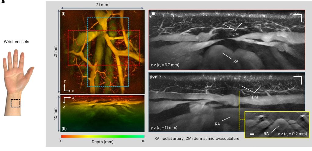
PAT images of wrist vasculature acquired in high-resolution scan mode. Wrist region, (i) x-y and (ii) x-z depth-to-colour encoded MIPs, (iii) x-z and (iv) y-z greyscale MIP slices of regions indicated by dashed red and blue rectangles in (i) showing fine dermal microvasculature (DM), radial artery (RA) and large wrist veins. Inset: x-z greyscale MIP showing cross-sectional view of the radial artery and adjacent veins in the plane indicated by the dashed yellow line in (iv). Huynh et al., Nature Communications, 2024.

A new hand-held scanner developed by UCL researchers and tested in a series of clinical trials on UCLH patients can generate highly detailed 3D photoacoustic images in just seconds, paving the way for their use in a clinical setting for the first time and offering the potential for earlier disease diagnosis.

In the study, published in Nature Biomedical Engineering, the UCL and UCLH team show their technology can deliver photoacoustic tomography (PAT) imaging scans to doctors in real time, providing them with accurate and intricate images of blood vessels, helping inform patient care.

Photoacoustic tomography imaging uses laser-generated ultrasound waves to visualise subtle changes (an early marker of disease) in the sub-millimetre-scale veins and arteries up to 15mm deep in human tissues.

However, up until now, existing PAT technology has been too slow to produce high-enough quality 3D images for use by clinicians.

The older PAT scanners took more than five minutes to take an image – by reducing that time to a few seconds or less, image quality is much improved and far more suitable for people who are frail or poorly.

The researchers say the new scanner could help to diagnose cancer, cardiovascular disease and arthritis in three to five years’ time, subject to further testing.

In this study, the team tested the scanner during pre-clinical tests on 10 UCLH patients with type-2 diabetes, rheumatoid arthritis or breast cancer, along with seven healthy volunteers. They also compared the PAT scans to regular clinical scans taken at UCLH. Larger scale trials of the device are ongoing at UCLH and UCL.

In three patients with type-2 diabetes, the scanner was able to produce detailed 3D images of the microvasculature in the feet, highlighting deformities and structural changes in the vessels. The scanner was also used to visualise the skin inflammation linked to breast cancer.

UCLH consultant radiologist Andrew Plumb, a senior author of the study and Chief Investigator of the clinical PAT studies, said: “One of the complications often suffered by people with diabetes is low blood flow in the extremities, such as the feet and lower legs, due to damage to the tiny blood vessels in these areas. But until now we haven’t been able to see exactly what is happening to cause this damage or characterise how it develops.

“In one of our patients, we could see smooth, uniform vessels in the left foot and deformed, squiggly vessels in the same region of the right foot, indicative of problems that may lead to tissue damage in future. Photoacoustic imaging could give us much more detailed information to facilitate early diagnosis, as well as better understand disease progression more generally.” Dr Plumb is also Associate Professor of Medical Imaging at UCL.

Patients were identified and recruited from a number of clinics at UCLH, including consultant rheumatologist Madhura Castelino, consultant interventional radiologist Conrad von Stempel and research staff Katerina Soteriou and Antonia Yeung who co-ordinated safe, timely scanning at UCLH and UCL on the new PAT scanner.

UCL Professor of Biomedical Photoacoustics Paul Beard, corresponding author, said: “We’ve come a long way with photoacoustic imaging in recent years, but there were still barriers to using it in the clinic.

“The breakthrough in this study is the acceleration in the time it takes to acquire images, which is between 100 and 1000 times faster than previous scanners.

“This speed avoids motion-induced blurring, providing highly-detailed images of a quality that no other scanner can provide. It also means that rather than taking five minutes or longer, images can be acquired in real time, making it possible to visualise dynamic physiological events.

“These technical advances make the system suitable for clinical use for the first time, allowing us to look at aspects of human biology and disease that we haven’t been able to before.

“Now more research is needed with larger groups of patients to confirm our findings.”

Professor Beard added that a key potential use for the new scanner was to assess inflammatory arthritis, which requires scanning all 20 finger joints in both hands. With the new scanner, this can be done in a few minutes – older PAT scanners take nearly an hour, which is too long for elderly, frail patients, he said.

Source: University College London Hospitals

Macrophage Discovery Could Lead to Treatments for Diseases Such as Lupus and COVID

A macrophage engulfing a yeast cell. Source: CC0

Scientistshave made an important breakthrough in understanding failures during the progression of inflammatory diseases and in doing so unearthed a potential new therapeutic target. The scientists report in Nature that an enzyme called Fumarate Hydratase is repressed in macrophages. These immune cells are already implicated in a range of diseases including Lupus, arthritis, sepsis and COVID.

Lead author Luke O’Neill, Professor of Biochemistry at Trinity said: “No-one has made a link from Fumarate Hydratase to inflammatory macrophages before and we feel that this process might be targetable to treat debilitating diseases like Lupus, which is a nasty autoimmune disease that damages several parts of the body including the skin, kidneys and joints.”

Joint first-author Christian Peace added: “We have made an important link between Fumarate Hydratase and immune proteins called cytokines that mediate inflammatory diseases. We found that when Fumarate Hydratase is repressed, RNA is released from mitochondria which can bind to key proteins ‘MDA5’ and ‘TLR7’ and trigger the release of cytokines, thereby worsening inflammation. This process could potentially be targeted therapeutically.”

Fumarate Hydratase was shown to be repressed in a model of sepsis, an often-fatal systemic inflammatory condition that can happen during bacterial and viral infections. Similarly, in blood samples from patients with Lupus, Fumarate Hydratase was dramatically decreased.

“Restoring Fumarate Hydratase in these diseases or targeting MDA5 or TLR7 therefore presents an exciting prospect for badly needed new anti-inflammatory therapies,” said Prof O’Neill.

Excitingly, this newly published work is accompanied by another publication by a group led by Professor Christian Frezza, now at the University of Cologne, and Dr Julien Prudent at the MRC Mitochondrial Biology Unit (MBU), who have made similar findings in the context of kidney cancer.

“Because the system can go wrong in certain types of cancer, the scope of any potential therapeutic target could be widened beyond inflammation,” added Prof O’Neill.

Source: Trinity College Dublin

Axial Spondyloarthritis Association of South Africa wins Excellence Award at EULAR PARE Assembly 2022

Maranda Van Dam, Chairperson of ASASA (left) accepting award, with Souzi Makri President of PARE (middle) and Nikos Sleeks, ELEANA Greece (right)

Maranda van Dam, Chairman of the Axial Spondyloarthritis Association of South Africa (ASASA) won an award of excellence at the EULAR PARE (People with Arthritis/Rheumatism across Europe) Congress 2022, for work performed by ASASA around axial spondyloarthritis (AxSpA) in South Africa. The PARE congress took place from 20 – 22 October 2022, in Brussels, and included a Representation Committee consisting of members of the PARE, of which van Dam is an associate.

The award was based on strides made by ASASA towards improving the quality of life of people living with AxSpa, as well as training done to build awareness in the medical fraternity around AxSpA in the country. With 36 posters entered into the awards by organisations across the globe, ASASA came out tops.

When asked about the award, van Dam said, “This was a real honour to represent South Africa at PARE. 2022 is also the first year that an African country was invited to attend PARE. Winning this award sheds light on our country and our unique problems. The delay to diagnosis of 10.8 years is just unacceptable. The access to the correct medication in both the private and public sector is also not sufficient for a debilitating, progressive disease that can lead to disability if left untreated.”

ASASA estimates that there are approximately 160 000 people suffering from the AxSpA in South Africa, with many of these sufferers undiagnosed. ASASA has made significant strides this year in the training of over 100 General Practitioners and over 250 optometrists around AxSpA diagnosis and the effects it can have on other parts of the body, like the eyes. In addition, ASASA, along with other partners, assisted in gathering data from South African respondents in the first ever live patient survey, called the International Map of Axial Spondyloarthritis (IMAS) survey, which is run by the Axial Spondyloarthritis International Federation that surveys people diagnosed with AxSpA and assesses the impact and burden that AxSpA has on the lives of patients, from their perspective.

Van Dam concluded, “There is still a lot we can do in South Africa and ASASA is busy growing its team of volunteers to help to build awareness around AxSpA in the country. We aim to continue to build support structures for patients in the country, as well as continually working with the medical fraternity, assisting with early diagnosis and access to treatment.”

New Drug Relieves Rheumatoid Arthritis Pain

A new drug, otilimab, has shown effectiveness in treating rheumatoid arthritis (RA). Otilimab is a human monoclonal antibody which inhibits granulocyte-macrophage colony-stimulating factor (GM-CSF). GM-CSF is a large driver of immune-mediated inflammatory conditions.

The drug is currently being tested on its ability to suppress inflammation, tissue damage and pain in RA sufferers.

A multicentre, dose-ranging trial conducted with the drug. Participants were administered subcutaneous injections with one of five different dosages of otilimab (22.5 mg, 45 mg, 90 mg, 135 mg, or 180 mg) or placebo weekly for five weeks. Thereafter, they received injections once every two weeks for one year. The results showed a rapid reduction in tenderness and swelling, and a very high reduction in pain.

The study was unusual in that it offered an escape arm. It is often difficult to recruit participants when they know they may be receiving a dummy injection, and so if, after 12 weeks the participants  on the placebo arm derived no benefit, they were transferred to the highest dose arm of 180mg.

Source: Medical Xpress

Journal information: Christopher D Buckley et al, Efficacy, patient-reported outcomes, and safety of the anti-granulocyte macrophage colony-stimulating factor antibody otilimab (GSK3196165) in patients with rheumatoid arthritis: a randomised, phase 2b, dose-ranging study, The Lancet Rheumatology (2020). DOI: 10.1016/S2665-9913(20)30229-0