Tag: amputation

Heparin Could be a New Cobra Venom Antidote

Cheap, available drug could help reduce impact of snakebites worldwide

Photo by Nivedh P on Unsplash

More than 100 000 people die from snake bites every year. Cobra antivenom is expensive and doesn’t treat the necrosis of flesh caused by the bite, which can lead to amputations. Now, Scientists at the University of Sydney and Liverpool School of Tropical Medicine have made a remarkable discovery: a commonly used blood thinner, heparin, can be repurposed as an inexpensive antidote for cobra venom.

“Our discovery could drastically reduce the terrible injuries from necrosis caused by cobra bites – and it might also slow the venom, which could improve survival rates,” said Professor Greg Neely, a corresponding author of the study from the University of Sydney.

Using CRISPR gene-editing technology to identify ways to block cobra venom, the team, which consisted of scientists based in Australia, Canada, Costa Rica and the UK, successfully repurposed heparin and related drugs and showed they can stop the necrosis caused by cobra bites.

The research is published on the front cover of Science Translational Medicine.

PhD student and lead author, Tian Du, also from the University of Sydney, said: “Heparin is inexpensive, ubiquitous and a World Health Organization-listed Essential Medicine. After successful human trials, it could be rolled out relatively quickly to become a cheap, safe and effective drug for treating cobra bites.”

The team used CRISPR to find the human genes that cobra venom needs to cause necrosis that kills the flesh around the bite. One of the required venom targets are enzymes needed to produce the related molecules heparan and heparin, which many human and animal cells produce. Heparan is on the cell surface and heparin is released during an immune response. Their similar structure means the venom can bind to both. The team used this knowledge to make an antidote that can stop necrosis in human cells and mice.

Unlike current antivenoms for cobra bites, which are 19th century technologies, the heparinoid drugs act as a ‘decoy’ antidote. By flooding the bite site with ‘decoy’ heparin sulfate or related heparinoid molecules, the antidote can bind to and neutralise the toxins within the venom that cause tissue damage.

Joint corresponding author, Professor Nicholas Casewell, Head of the Centre for Snakebite Research & Interventions at Liverpool School of Tropical Medicine, said: “Snakebites remain the deadliest of the neglected tropical diseases, with its burden landing overwhelmingly on rural communities in low- and middle-income countries.

“Our findings are exciting because current antivenoms are largely ineffective against severe local envenoming, which involves painful progressive swelling, blistering and/or tissue necrosis around the bite site. This can lead to loss of limb function, amputation and lifelong disability.”

Snakebites kill up to 138 000 people a year, with 400 000 more experiencing long-term consequences of the bite. While the number affected by cobras is unclear, in some parts of India and Africa, cobra species account for most snakebite incidents.

Working in the Dr John and Anne Chong Laboratory for Functional Genomics at the Charles Perkins Centre, Professor Neely’s team takes a systematic approach to finding drugs to treat deadly or painful venoms. It does this using CRISPR to identify the genetic targets used by a venom or toxin inside humans and other mammals. It then uses this knowledge to design ways to block this interaction and ideally protect people from the deadly actions of these venoms.

This approach was used to identify an antidote to box jellyfish venom by the team in 2019.

Source: University of Sydney

Össur South Africa Extends its Range of Non-invasive Prosthetics with Naked Prosthetics for Finger and Partial-hand Amputations

Össur South Africa has announced the availability of Naked Prosthetics to the local market. This range of custom-made prostheses, precisely tailored to the user’s amputation and individual hand structure, positively impacts those with finger and partial-hand amputations by providing functional finger prostheses of high quality.

“Partial hand limb loss is the most prevalent of upper limb loss, with over 90% of upper limb amputations involving the fingers. Finger and partial-hand amputations also accounts for a significant number of amputations each year,” says Ernst van Dyk, Managing Director, Össur South Africa.

Whilst more common amongst working age men, finger and partial-hand amputations occurs regardless of gender or age. “The lack of mobility resulting from a finger and partial-hand amputation is not limited to the area of amputation only. Many amputees experience loss of mobility beyond the area of amputation,” stresses van Dyk. No fewer than 5% experience a resultant impairment of the entire body and as many as 75% of heavy manual labourers are unable to return to work.

“With Naked Prosthetics we are dedicated to positively impacting the lives of finger and partial-hand amputees. We aim to provide them with functional, high-quality solutions that seamlessly integrate into their lives and empower them to not only resume employment but, as importantly, to engage in the activities they love, thereby assisting them to live a life without limitations,” says van Dyk.

Naked Prosthetics’ innovative solutions, the result of strong research and development (R&D) efforts and manufacturing capabilities, has been recognised by Business Insider as one of the medical technologies that are changing people’s lives[1]. It currently offers four custom-designed devices that are fabricated to within millimetres of a patient’s unique anatomy to mimic the complex motion of a finger.

  • The PIPDriver is a body-controlled prosthesis designed for a finger amputation or limb difference on the proximal or distal phalanx. Its design is anatomically adapted to the proximal and distal interphalangeal joints for intuitive and natural movements. Benefits include improved functionality for everyday activities. It is easy to clean and care for, easy to put on and take off and has a cage-like structure that protects the residual finger. Its slim and smooth design allows the prosthesis to be worn on two or more adjacent fingers. It also includes a conductive tip option for touchscreen operation.
  • The MCPDriver is a body-driven prosthesis designed for a finger amputation or limb difference on the MCP joint (also known as the knuckle) of the index, middle, ring, and/or the little finger. It restores the original finger length, thereby helping to imitate natural gripping patterns and excels at restoring pinch, key, cylindrical and power grasps as well as grip stability. Its durable stainless-steel linkages and robust components allow the user to return to a highly demanding lifestyle. Benefits include a silicone pad that cushions the backplate for improved comfort, interchangeable silicone adjustment inserts that can be used to vary the volume and adjusting discs to obtain the best possible fit. Its natural abduction and adduction allow for intuitive use. As a result, the acclimatisation time after the initial fitting can be considerably reduced. It also includes the conductive tip option for touchscreen operation.
  • The ThumbDriver is a body-controlled prosthesis designed for an amputation or limb difference on the MCP joint of the thumb. It can restore two and three-point grips, enable secure gripping patterns with medium to large diameters and improve fine motor functions and skills. It features an adjustable preflex option that allows you to adapt the prosthesis according to the requirements of the task at hand. As a result, functional gripping patterns can be more easily attained.
  • The GripLock Finger is a passive and positionable prosthetic finger designed for a finger amputation or limb difference on the MCP joint of the index, middle, ring, and/or little finger. It is intended for use in conjunction with a custom-made socket adapted by a certified prosthetist. You can flex the finger to various degrees with your other hand or on a hard surface. Subsequently, you can release and fully extend the GripLock Finger by pressing the latch (lever arm) on the back or flexing the finger beyond the last locking position. It restores the original length, supports the use of both hands, prevents a misalignment of the metacarpal bone and provides a valuable tool to master everyday activities.  GripLock Fingers can be combined with our MCPDriver, PIPDriver, and/or ThumbDriver.

Says Kai, a trained plant and machine operator who suffered the loss of his forefinger, middle and ring finger after a work-related accident. “Thanks to the precise adaptation to my individual anatomical conditions, the prosthesis is an irreplaceable everyday companion for me. When I come home at night, I take off the prosthesis in seconds – just like you kick off your shoes after a long day at work. I think it’s important to convey to other people in similar situations that a work accident like mine doesn’t have to mean the end of the world. You can come to terms with many situations and end up living a normal life.”

Similarly, Cara (an active member of the Finger and Partial Hand Amputee Peer & Support Group), lost two and a half fingers on her left hand due to an unforeseen accident. Prior to her accident, Cara was an avid yogi and enjoyed practicing inversions (yoga poses where the heart is higher from the ground than the head) and handstands. “I spent a year doing physical therapy to regain strength in my left hand, but I still felt as though I was struggling to hold and grip my mat as I practiced yoga,” she recalls. Every time she tried to balance her weight, she would fall backwards due to the lack of grip and support. Within one week of receiving her Naked Prosthetics PIPDrivers, Cara was able to hold a side plank during yoga. “You may feel hopeless in the moment, but it does get better. And you will be surprised at what you could learn. I am a different person now and I grew from the experience.”

“We are committed to helping digit amputees discover innovative and life-changing solutions. It’s all about function and getting people back to living full lives, without limitations,” continues van Dyk. “We believe our range of technologically advanced and custom-made prostheses helps to achieve exactly this and we are excited to be able to offer it to local amputees.”

To find out more, please visit: https://www.ossur.com/en-za/prosthetics/np-devices

[1] Naked Prosthetics’ Technology Recognized – The O&P EDGE Magazine (opedge.com)

Brains do Not Actually ‘Rewire’ Themselves, Scientists Argue

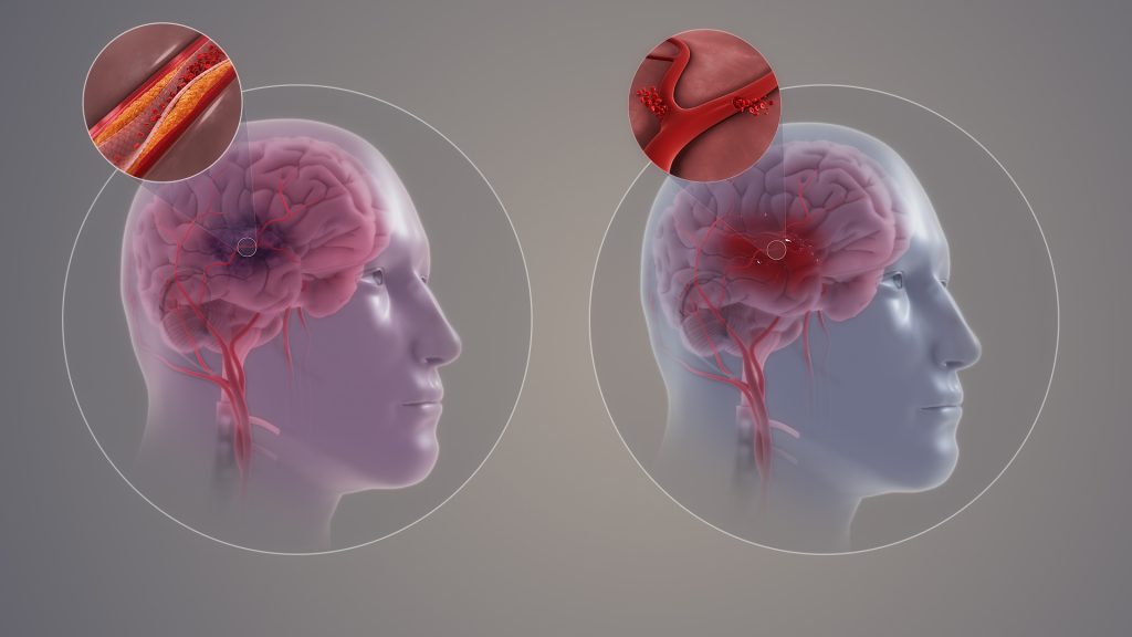
Ischaemic and haemorrhagic stroke. Credit: Scientific Animations CC4.0

Contrary to the commonly-held view, the brain does not have the ability to rewire itself to compensate for conditions such as stroke, loss of sight or an amputation, say scientists in the journal eLife.

Professors Tamar Makin of Cambridge University and John Krakauer of Johns Hopkins University argue that the notion that the brain, in response to injury or deficit, can reorganise itself and repurpose particular regions for new functions, is fundamentally flawed – despite being commonly cited in scientific textbooks. Instead, they argue that what is occurring is merely the brain being trained to utilise already existing, but latent, abilities.

One of the most common examples given is where a person loses their sight – or is born blind – and the visual cortex, previously specialised in processing vision, is rewired to process sounds, allowing the individual to use a form of ‘echolocation’ to navigate a cluttered room. Another common example is of people who have had a stroke and are initially unable to move their limbs repurposing other areas of the brain to allow them to regain control.

Krakauer, Director of the Center for the Study of Motor Learning and Brain Repair at Johns Hopkins University, said: “The idea that our brain has an amazing ability to rewire and reorganise itself is an appealing one. It gives us hope and fascination, especially when we hear extraordinary stories of blind individuals developing almost superhuman echolocation abilities, for example, or stroke survivors miraculously regaining motor abilities they thought they’d lost.

“This idea goes beyond simple adaptation, or plasticity – it implies a wholesale repurposing of brain regions. But while these stories may well be true, the explanation of what is happening is, in fact, wrong.”

In their article, Makin and Krakauer look at a ten seminal studies that purport to show the brain’s ability to reorganise. They argue, however, that while the studies do indeed show the brain’s ability to adapt to change, it is not creating new functions in previously unrelated areas – instead it’s utilising latent capacities that have been present since birth.

For example, a 1980s study by Professor Michael Merzenich at University of California, San Francisco looked at what happens when a hand loses a finger. The hand has a particular representation in the brain, with each finger appearing to map onto a specific brain region. Remove the forefinger, and the area of the brain previously allocated to this finger is reallocated to processing signals from neighbouring fingers, argued Merzenich – in other words, the brain has rewired itself in response to changes in sensory input.

Not so, says Makin, whose own research provides an alternative explanation.

In a study published in 2022, Makin used a nerve blocker to temporarily mimic the effect of amputation of the forefinger in her subjects. She showed that even before amputation, signals from neighbouring fingers mapped onto the brain region ‘responsible’ for the forefinger — in other words, while this brain region may have been primarily responsible for process signals from the forefinger, it was not exclusively so. All that happens following amputation is that existing signals from the other fingers are ‘dialled up’ in this brain region.

Makin, from the Medical Research Council (MRC) Cognition and Brain Sciences Unit at the University of Cambridge, said: “The brain’s ability to adapt to injury isn’t about commandeering new brain regions for entirely different purposes. These regions don’t start processing entirely new types of information. Information about the other fingers was available in the examined brain area even before the amputation, it’s just that in the original studies, the researchers didn’t pay much notice to it because it was weaker than for the finger about to be amputated.”

Another compelling counterexample to the reorganisation argument is seen in a study of congenitally deaf cats, whose auditory cortex appears to be repurposed to process vision. But when they are fitted with a cochlear implant, this brain region immediately begins processing sound once again, suggesting that the brain had not, in fact, rewired.

Examining other studies, Makin and Krakauer found no compelling evidence that the visual cortex of individuals that were born blind or the uninjured cortex of stroke survivors ever developed a novel functional ability that did not otherwise exist.

Makin and Krakauer do not dismiss stories such as blind people navigating using hearing, or individuals who have experienced a stroke regain their motor functions. They argue instead that rather than completely repurposing regions for new tasks, the brain is enhancing or modifying its pre-existing architecture — and it is doing this through repetition and learning.

Understanding the true nature and limits of brain plasticity is crucial, both for setting realistic expectations for patients and for guiding clinical practitioners in their rehabilitative approaches, they argue.

Makin added: “This learning process is a testament to the brain’s remarkable – but constrained – capacity for plasticity. There are no shortcuts or fast tracks in this journey. The idea of quickly unlocking hidden brain potentials or tapping into vast unused reserves is more wishful thinking than reality. It’s a slow, incremental journey, demanding persistent effort and practice. Recognising this helps us appreciate the hard work behind every story of recovery and adapt our strategies accordingly.

“So many times, the brain’s ability to rewire has been described as ‘miraculous’ – but we’re scientists, we don’t believe in magic. These amazing behaviours that we see are rooted in hard work, repetition and training, not the magical reassignment of the brain’s resources.”

The original text of this story is licensed under a Creative Commons Licence.

Source: University of Cambridge

Understanding Mechanisms Driving Bone Density Loss in Amputee Soldiers

Photo by MART PRODUCTION

Combat-related injuries to bone are common in military personnel and can lead to pain and disability. Results from a new study in the Journal of Bone and Mineral Research suggest that amputations for such injuries may negatively affect bone mass.

Traumatic amputation from combat injuries has the potential to lead to osteoporosis through not only systemic inflammation and hormonal changes but also altered loading. Although a documented long-term complication of lower limb amputation is osteoporosis, this is often observed in older less active subjects with comorbidities, thus it is unknown whether this is secondary to systemic changes or changes to the loading environment.

In the study of 575 male adult UK military personnel with combat-related traumatic injuries and 562 without such injuries, veterans who sustained traumatic amputations often had low bone density in the hip region. Changes in bone health appeared to be mechanically driven rather than systemic and were only evident in those with lower limb amputations.

“We hope these results will drive further research into ways to reverse bone mineral density changes,” said co-author Group Captain Alex Bennett, Defence Professor of Rehabilitation, Defence Medical Rehabilitation Centre. “We need to investigate the role of prosthetics and exercise in reversing bone mineral density loss to reduce the longer-term risk of hip fracture. Because systemic treatments like bisphosphonates are not indicated in this young population with bone mineral density loss, it is important to understand other ways to reduce their hip fracture risk.”

Source: Wiley

Oldest Known Successful Amputation Dates Back 31 000 Years

Mariano, Public domain, via Wikimedia Commons

At a site in Borneo, archaeologists have unearthed the oldest case of surgical amputation to date, a remarkable feat of prehistoric medical practice.

Published in Nature, the researchers describe the skeletal remains of a young adult found in a cave in Borneo, who had part of the left lower leg and left foot amputated, probably as a child, at least 31 000 years ago. The person lived for at least another six to nine years after the procedure, surviving into adulthood where they died from an unknown cause, possibly in their 20s.

The prehistoric surgery is a remarkable feat; preventing infections is difficult even in modern surgical amputations.

Study co-lead author, Dr Melandri Vlok, at University of Sydney said the find is “incredibly exciting and unexpected.”

“The discovery implies that at least some modern human foraging groups in tropical Asia had developed sophisticated medical knowledge and skills long before the Neolithic farming transition,” said bioarchaeologist Dr Vlok, an expert in ancient skeletons.

The skeleton of the young adult was carefully buried within LiangTebo cave – located Borneo in East Kalimantan, in a limestone karst area home to some of the world’s earliest dated rock art.

The bones were uncovered by archaeologists just days before borders closed for the COVID pandemic in March 2020. Dr Vlok was invited to study the bones when they were brought back to Australia.

“No one told me they had not found the left foot in the grave,” Dr Vlok said. “They kept it hidden from me to see what I would find.”

As Dr Vlok laid the bones out, the left leg looked withered, and was the size of a child’s, but the individual was an adult. She unwrapped the part of the leg that contained the stump and noticed the cut was clean, well healed and had no evidence of any infection. “The chances the amputation was an accident was so infinitely small,” Dr Vlok said. “The only conclusion was this was stone age surgery.”

Dr Vlok ran to the office to tell her research colleagues what she had found. “I told them I thought it looked like a surgical amputation,” she said. “It wasn’t until then that they said they already knew the foot was missing.” Dr Vlok had just confirmed their suspicions. The foot was never placed in the grave to begin with.

An accident in difficult terrain

While the cause for the amputation was unclear, the individual also had a very well healed neck fracture and trauma to their collar bone that may have occurred during the same event, said Dr Vlok.

“An accident, such as a rock fall may have caused the injuries, and it was clearly recognised by the community that the foot had to be taken off for the child to survive,” she said.

The location of the cave is surrounded by extremely rugged terrain, and accessing the site was challenging, making the individual’s survival after the the surgery even more remarkable. The finding will provide even more insight into prehistoric medicine, the researchers said.

Source: University of Sydney

A Soft Robotic Design for Diabetic Amputee Pain Relief

Proof-of-concept rendering (left) and photo (right) of the prototype of the new microfluidics-enabled soft robotic prosthesis for lower limb amputees.
Credit: Waterloo Microfluidics Laboratory at University of Waterloo

Diabetic amputations often involve neuropathy, and patients detect damage resulting from an ill-fitting prosthesis, leading to further amputation. To solve this, in Biomicrofluidics, scientists described a new type of prosthetic using microfluidics-enabled soft robotics that reduces skin ulcerations and pain in patients who have had an amputation between the ankle and knee.

More than 80% of lower-limb amputations are due to diabetic foot ulcers, and the lower limb is known to swell at unpredictable times, resulting in volume changes of 10% or more.

Typically, the prosthesis used after amputation includes fabric and silicone liners that can be added or removed to improve fit. The amputee needs to manually change the liners, but neuropathy leading to poor sensation makes this difficult and can lead to more damage to the remaining limb.

“Rather than creating a new type of prosthetic socket, the typical silicon/fabric limb liner is replaced with a single layer of liner with integrated soft fluidic actuators as an interfacing layer,” said author Carolyn Ren, from the University of Waterloo. “These actuators are designed to be inflated to varying pressures based on the anatomy of the residual limb to reduce pain and prevent pressure ulcerations.”

The scientists started off with pneumatic actuators to adjust the pressure of the prosthetic socket, but it was quite heavy.

To reduce weight, the group miniaturised the actuators, designing a microfluidic chip with 10 integrated pneumatic valves to control each actuator. The full system is controlled by a miniature air pump and two solenoid valves that provide air to the microfluidic chip. The control box is small and light enough to be worn as part of the prosthesis.

Prosthetics experts provided a detailed map of desired pressures for the prosthetic socket. The group carried out extensive measurements of the contact pressure provided by each actuator and compared these to the desired pressure for a working prosthesis.

All of the actuators produced the right pressures suggesting the new device will work well in the field, with the next step being a more accurate biological model.

The group plans additional research to integrate pressure sensors directly into the prosthetic liner, perhaps using newly available knitted soft fabric that incorporates pressure sensing material.

Source: American Institute of Physics

New Surgery Improves Prosthetic Use and Reduces Pain

A new type of surgery that links muscles together may improve the precision of prosthetic use and also relieve pain.

In typical amputations, the muscle pairs (such as triceps and biceps) that work together to control the joints are severed. However, an MIT team has discovered that reconnecting these muscles that are in an agonistic-antagonistic (‘push-pull’) relationship improves the sensory feedback and thus precision of the affected limb.

“When one muscle contracts, the other one doesn’t have its antagonist activity, so the brain gets confusing signals,” explained Srinivasan, a former member of the Biomechatronics group now working at MIT’s Koch Institute for Integrative Cancer Research. “Even with state-of-the-art prostheses, people are constantly visually following the prosthesis to try to calibrate their brains to where the device is moving.”

The 15 patients who received the AMI surgery were able to flex their prosthetic ankle joint with more precision than those without it, who were only able to fully extend or flex their joint.

“Through surgical and regenerative techniques that restore natural agonist-antagonist muscle movements, our study shows that persons with an AMI amputation experience a greater phantom joint range of motion, a reduced level of pain, and an increased fidelity of prosthetic limb controllability,” says Hugh Herr, a professor of media arts and sciences, head of the Biomechatronics group in the Media Lab, and the senior author of the paper.

The surgery also had a completely unexpected benefit: the reduction of pain in the amputated area, which can be from neuromas or phantom limb pain. Phantom limb pain can occur in 80% of amputess. Six of the 15 AMI patients reported zero pain. This may be significant as in the five centuries since phantom limb pain was first described, there has not been much advancement in the understanding of it.

“Our study wasn’t specifically designed to achieve this, but it was a sentiment our subjects expressed over and over again. They had a much greater sensation of what their foot actually felt like and how it was moving in space,” Srinivasan says. “It became increasingly apparent that restoring the muscles to their normal physiology had benefits not only for prosthetic control, but also for their day-to-day mental well-being.”

To treat patients who had received the traditional amputation surgery, the team is also working on using muscle grafts to create a ‘regenerative AMI’ procedure that restores the effect of agonist and antagonist muscles.

Source: Medical Xpress

Journal information: Shriya S. Srinivasan el al., “Neural interfacing architecture enables enhanced motor control and residual limb functionality postamputation,” PNAS (2021). www.pnas.org/cgi/doi/10.1073/pnas.2019555118