Tag: ALS

New Brain–computer Interface Allows Man with ALS to ‘Speak’ Again

A new brain-computer interface (BCI) developed at UC Davis Health translates brain signals into speech with up to 97% accuracy – the most accurate system of its kind. The researchers implanted sensors in the brain of a man with severely impaired speech due to amyotrophic lateral sclerosis (ALS). The man was able to communicate his intended speech within minutes of activating the system.

A study about this work was published in the New England Journal of Medicine.

ALS, also known as Lou Gehrig’s disease, affects the nerve cells that control movement throughout the body. The disease leads to a gradual loss of the ability to stand, walk and use one’s hands. It can also cause a person to lose control of the muscles used to speak, leading to a loss of understandable speech.

The new technology is being developed to restore communication for people who can’t speak due to paralysis or neurological conditions like ALS. It can interpret brain signals when the user tries to speak and turns them into text that is ‘spoken’ aloud by the computer.

“Our BCI technology helped a man with paralysis to communicate with friends, families and caregivers,” said UC Davis neurosurgeon David Brandman. “Our paper demonstrates the most accurate speech neuroprosthesis (device) ever reported.”

Brandman is the co-principal investigator and co-senior author of this study. He is an assistant professor in the UC Davis Department of Neurological Surgery and co-director of the UC Davis Neuroprosthetics Lab.

The new BCI breaks the communication barrier

When someone tries to speak, the new BCI device transforms their brain activity into text on a computer screen. The computer can then read the text out loud.

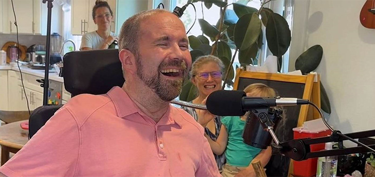
To develop the system, the team enrolled Casey Harrell, a 45-year-old man with ALS, in the BrainGate clinical trial. At the time of his enrolment, Harrell had weakness in his arms and legs (tetraparesis). His speech was very hard to understand (dysarthria) and required others to help interpret for him.

In July 2023, Brandman implanted the investigational BCI device. He placed four microelectrode arrays into the left precentral gyrus, a brain region responsible for coordinating speech. The arrays are designed to record the brain activity from 256 cortical electrodes.

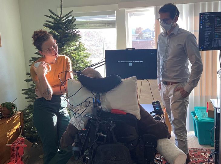
“We’re really detecting their attempt to move their muscles and talk,” explained neuroscientist Sergey Stavisky. Stavisky is an assistant professor in the Department of Neurological Surgery. He is the co-director of the UC Davis Neuroprosthetics Lab and co-principal investigator of the study. “We are recording from the part of the brain that’s trying to send these commands to the muscles. And we are basically listening into that, and we’re translating those patterns of brain activity into a phoneme – like a syllable or the unit of speech – and then the words they’re trying to say.”

Casey Harrell with his personal assistant Emma Alaimo and UC Davis neuroscientist Sergey Stavisky

Faster training, better results

Despite recent advances in BCI technology, efforts to enable communication have been slow and prone to errors. This is because the machine-learning programs that interpreted brain signals required a large amount of time and data to perform.

“Previous speech BCI systems had frequent word errors. This made it difficult for the user to be understood consistently and was a barrier to communication,” Brandman explained. “Our objective was to develop a system that empowered someone to be understood whenever they wanted to speak.”

Harrell used the system in both prompted and spontaneous conversational settings. In both cases, speech decoding happened in real time, with continuous system updates to keep it working accurately.

The decoded words were shown on a screen. Amazingly, they were read aloud in a voice that sounded like Harrell’s before he had ALS. The voice was composed using software trained with existing audio samples of his pre-ALS voice.

At the first speech data training session, the system took 30 minutes to achieve 99.6% word accuracy with a 50-word vocabulary.

“The first time we tried the system, he cried with joy as the words he was trying to say correctly appeared on-screen. We all did,” Stavisky said.

In the second session, the size of the potential vocabulary increased to 125 000 words. With just an additional 1.4 hours of training data, the BCI achieved a 90.2% word accuracy with this greatly expanded vocabulary. After continued data collection, the BCI has maintained 97.5% accuracy.

“At this point, we can decode what Casey is trying to say correctly about 97% of the time, which is better than many commercially available smartphone applications that try to interpret a person’s voice,” Brandman said. “This technology is transformative because it provides hope for people who want to speak but can’t. I hope that technology like this speech BCI will help future patients speak with their family and friends.”

The study reports on 84 data collection sessions over 32 weeks. In total, Harrell used the speech BCI in self-paced conversations for over 248 hours to communicate in person and over video chat.

“Not being able to communicate is so frustrating and demoralising. It is like you are trapped,” Harrell said. “Something like this technology will help people back into life and society.”

“It has been immensely rewarding to see Casey regain his ability to speak with his family and friends through this technology,” said the study’s lead author, Nicholas Card. Card is a postdoctoral scholar in the UC Davis Department of Neurological Surgery.

“Casey and our other BrainGate participants are truly extraordinary. They deserve tremendous credit for joining these early clinical trials. They do this not because they’re hoping to gain any personal benefit, but to help us develop a system that will restore communication and mobility for other people with paralysis,” said co-author and BrainGate trial sponsor-investigator Leigh Hochberg. Hochberg is a neurologist and neuroscientist at Massachusetts General Hospital, Brown University and the VA Providence Healthcare System.

Brandman is the site-responsible principal investigator of the BrainGate2 clinical trial. The trial is enrolling participants. To learn more about the study, visit braingate.org or contact braingate@ucdavis.edu.

Source: UC Davis Health

A Genetic Treatment for ALS That Restores Key Protein May Be Possible

DNA repair
Source: Pixabay/CC0

In virtually all persons with amyotrophic lateral sclerosis (ALS) and in up to half of all cases of Alzheimer’s disease (AD) and frontotemporal dementia, a protein called TDP-43 is lost from its normal location in the nucleus of the cell. In turn, this triggers the loss of stathmin-2, a protein crucial to regeneration of neurons and the maintenance of their connections to muscle fibres.

Writing in Science, a team of scientists demonstrate that stathmin-2 loss can be rescued using designer DNA drugs that restore normal processing of protein-encoding RNA.

“With mouse models we engineered to misprocess their stathmin-2 encoding RNAs, like in these human diseases, we show that administration of one of these designer DNA drugs into the fluid that surrounds the brain and spinal cord restores normal stathmin-2 levels throughout the nervous system,” said senior study author Don Cleveland, PhD, Distinguished Professor of Medicine, Neurosciences and Cellular and Molecular Medicine at University of California San Diego School of Medicine.

Cleveland is broadly credited with developing the concept of designer DNA drugs, which act to either turn on or turn off genes associated with many degenerative diseases of the aging human nervous system, including ALS, AD, Huntington’s disease and cancer.

Several designer DNA drugs are currently in clinical trials for multiple diseases. One such drug has been approved to treat a childhood neurodegenerative disease called spinal muscular atrophy.

The new study builds upon ongoing research by Cleveland and others regarding the role and loss of TDP-43, a protein associated with ALS, AD and other neurodegenerative disorders. In ALS, TDP-43 loss impacts the motor neurons that innervate and trigger contraction of skeletal muscles, causing them to degenerate, eventually resulting in paralysis.

“In almost all of instances of ALS, there is aggregation of TDP-43, a protein that functions in maturation of the RNA intermediates that encode many proteins. Reduced TDP-43 activity causes misassembly of the RNA-encoding stathmin-2, a protein required for maintenance of the connection of motor neurons to muscle,” said Cleveland.

“Without stathmin-2, motor neurons disconnect from muscle, driving paralysis that is characteristic of ALS. What we have now found is that we can mimic TDP-43 function with a designer DNA drug, thereby restoring correct stathmin-2 RNA and protein level in the mammalian nervous system.”

Specifically, the researchers edited genes in mice to contain human STMN2 gene sequences and then injected antisense oligonucleotides – small DNA or RNA pieces that can bind to specific RNA molecules, blocking their ability to make a protein or changing how their final RNAs are assembled – into cerebral spinal fluid. The injections corrected STMN2 pre-mRNA misprocessing and restored stathmin-2 protein expression fully independent of TDP-43 function.

“Our findings lay the foundation for a clinical trial to delay paralysis in ALS by maintaining stathmin-2 protein levels in patients using our designer DNA drug,” Cleveland said.

Source: University of California – San Diego

Proportions of Immune Cells can Predict ALS Progression

Source: Pixabay CC0

By measuring the proportions of certain immune cells in the cerebrospinal fluid when diagnosing amyotrophic lateral sclerosis (ALS), it is possible to predict the disease’s speed of progression, according to a study from Karolinska Institutet published in Nature Communications.

ALS is a rare, but fatal disease that affects the nerve cells and leads to paralysis of voluntary muscles and death. This new research offers a way to predict the course of the disease in ALS patients.

Between March 2016 and March 2020, researchers collected fresh blood and cerebrospinal fluid from 89 patients in Stockholm who had recently been diagnosed with ALS, and followed-up until October 2020.

The study shows that a high proportion of so-called effector T cells are associated with a low survival rate. At the same time, a high proportion of activated regulatory T cells indicate a protective role against the rapid disease progression. The findings provide new evidence for the involvement of T cells in the course of the disease and show that certain types of effector T cells accumulate in the cerebrospinal fluid of ALS patients.

“The study could contribute to the development of new treatments that target immune cells to slow down the course of the disease,” says study first author Solmaz Yazdani, a doctoral student at the Institute of Environmental Medicine at Karolinska Institutet.

The next step in her research is to study how T cells contribute to the course of the disease.

“We have plans to collect samples from these individuals to study changes in the immune cells over time. In addition, we want to study effector T cells in more detail to understand their role in ALS.”

Source: Karolinska Institutet