Tag: age-related macular degeneration

Why Some Patients Respond Poorly to Wet Macular Degeneration Treatment
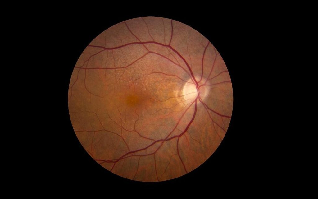
Retina showing reticular pseudodrusen. Although they can infrequently appear in individuals with no other apparent pathology, their highest rates of occurrence are in association with age-related macular degeneration (AMD), for which they hold clinical significance by being highly correlated with end-stage disease sub-types, choroidal neovascularisation and geographic atrophy. Credit: National Eye Institute

A new study from researchers at Wilmer Eye Institute, Johns Hopkins Medicine explains not only why some patients with wet age-related macular degeneration (or “wet” AMD) fail to have vision improvement with treatment, but also how an experimental drug could be used with existing wet AMD treatments to save vision.

Wet AMD, one of two kinds of AMD, is a progressive eye condition caused by an overgrowth of blood vessels in the retina. Such blood vessels – caused by an overexpression of a protein known as VEGF that leads to blood vessel growth – then leak fluid or bleed and damage the retina, causing vision loss.

Despite the severe vision loss often experienced by people with wet AMD, less than half of patients treated with monthly eye injections, known as anti-VEGF therapies, show any major vision improvements. Additionally, for those who do benefit with improved vision, most will lose those gains over time.

Now, in the full report published November 4 in the Proceedings of the National Academy of Sciences, the Wilmer-led team of researchers share how such anti-VEGF therapies may actually contribute to lack of vision improvements by triggering the overexpression of a second protein. Known as ANGPTL4, the protein is similar to VEGF, as it can also stimulate overproduction of abnormal blood vessels in the retina.

“We have previously reported that ANGPTL4 was increased in patients who did not respond well to anti-VEGF treatment,” says Akrit Sodhi, MD, PhD, corresponding author and associate professor of ophthalmology at the Johns Hopkins University School of Medicine and the Wilmer Eye Institute. “What we saw in this paper was a paradoxical increase of ANGPTL4 in patients that received anti-VEGF injections – the anti-VEGF therapy itself turned on expression of this protein.”

The team compared VEGF and ANGPTL4 levels in the eye fluid of 52 patients with wet AMD at various stages of anti-VEGF treatment. Prior to anti-VEGF injections, patients with wet AMD had high levels of ANGPTL4 and VEGF proteins. After treatment, their VEGF levels predictably decreased, yet ANGPTL4 levels rose higher, indicating ANGPTL4 remained active following the anti-VEGF injections and the treatments contributed to an increase in ANGPTL4. Such ANGPTL4 activity can lead to blood vessel overgrowth and lack of vision improvement.

The team then investigated ways to bridge the gap between patients with increased ANGPTL4 following anti-VEGF treatments by testing the experimental drug 32-134D in mice with wet AMD. The drug decreases levels of a third protein, HIF-1, known to be involved in wet AMD and diabetic eye disease for its role in switching on VEGF production. Researchers believed the HIF-inhibitor 32-134D would have a similar effect on ANGPTL4 following anti-VEGF treatment, since ANGPTL4 production is also turned on by HIF-1.

In mice treated with 32-134D, the team observed a decrease in HIF-1 levels and VEGF, as well as decreased levels of ANGPTL4 and blood vessel overgrowth. Mice treated only with anti-VEGF therapies corroborated the team’s findings in human patients: levels of VEGF were lower, yet ANGPTL4 levels rose, preventing anti-VEGF therapies from fully working to prevent blood vessel growth (and vision loss). Researchers also determined that combining 32-134D with anti-VEGF treatments prevented the increase in HIF-1, VEGF and ANGPTL4. This treatment combination was more effective than either drug alone, showing promise for treating wet AMD.

“This work exposes a way to improve anti-VEGF therapy for all patients and potentially help a subset of patients with wet AMD who still lose vision over time despite treatment,” Sodhi says. “Our hope is that this [project] will further the three goals we have related to wet AMD: make current therapies as effective as possible, identify new therapies, and prevent people from ever getting wet AMD.”

Source: John Hopkins Medicine

In New Study, Supplements Slow Late-stage Age-related Macular Degeneration

Retina showing reticular pseudodrusen. Although they can infrequently appear in individuals with no other apparent pathology, their highest rates of occurrence are in association with age-related macular degeneration (AMD), for which they hold clinical significance by being highly correlated with end-stage disease sub-types, choroidal neovascularisation and geographic atrophy. Credit: National Eye Institute

In a new analysis of data, researchers at the National Institutes of Health (NIH) have found that taking a daily supplement containing antioxidant vitamins and minerals slows progression of late-stage dry age-related macular degeneration (AMD), potentially helping people with late-stage disease preserve their central vision.

Age-related macular degeneration affects the macula, the part of the retina that provides central vision.

Researchers reviewed the original retinal scans of participants in the Age-Related Eye Diseases Studies (AREDS and AREDS2) and found that, for people with late-stage dry AMD, taking the antioxidant supplement slowed expansion of geographic atrophy regions towards the central foveal region of the retina. The study was published in the journal Ophthalmology.

“We’ve known for a long time that AREDS2 supplements help slow the progression from intermediate to late AMD. Our analysis shows that taking AREDS2 supplements can also slow disease progression in people with late dry AMD,” said Tiarnan Keenan, MD, PhD, of NIH’s National Eye Institute (NEI) and lead author of the study. “These findings support the continued use of AREDS2 supplements by people with late dry AMD.”

In their new analysis, the researchers reviewed the original retinal scans of participants in the AREDS (total 318 participants, 392 eyes) and AREDS2 (total 891 participants, 1210 eyes) trials who developed dry AMD, calculating the position and expansion rate of their regions of geographic atrophy. For those people who developed geographic atrophy in their central vision, the supplements had little benefit. But for the majority who developed geographic atrophy far from the fovea, the supplements slowed the rate of expansion towards the fovea by approximately 55% over an average of three years.

In early and intermediate AMD, the light-sensing retina at the back of the eye develops small yellow deposits of fatty proteins called drusen. When the disease progresses to the late stage, people can develop leaky blood vessels (“wet” AMD) or can lose regions of light-sensitive cells in the retina (“dry” AMD). The geographic atrophy in these regions slowly expands over time, causing people to progressively lose their central vision.

The original AREDS trial found that a supplement formula containing antioxidants (vitamin C, E, and beta-carotene), along with zinc and copper, could slow progression of intermediate to late-stage AMD. The subsequent AREDS2 trial found that substituting the antioxidants lutein and zeaxanthin for beta-carotene improved the efficacy of the supplement formula and eliminated certain risks. At the time, neither trial detected any further benefit once participants had developed late-stage disease.

However, that original analysis did not account for a phenomenon in the dry form of late AMD called “foveal sparing.”  While all regions of the retina are sensitive to light, the region that gives us the highest acuity central vision is called the fovea. Many people with dry AMD first develop geographic atrophy outside this foveal region, and they only lose their central vision when the geographic atrophy regions expand into the foveal area.

“Our high acuity central vision is essential for tasks like reading and driving. Given that there are few therapeutic options for people with late-stage dry AMD to retain or restore their vision, antioxidant supplementation is a simple step that may slow central vision loss, even for those with late disease,” Keenan said. “We plan to confirm these findings in a dedicated clinical trial in the near future.”

Learn more about AREDS and AREDS2

Source: NIH/National Eye Institute

Trial of Minocycline for Dry Age-related Macular Degeneration Flops

Retina showing reticular pseudodrusen. Although they can infrequently appear in individuals with no other apparent pathology, their highest rates of occurrence are in association with age-related macular degeneration (AMD), for which they hold clinical significance by being highly correlated with end-stage disease sub-types, choroidal neovascularisation and geographic atrophy. Credit: National Eye Institute

The drug minocycline, an antibiotic that also decreases inflammation, failed to slow vision loss or expansion of geographic atrophy in people with dry age-related macular degeneration (AMD), according to a phase II clinical study at the National Eye Institute (NEI), part of the National Institutes of Health.

Dry AMD affects the macula, the part of the retina that allows for clear central vision. In people with dry AMD, patches of photoreceptors and their nearby support cells begin to die off, leaving regions known as geographic atrophy. Over time, these regions expand, causing people to lose more and more of their central vision.

Microglia, immune cells that help maintain tissue and clear up debris, are present at higher levels around damaged retinal regions in people with dry AMD than in people without AMD. Scientists have suggested that inflammation – and particularly microglia – may be driving the expansion of geographic atrophy regions.

This study, led by Tiarnan Keenan, MD, PhD, a Stadtman Tenure-Track Investigator at the NEI’s Division of Epidemiology and Clinical Applications, tested whether inhibiting microglia with minocycline might help slow geographic atrophy expansion and its corresponding vision loss.

The trial enrolled 37 participants at the NIH Clinical Center in Bethesda, Maryland, and at the Bristol Eye Hospital, United Kingdom.

After a nine-month period where the researchers tracked each participant’s rate of geographic atrophy expansion, the participants took twice-daily doses of minocycline for two years.

The researchers compared each participant’s rate of geographic atrophy expansion while taking minocycline to their baseline rate, and found there was no difference in geographic atrophy expansion rate or vision loss with minocycline.

Previous studies have shown that minocycline can help reduce inflammation and microglial activity in the eye, including the retina.

The drug has shown beneficial effects for conditions such as diabetic retinopathy, but has not previously been tested for dry AMD.

Source: NIH/National Eye Institute

Eyedrops instead of Injections for Age-related Macular Degeneration

Retina showing reticular pseudodrusen. Although they can infrequently appear in individuals with no other apparent pathology, their highest rates of occurrence are in association with age-related macular degeneration (AMD), for which they hold clinical significance by being highly correlated with end-stage disease sub-types, choroidal neovascularisation and geographic atrophy. Credit: National Eye Institute

A new compound potentially could offer an alternative to injections for the millions of people who suffer from wet age-related macular degeneration (AMD). The condition causes vision loss due to the uncontrolled growth and leakage of blood vessels in the back of the eye. A new paper in Cell Reports Medicine finds that a small-molecule inhibitor can reverse damage from AMD and promote regenerative and healing processes.

The drug can also be delivered via eyedrops – an improvement over current treatments for AMD, which require repeated injections into the eye.

“The idea was to develop something that can be more patient-friendly and doesn’t require a visit to the doctor’s office,” said lead researcher Yulia Komarova, associate professor of pharmacology at University of Illinois Chicago.

Komarova’s compound targets the protein End Binding-3 in endothelial cells, which line the inside of blood vessels. In the new study, the researchers looked at whether inhibiting EB3 function could stop the damaging leakage associated with wet AMD.

Using computational drug design methods, the team developed a small molecule drug, End Binding-3 inhibitor (EBIN), that could be delivered externally via eyedrops instead of by injection. They then tested its effectiveness in animal models of wet AMD, finding that twice-daily treatment reduced eye damage within 2–3 weeks.

Further investigation found that the inhibitor worked by rolling back aging-related genetic modifications. Aging causes inflammation and hypoxia in the eye that leads to changes in gene expression associated with the cellular effects and symptoms of wet AMD. Komarova and colleagues found that the EB3 inhibitor they developed reversed these epigenetic changes, restoring gene expression to a normal, healthy state.

“We reduce the effects of the stressor on endothelial cells and we improve regenerative processes, accelerating healing,” Komarova said. “That can be tremendous for the function of the cells.”

Because blood vessel leakage and hypoxic stress also drive many other medical conditions, Komarova’s group is interested in testing the inhibitor in models of acute lung injury, diabetic retinopathy, stroke, heart disease and even the general effects of aging on the brain. They are also exploring whether an implantable lens, similar to a contact lens, could deliver the drug to the eye more effectively than eyedrops.

Source: University of Illinois Chicago

Type of Macular Degeneration Linked to Cardiovascular Disease

Credit: National Eye Institute

Patients with a certain subtype of age-related macular degeneration (AMD) are at significant risk for cardiovascular disease and stroke, according to new research published in Retina.

“For the last three decades researchers have suggested an association between AMD and cardiovascular disease, but there has been no conclusive data on this until now. Our retinal team answered this important question by focusing on two different varieties of AMD that can be seen with advanced retinal imaging. We discovered that only one form of AMD, that with subretinal drusenoid deposits, is tightly connected to high-risk vascular diseases, and the other form, known as drusen, is not,” explained lead author R. Theodore Smith, MD, PhD, Professor at Mount Sinai. “If ophthalmologists diagnose or treat someone with the specific subretinal drusenoid deposits form of AMD, but who otherwise seems well, that patient may have significant undetected heart disease, or possibly carotid artery stenosis that could result in a stroke. We foresee that in the future, as an improved standard of care, such patients will be considered for early referral to a cardiologist for evaluation and possibly treatment.”

AMD is the leading cause of visual impairment and blindness over the age of 65. Drusen is one major form of early AMD: small yellow cholesterol deposits form in a layer under the retina, depriving it of blood and oxygen, leading to vision loss. Drusen formation can be slowed by appropriate vitamin supplementation.

The other major form of early AMD is the presence of subretinal drusenoid deposits (SDD), which is lesser known, which needs advanced retinal imaging to detect. These deposits are also made of fatty lipids and other materials, but form in a different layer beneath the light sensitive retina cells, where they are also associated with vision loss. There is no known treatment for SDD at present.

Mount Sinai researchers analysed 126 patients with AMD, using optical coherence tomography (OCT) which captures high-resolution cross-sectional scans of the retina. Patients also answered health history questionnaires. Of the patients on the study, 62 had SDD and 64 had drusen; 51 of the 126 total patients (40%) reported having cardiovascular disease or a past stroke, and most (66%) of those patients had SDD. By contrast, of the 75 patients who did not have known heart disease or stroke, relatively few (19%) had SDD. The odds of patients with cardiovascular disease or stroke having SDD was three times than in patients without.

The researchers suggested that the underlying cardiovascular disease likely compromises blood circulation in the eye, leading to the SDDs beneath the retina.

“We believe poor ocular circulation that causes SDDs is a manifestation of underlying vascular disease. This has important public health implications and can facilitate population screening and disease detection with major impact,” explained author Jagat Narula, MD, PhD, Associate Dean of Global Affairs and Professor of Medicine (Cardiology), and Radiology, at the Icahn School of Medicine at Mount Sinai. “Seen in an eye clinic, such patients should be prompted to see a cardiologist. On the other hand, if clinically substantiated in prospective studies, SDDs could emerge as a risk marker for underlying vascular disease in asymptomatic patients in primary care or a cardiology clinic. The temporal relationship between SDDs and macrovascular disease will also need to be established in prospective studies which are currently in progress.”

Analysis of patient blood samples revealed genetic risk factors may also play a role in SDD cases in addition to vascular causes. Specifically, they found that the ARMS2 gene acted independently of vascular disease to cause SDD in some patients.

“This study further demonstrates that AMD is not a single condition or an isolated disease, but is often a signal of systemic malfunction which could benefit from targeted medical evaluation in addition to localised eye care,” noted Richard B. Rosen, MD, Chief of the Retina Service for the Mount Sinai Health System. “It helps bring us one step closer to unraveling the mystery of this horrible condition which robs so many patients of the pleasure of good vision during their later years. “

Source: The Mount Sinai Hospital / Mount Sinai School of Medicine

Supplementation Effective in Slowing Age-related Macular Degeneration

Credit: National Eye Institute

A pair of major studies established that dietary supplements can slow progression of age-related macular degeneration (AMD). In a new report published in JAMA Ophthalmology, scientists went through 10 years of Age-Related Eye Disease Studies (AREDS2) data and showed that the AREDS2 formula, which substituted antioxidants lutein and zeaxanthin for beta-carotene, not only reduces risk of lung cancer due to beta-carotene, but is also more effective at reducing risk of AMD progression, compared to the original formula.

“Because beta-carotene increased the risk of lung cancer for current smokers in two NIH-supported studies, our goal with AREDS2 was to create an equally effective supplement formula that could be used by anyone, whether or not they smoke,” said Emily Chew, MD, lead author of the study report. “This 10-year data confirms that not only is the new formula safer, it’s actually better at slowing AMD progression.”

AMD is a degenerative disease of the retina, the light-sensitive tissue at the back of the eye. Progressive death of retinal cells in the macula, the part of the retina that provides clear central vision, eventually leads to blindness. Treatment can slow or reverse vision loss; however, no cure for AMD exists.

The original AREDS study, launched in 1996, showed that a dietary supplement formulation (50 mg vitamin C, 400 international units vitamin E, 2mg copper, 80mg zinc, and 15mg beta-carotene) could significantly slow the progression of AMD from moderate to late disease. However, two concurrent studies also revealed that people who smoked and took beta-carotene had a significantly higher risk of lung cancer than expected.

In AREDS2, begun in 2006, Dr Chew and colleagues compared the beta-carotene formulation to one with 10 mg lutein and 2 mg zeaxanthin instead. Like beta-carotene, lutein and zeaxanthin are antioxidants with activity in the retina. The beta-carotene-containing formation was only given to participants who had never smoked or who had quit smoking.

At the end of the five-year AREDS2 study period, the researchers concluded that lutein and zeaxanthin did not increase risk for lung cancer, and that the new formation could reduce the risk of AMD progression by about 26%. After the completion of the five-year study period, the study participants were all offered the final AREDS2 formation that included lutein and zeaxanthin instead of beta-carotene.

In this new report, the researchers followed up with 3883 of the original 4203 AREDS2 participants an extra five years from when the AREDS2 study ended in 2011, collecting information AMD progression, and lung cancer diagnosis. Even though all the participants had switched to the formula containing lutein and zeaxanthin after the end of the study period, the follow up study continued to show that beta-carotene increased risk of lung cancer for people who had ever smoked by nearly double. No increased risk for lung cancer was seen in those receiving lutein/zeaxanthin. In addition, after 10 years, the group originally assigned to receive lutein/zeaxanthin had an additional 20% reduced risk of progression to late AMD compared to those originally assigned to receive beta-carotene.

“These results confirmed that switching our formula from beta-carotene to lutein and zeaxanthin was the right choice,” said Dr Chew.

Source: NIH/National Eye Institute