Tag: adipose tissue

Immune Dysfunction as a Possible Aspect of PCOS Pathology

Photo by Sora Shimazaki on Pexels

The study “Androgens Modulate the Immune Profile in a Mouse Model of Polycystic Ovary Syndrome” sheds light on immune dysfunction as a possible aspect of PCOS pathology, which could constitute a novel target for therapies.

The study shows that hyperandrogenism, a key characteristic of polycystic ovary syndrome (PCOS), affects immune cell populations in reproductive, metabolic, and immunological tissues in a PCOS-like mouse model. These findings are of great importance as it is known that immune dysfunction is an essential part of reproductive complications and metabolic disease, which are very common among women with PCOS. 

However, as the study shows that hyperandrogenism affects different tissues in unique ways, any possible treatments would have to be carefully tailored to target specific tissue dysfunctions. Since concurrent treatment with an androgen receptor antagonist prevented many changes in the mouse model we used in our study, combination therapies that include both anti-androgens and other drugs that target specific altered immune pathways could be explored.

Alteration of immune cells in adipose tissue

One of the most fascinating findings was the clear alterations of immune cells in adipose tissue, despite an unaltered fat mass of the androgen exposed mice. It is well known that immune cells in adipose tissue contribute to insulin resistance in overweight and obese individuals, but here we have an insulin resistant mouse model that mimics normal weight women with PCOS. The impact of androgens on immune cells in adipose tissue is therefore very interesting considering the high prevalence of insulin resistance and type-2 diabetes among normal weight women with PCOS. Another rather surprising finding was the drastic decrease of eosinophils in the uterus, as these don’t express the androgen receptor. This shows that androgens play a broader and more complex role in modulating the immune environment than only through direct androgen receptor activation on immune cells. 

The next step will be to dissect the underlying mechanisms of the immune alterations, and to assess if these do contribute to the reproductive complications and metabolic comorbidities of PCOS. This will include further characterisation of immune cell changes to understand how their function could be affected and link these to reproductive and metabolic mechanisms. We also want to understand how the effect of androgens on eosinophils and mature NK cells is mediated, since neither of them expresses the androgen receptor. 

Source: Karolinska Institutet

Most Artificial Sweeteners Linked to Abdominal and Intramuscular Fat Increases

Source: Pixabay CC0

Artificial sweeteners have once again returned to the headlines with the WHO listing them as a possible carcinogen, Now, a long-term study on artificial sweeteners in diets published in the International Journal of Obesity has shown that, ironically, nearly all of them are linked to increased adiposity.

In the two decade long study, University of Minnesota researchers examined people’s regular dietary intake, with a focus on non-nutritive sweeteners commonly found in artificial sweeteners. They found that long-term consumption of aspartame, saccharin and diet beverages were linked to increased abdominal and intramuscular adiposity. However, the study found no significant association between the artificial sweetener sucralose and these measures of fat volume.

“This study showed that habitual, long-term intake of total and individual artificial sweetener intakes are related to greater volumes of adipose tissue, commonly known as body fat,” said Brian Steffen, PhD, MSCR, a professor in the Department of Surgery at the U of M Medical School and co-investigator on the funded grant. “This was found even after accounting for other factors, including how much a person eats or the quality of one’s diet.”

The study’s findings raise concerns about the recommendations from the American Diabetes Association and the American Heart Association that promote the replacement of added sugars with artificial sweeteners. Based on their results, the researchers recommend considering alternative approaches, as long-term artificial sweetener consumption may have potential health consequences.

“This is an especially timely study, given the World Health Organization’s recent warning of the potential health risks of aspartame,” said Lyn Steffen, PhD, MPH, a professor in the School of Public Health and principal investigator on the study. “These findings underscore the importance of finding alternatives to artificial sweeteners in foods and beverages, especially since these added sweeteners may have negative health consequences.”

The researchers say that more studies are needed to better understand the connection between artificial sweetener intake and increased body fat. Further research is warranted to explore the underlying mechanisms and gain clearer insights into how dietary habits affect metabolic health.

Source: University of Minnesota Medical School

Muscle Adiposity may Indicate Cognitive Decline in Aging

Photo by Kindel Media on Pexels

New research reveals that the level of muscle adiposity (fat content) may indicate a person’s likelihood of experiencing cognitive decline as they age. In the study published in the Journal of the American Geriatrics Society, a five-year increase in fat stored in the thigh muscle was a risk factor for cognitive decline.

This risk was independent of total weight, other fat deposits, and muscle characteristics (such as muscle strength or mass) and also independent of traditional dementia risk factors.

Investigators assessed muscle fat in 1634 adults 69–79 years of age at years 1 and 6 and evaluated their cognitive function at years 1, 3, 5, 8, and 10. Increases in muscle adiposity from year 1 to year 6 were associated with faster and more cognitive decline over time. The findings were similar for Black and White men and women.

“Our data suggest that muscle adiposity plays a unique role in cognitive decline, distinct from that of other types of fat or other muscle characteristics,” said corresponding author Caterina Rosano, MD, MPH, of the University of Pittsburgh’s School of Public Health. “If that is the case, then the next step is to understand how muscle fat and the brain ‘talk’ to each other, and whether reducing muscle adiposity can also reduce dementia risk.”

Source: Wiley

How Immune Cells Fight Infection Using Body Fat

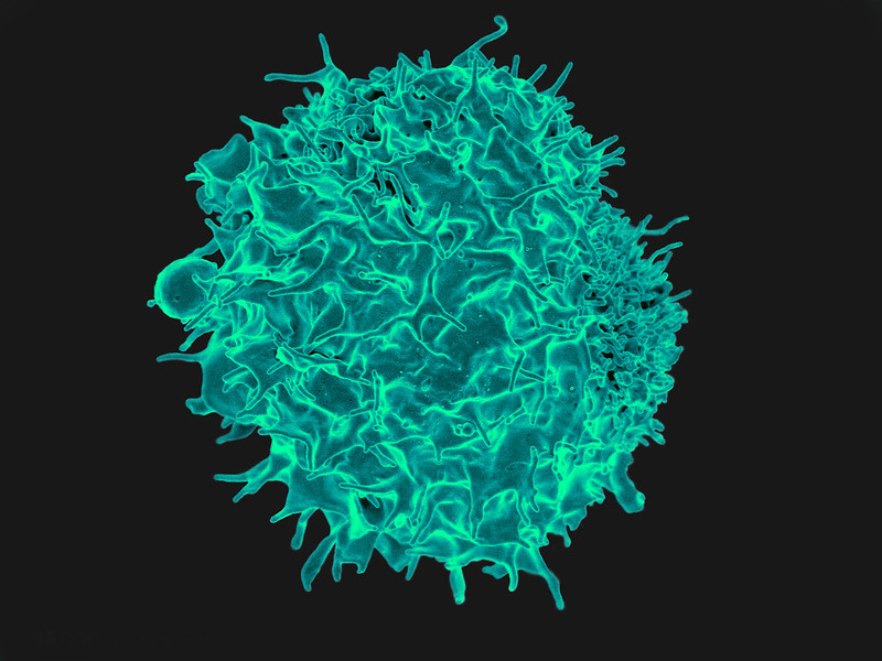
T lymphocyte. Credit: NIH/NIAID

A new study from the University of East Anglia and Quadram Institute sheds light on how our immune cells make use of body fat to fight infection. The research, published today in Nature Communications, could lead to new approaches to treating people with bacterial infections.

The work could one day help treat infections in vulnerable and older people, the researchers said. The team studied Salmonella bacteria and tracked fatty acid movement and consumption in live stem cells. They then examined the immune response to Salmonella bacterial infection, by analysing liver damage.

They uncovered how blood stem cells respond to infection, by acquiring high energy fatty acids from the body’s fat stores. In the bone marrow where blood stem cells are resident, infection signals drive adipocytes to release their fat stores as fatty acids into the blood.

And they identified that these high energy fatty acids are then taken up by blood stem cells, effectively feeding the stem cells and enabling them to make millions of Salmonella-fighting white blood cells. The researchers also identified the mechanism by which the fatty acids are transferred and discusses the potential impact this new knowledge could have on future treatment of infection.

Dr Stuart Rushworth, from UEA’s Norwich Medical School, said: “Our results provide insight into how the blood and immune system is able to respond to infection.

“Fighting infection takes a lot of energy and fat stores are huge energy deposits, which provide the fuel for the blood stem cells to power up the immune response.

“Working out the mechanism through which this ‘fuel boost’ works gives us new ideas on how to strengthen the body’s fight against infection in the future.”

Dr Naiara Beraza, from the Quadram institute, said: “Our results allow us to understand how our immune system uses fat to fuel the response to infection. Defining these mechanisms will enable us to develop new therapeutics to treat infections in the liver.”

Source: University of East Anglia

Stress Signal From Fat Cells Induces Protective Effect in Heart

Source: Unsplash CC0

A stress signal sent from fat cells to the heart could be protective against obesity-induced cardiac damage, according to new research. 

This might help explain the ‘obesity paradox’, where obese individuals have better short- and medium-term cardiovascular disease prognoses compared with those who are normal weight, but have worse long-term outcomes.

“The mechanism we have identified here could be one of many that protects the heart in obesity,” said study leader Philipp E. Scherer, PhD, Professor of Internal Medicine and Cell Biology at UTSW who has long studied fat metabolism.

Study co-leader Clair Crewe, PhD, Assistant Instructor of Internal Medicine at UTSW, explained that the metabolic stress of obesity gradually makes fat tissue dysfunctional, causing shrinkage and death of its mitochondria. This unhealthy fat loses the ability to store lipids generated by excess calories in food, causing lipotoxicity and poisoning other organs. However some organs, including the heart, preemptively defend against lipotoxicity. How the heart actually senses fat’s dysfunctional state has been unknown so far.

The researchers used a genetic technique to speed the loss of mitochondrial mass and function in mice. The mice were fed a high-fat diet and became obese, and their fat cells began sending out extracellular vesicles filled with small pieces of dying mitochondria. Some of these mitochondrial snippets travelled through the bloodstream to the heart and triggered oxidative stress.

Cardiaccells produce a flood of protective antioxidant molecules to counteract this stress, and this protective backlash was so strong that when the scientists injected mice with extracellular vesicles filled with mitochondrial snippets and then induced a heart attack, the animals had significantly less damage to their hearts compared with mice that didn’t receive an injection.

Fat tissue from obese human patients showed that these cells also release mitochondria-filled extracellular vesicles.

The heart and other organs in obese individuals are eventually overwhelmed by lipotoxic effects, resulting in a number of obesity’s comorbidities. If the protective mechanism identified in this study could be artificially generated, it could result in new ways of treating obesity’s negative consequences. This might even be adapted to treat normal weight individuals.

“By better understanding the distress signal from fat,” Dr Crewe said, “we may be able to harness the mechanism to improve heart health in obese and non-obese individuals alike.”

The team’s findings were published in Cell Metabolism.

Source: UT Southwestern Medical Center

A Specific Type of Fat Cell Responds to Insulin

Source: Pixabay

While it was known that fat cells can influence insulin sensitivity, researchers have recently discovered that there are three different subtypes of mature fat cells in white adipose tissue and that it is only one of these, called AdipoPLIN, that responds to insulin. The findings, which were published in Cell Metabolism, may have implications for the treatment of metabolic diseases such as Type 2 diabetes. 

“These findings increase our knowledge about the function of fat tissue,” said co-corresponding author Niklas Mejhert, researcher at the Department of Medicine, Huddinge, at Karolinska Institutet. “They show that the overall capacity of fat tissue to respond to insulin is determined by the proportion and function of a specific fat cell subtype. This could have implications for diseases such as obesity, insulin resistance and Type 2 diabetes.”

The researchers identified 18 classes of cells that form clusters in white adipose tissue in humans. Of these, three constituted mature fat cells with distinct phenotypes.

To determine if a specific function was linked to the fat cell subtypes, the researchers measured how these subtypes in four people reacted to short-term increases in insulin levels. They found that insulin activated the gene expression in the AdipoPLIN subtype but did not affect the other two subtypes. The response to insulin stimulation was also proportional to the individual’s whole-body insulin sensitivity.

A challenge to the prevailing view
“Our findings challenge the current view of insulin resistance as a generally reduced response to insulin in the fat cells,” said co-corresponding author Mikael Rydén, professor in the same department. “Instead, our study suggests that insulin resistance, and possibly type 2 diabetes, could be due to changes in a specific subtype of fat cells. This shows that fat tissue is a much more complex tissue than previously thought. Like muscle tissue, people have several types of fat cells with different functions, which opens up for future interventions targeted at different fat cell types.”

The researchers employed spatial transcriptomics, which generates information about tissue organisation via microscopy and gene expression via RNA sequencing.

”This study is unique in that it is the first time we’ve applied spatial transcriptomics to fat tissue, which has a special set of characteristics and composition,” said third corresponding author Patrik Ståhl. “We are very happy that the technology continues to contribute to solving biologically complex questions in an increasing number of research areas.”

Source: Karolinska Institute