Tag: 7/11/22

Brain Changes in Autism More Widely Spread than Previously Believed

In autism, brain changes are spread throughout the cerebral cortex instead of in areas thought affect social behaviour and language, according to a new study that significantly refines scientists’ understanding of how autism spectrum disorder (ASD) progresses at the molecular level.

The study, published in Nature, represents a comprehensive effort to characterise ASD at the molecular level. While neurological disorders like Alzheimer’s disease or Parkinson’s disease have well-defined pathologies, autism and other psychiatric disorders have had a lack of defining pathology, challenging efforts to develop more effective treatments.  

The new study finds brain-wide changes in virtually all of the 11 cortical regions analysed, regardless of whether they are higher critical association regions – those involved in functions such as reasoning, language, social cognition and mental flexibility – or primary sensory regions.  

“This work represents the culmination of more than a decade of work of many lab members, which was necessary to perform such a comprehensive analysis of the autism brain,” said study author Dr Daniel Geschwind, the Gordon and Virginia MacDonald Distinguished Professor of Human Genetics, Neurology and Psychiatry at UCLA. “We now finally are beginning to get a picture of the state of the brain, at the molecular level, of the brain in individuals who had a diagnosis of autism. This defines a molecular pathology, which similar to other brain disorders such as Parkinson’s, Alzheimer’s and stroke, provides a key starting point for understanding the disorder’s mechanisms, which will inform and accelerate development of disease-altering therapies.” 
 
Just over a decade ago, Geschwind led the first effort to identify autism’s molecular pathology by focusing on two brain regions, the temporal lobe and the frontal lobe. Those regions were chosen because they are higher order association regions involved in higher cognition – especially social cognition, which is disrupted in ASD.  
 
For the new study, researchers examined gene expression in 11 cortical regions by sequencing RNA from each of the four main cortical lobes. They compared brain tissue samples obtained after death from 49 people with ASD against 54 controls individuals.  
 
While each profiled cortical region showed changes, the largest changes in RNA levels were in the visual cortex and the parietal cortex, which processes information like touch, pain and temperature. The researchers said this may reflect the sensory hypersensitivity that is frequently reported in people with ASD. Researchers found strong evidence that the genetic risk for autism is enriched in a specific group of genes expressed in neurons that has lower expression across the brain, indicating that these correlated RNA changes in the brain are likely the cause of ASD rather than a result of the disorder. 

One of the next steps is to determine whether researchers can use computational approaches to develop therapies based on reversing gene expression changes the researchers found in ASD, Geschwind said, adding that researchers can use organoids to model the changes in order to better understand their mechanisms.  

Source: University of California, Los Angeles

Study Reveals a Possible Secret to Viral Infection Resistance in Humans

Colourised scanning electron microscope image of a natural killer cell. Credit: National Institutes of Health

Studying resistance to viral infections in humans is difficult because it’s virtually impossible to disentangle resisting being infected from simply not being exposed. By studying women who were accidentally exposed to hepatitis C (HCV) over 40 years ago, scientists in Ireland have uncovered a secret that may explain why some people are able to resist viral infections.

The extraordinary work, published in Cell Reports Medicine, has wide-ranging implications from improving our fundamental understanding of viral resistance to the potential design of therapies to treat infected people.

From 1977–79, several thousand women in Ireland were exposed to the hepatitis C virus through contaminated anti-D, a medication made using plasma from donated blood and given to Rhesus negative women who are pregnant with a Rhesus positive foetus. The medication prevents the development of antibodies that could be dangerous in subsequent pregnancies. Some of the anti-D used during the 1977–79 period was contaminated with hepatitis C.

Infected women fell into three groups: those who were chronically infected; those who cleared the infection with an antibody response; and those who appeared protected against infection but yet produced no antibodies against hepatitis C.

“We hypothesised that women who seemed to resist HCV infection must have an enhanced innate immune response, which is the ancient part of the immune system that acts as a first line of defence,” said senior author Cliona O’Farrelly, Professor of Comparative Immunology in Trinity’s School of Biochemistry and Immunology.

“To test this we needed to make contact with women exposed to the virus over forty years ago and ask them to help us by allowing us to study their immune systems to hunt for scientific clues that would explain their differing responses.

“After a nationwide campaign over 100 women came forward and we have gained some unique and important insights. That so many women – many of whom have lived with medical complications for a long time – were willing to help is testament to how much people want to engage with science and help pursue research with the potential to make genuine, positive impacts on society. We are deeply grateful to them.”

The scientists ultimately recruited almost 40 women from the resistant group, alongside 90 women who were previously infected.

In collaboration with the Institut Pasteur in Paris they then invited almost 20 women in each group to donate a blood sample that they stimulated with molecules that mimic viral infection and lead to activation of the innate immune system.

“By comparing the response of the resistant women to those who became infected, we found that resistant donors had an enhanced type I interferon response after stimulation,” said first author Jamie Sugrue, PhD Candidate. Type I interferons are a key family of antiviral immune mediators that play an important role in defence against viruses including hepatitis C and SARS-CoV-2, or COVID.

“We think that the increased type I interferon production by our resistant donors, seen now almost 40 years after the original exposure to hepatitis C, is what protected them against infection.

“These findings are important as resistance to infection is very much an overlooked outcome following viral outbreak, primarily because identifying resistant individuals is very difficult – since they do not become sick after viral exposure, they wouldn’t necessarily know that they were exposed. That’s why cohorts like this, though tragic in nature, are so valuable – they provide a unique opportunity to study the response to viral infections in an otherwise healthy population.”

The lab’s efforts are now focused on leveraging these biological findings to unpick the genetics of viral resistance in the HCV donors. Their work on HCV resistance has already helped ignite international interest in resistance to other viral infections such as SARS-CoV-2.

The O’Farrelly lab has expanded its search for virus-resistant individuals by joining in the COVID human genetic effort and by recruiting members of the public who have been heavily exposed to SARS-CoV-2 but never developed an infection.

Source: Trinity College Dublin

A Shot of Vitamin C Gives Dendritic Cells a Potent Cancer-fighting Boost

Vitamin C pills and orange
Photo by Diana Polekhina on Unsplash

New research published in Nucleic Acids Research has shown that vitamin C improves the immunogenic properties of dendritic cells, activating genes involved in the immune response. This discovery could help the development of potent new dendritic cell-based immunotherapies.

Since the onset of anticancer cell therapies, many types of immune cells have been used. The best-known of these cell therapies use lymphocytes, as in the highly successful CAR-T therapies. Recently, researchers have to turned to dendritic cells, known as the ‘master regulators of the immune system‘, for their ability to uptake and present antigens to the T-lymphocytes and induce an antigen-specific potent immune activation. This approach entails loading dendritic cells with specific antigens to create immune memory to make dendritic cell (DC)-vaccines.

To study dendritic cells in the lab, researchers differentiate them from monocytes using a particular set of molecular signalling. This differentiation is accomplished through a complex set of gene activation processes in the nucleus, mostly thanks to the activity of the chromatin remodelling machinery spearheaded by the TET family of demethylases, proteins that act upon the DNA epigenetic marks.

Vitamin C was already known to interact with several TET proteins to enhance its activity, but the specific mechanism was still poorly understood in human cells. In this study, a team lead by Dr Esteban Ballestar hypothesised that treating monocytes in vitro while differentiating into dendritic cells, would help the resulting cells be more mature and active.

The results obtained show that vitamin C treatment triggers an extensive demethylation at NF- kB/p65 binding sites compared with non-treated cells, promoting the activity of genes involved in antigen presentation and immune response activation. Vitamin C was also found to increase the communication of the resulting dendritic cells with other components of the immune system and stimulates the proliferation of antigen-specific T cells.

The researchers proved that vitamin C-stimulated dendritic cells loaded with antigens specific for the SARS-CoV-2 virus were able to activate T cells in vitro more efficiently than non-treated cells.

Overall, these new findings support the hypothesis that treating monocyte-derived dendritic cells with vitamin C may help generate more effective DC-vaccines. After consolidating these results in preclinical models and, hopefully, in clinical trials, a new generation of cell therapies based on dendritic cells may be used in the clinic to fight cancer more efficiently.

Source: Josep Carreras Leukaemia Research Institute

World First Trial of Lab-grown Red Blood Cells for Transfusion

https://www.pexels.com/photo/a-close-up-shot-of-bags-of-blood-4531306/
Photo by Charlie-Helen Robinson on Pexels

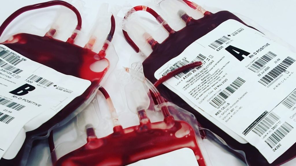
In a world first, researchers have launched a clinical trial of lab-grown red blood cells for transfusion into another person. These manufactured blood cells were grown from stem cells from donors, for transfusion into volunteers in the RESTORE randomised controlled clinical trial.

If our trial is successful, it will mean that patients who currently require regular long-term blood transfusions will need fewer transfusions in future, helping transform their care

Professor Cedric Ghevaert, chief investigator

If the technique is proven safe and effective, manufactured blood cells could in time revolutionise treatments for people with blood disorders such as sickle cell and rare blood types. It can be difficult to find enough well-matched donated blood for some people with these disorders.

To produce the lab-grown blood cells, stem cells are first magnetically extracted from a normal 470ml blood donation. These stem cells are then coaxed into becoming red blood cells. Over the three week process, an initial pool of about half a million stem cells generates 50 billion red blood cells.

Chief Investigator Professor Cedric Ghevaert, Professor in Transfusion Medicine and Consultant Haematologist at the University of Cambridge and NHS Blood and Transplant, said: “We hope our lab grown red blood cells will last longer than those that come from blood donors. If our trial, the first such in the world, is successful, it will mean that patients who currently require regular long-term blood transfusions will need fewer transfusions in future, helping transform their care.”

Professor Ashley Toye, Professor of Cell Biology at the University of Bristol and Director of the NIHR Blood and Transplant Unit in red cell products, said: “This challenging and exciting trial is a huge stepping stone for manufacturing blood from stem cells. This is the first-time lab grown blood from an allogeneic donor has been transfused and we are excited to see how well the cells perform at the end of the clinical trial.”

The trial is studying the lifespan of the lab grown cells compared with infusions of standard red blood cells from the same donor. The lab-grown blood cells are all fresh, so the trial team expect them to perform better than a similar transfusion of standard donated red cells, which contains cells of varying ages.

Additionally, if manufactured cells last longer in the body, patients who regularly need blood may not need transfusions as often. That would reduce iron overload from frequent blood transfusions, which can lead to serious complications.

The trial is the first step towards making lab grown red blood cells available as a future clinical product. For the foreseeable future, manufactured cells could only be used for a very small number of patients with very complex transfusions needs. NHSBT continues to rely on the generosity of donors.

Co-Chief Investigator Dr Rebecca Cardigan, Head of Component Development NHS Blood and Transplant and Affiliated Lecturer at the University of Cambridge, said: “It’s really fantastic that we are now able to grow enough red cells to medical grade to allow this trial to commence. We are really looking forward to seeing the results and whether they perform better than standard red cells.”

Thus far, two people have been transfused with the lab grown red cells. They are well and healthy, and were closely monitored with no untoward side effects were reported. The amount of lab grown cells being infused varies but is around 5-10mls.

Donors were recruited from NHSBT’s blood donor base. They donated blood to the trial and stem cells were separated out from their blood. These stem cells were then grown to produce red blood cells in a laboratory at NHS Blood and Transplant’s Advanced Therapies Unit in Bristol. The recipients of the blood were recruited from healthy members of the NIHR BioResource.

A minimum of 10 participants will receive two mini transfusions at least four months apart, one of standard donated red cells and one of lab grown red cells, to see if the young lab-made red blood cells last longer than cells made in the body.

Further trials are needed before clinical use, but this research marks a significant step in using lab grown red blood cells to improve treatment for patients with rare blood types or people with complex transfusion needs.

Source: University of Cambridge