Tag: 6/9/22

Blood Type may Point to Early-onset Stroke Risk
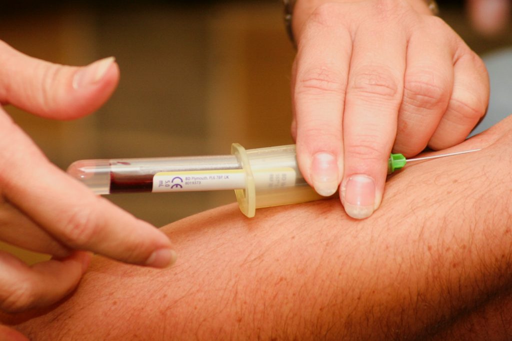
Blood sample being drawn
Photo by Hush Naidoo Jade Photography on Unsplash

Blood type may be associated with early-onset stroke risk, according to a new meta-analysis, which was published in the journal Neurology. The meta-analysis included all available data from genetic studies focusing on ischaemic strokes in adults under age 60.

“The number of people with early strokes is rising. These people are more likely to die from the life-threatening event, and survivors potentially face decades with disability. Despite this, there is little research on the causes of early strokes,” said study co-principal investigator Steven J. Kittner, MD, MPH, Professor of Neurology at University of Maryland School of Medicine (UMSOM).

He and his colleagues performed a meta-analysis of 48 studies on genetics and ischaemic stroke that included 17 000 stroke patients and nearly 600 000 healthy controls. They looked for genetic variants associated with a stroke and found a link between early-onset stroke (before age 60) and the area of the chromosome that includes the gene that determines whether a blood type is A, AB, B, or O.

The study found that people with early stroke were more likely to have blood type A and less likely to have blood type O, compared to people with late stroke and people who never had a stroke. Both early and late stroke were also more likely to have blood type B compared to controls. After adjusting for sex and other factors, researchers found that, compared to those with other blood types, blood type A had a 16% higher risk while blood type O had a 12% lower risk.

“Our meta-analysis looked at people’s genetic profiles and found associations between blood type and risk of early-onset stroke. The association of blood type with later-onset stroke was much weaker than what we found with early stroke,” said study co-principal investigator Braxton D. Mitchell, PhD, MPH, Professor of Medicine at UMSOM.

The researchers emphasised that the increased risk was very modest and that those with type A blood should not worry about having an early-onset stroke or engage in extra screening or medical testing based on this finding.

“We still don’t know why blood type A would confer a higher risk, but it likely has something to do with blood-clotting factors like platelets and cells that line the blood vessels as well as other circulating proteins, all of which play a role in the development of blood clots,” said Dr Kittner. Previous studies suggest that those with an A blood type have a slightly higher risk of developing blood clots in the legs known as deep vein thrombosis. “We clearly need more follow-up studies to clarify the mechanisms of increased stroke risk,” he added.

A limitation of the study was the relative lack of diversity among participants, with only 35% of the participants having non-European ancestry.

Source: University of Maryland School of Medicine

Early Sensing of Malaria in the Brain Leads to Cerebral Malaria

Colourised scanning electron micrograph of red blood cell infected with malaria parasites, which are colourised in blue. The infected cell is in the centre of the image area. To the left are uninfected cells with a smooth red surface. Credit: National Institute of Allergy and Infectious Diseases, NIH

A recent study published in PNAS revealed that endothelial cells in the brain are able to sense the infection by the malaria parasite at an early phase, triggering the inflammation underlying cerebral malaria. This discovery identified new targets for adjuvant therapies that could restrain brain damage in initial phases of the disease and avoid neurological sequelae.

Cerebral malaria is a severe complication of infection with Plasmodium falciparum, the most lethal of the parasites causing malaria. This form of the disease manifests through impaired consciousness and coma and affects mainly children under 5, being one of the main causes of death in this age group in countries of Sub-Saharan Africa. Survivors are frequently affected by debilitating neurological sequelae, such as motor deficits, paralysis, and speech, hearing, and visual impairment.

To prevent certain molecules and cells from reaching the brain, which would disturb its normal functioning, endothelial cells forming a tight barrier between the blood and this organ. Cerebral malaria results from an unrestrained inflammatory response to infection which leads to significant alterations in this barrier and, consequently, neurological complications.

Over the last years, specialists in this field have turned their attention to a molecule, named interferon-β, which seems to be associated with this pathological process. So called for interfering with viral replication, this highly inflammatory molecule has two sides: it can either be protecting or cause tissue destruction. It is known, for example, that despite its antiviral role in COVID-19, at a given concentration and phase of infection, it can cause lung damage. A similar dynamic is thought to occur in cerebral malaria. However, we still don’t know what leads to the secretion of interferon-β, nor the main cells involved.

The present study revealed that endothelial cells in the brain play a crucial role, being able to sense the infection by the malaria parasite at an early phase. These detect the infection through an internal sensor which triggers a cascade of events, starting with the production of interferon-β. Next, they release a signalling molecule that attracts cells of the immune system to the brain, initiating the inflammatory process.

To reach these conclusions, researchers used mice that mimic several symptoms described in human malaria and a genetic manipulation system that allowed them to delete this sensor in several types of cells. When they deleted this sensor in brain endothelial cells, the animals’ symptoms were not as severe with lower mortality. They then realised these brain cells contributed greatly to the pathology of cerebral malaria. “We thought brain endothelial cells acted in a later phase, but we ended up realising that they are participants from the very beginning”, explained Teresa Pais, a post-doctoral researcher at the IGC and first author of the study. “Normally we associate this initial phase of the response to infection with cells of the immune system. These are already known to respond, but cells of the brain, and maybe other organs, also have this ability to sense the infection because they have the same sensors.”

But what really surprised the researchers was the factor activating the sensor and triggering this cell response. This factor is nothing more nothing less than a by-product of the activity of the parasite. Once in the blood, the parasite invades the host’s red blood cells, where it multiplies. Here, it digests haemoglobin, a protein that transports oxygen, to get nutrients. During this process, a molecule named haeme is formed and it can be transported in tiny particles in the blood that are internalised by endothelial cells. When this happens, haeme acts as an alarm for the immune system. “We weren’t expecting that haeme could enter cells this way and activate this response involving interferon-β in endothelial cells”, the researcher confessed.

This six-year project allowed the researchers to identify a molecular mechanism that is critical for the destruction of brain tissue during infection with the malaria parasite and, with that, new therapeutic targets. “The next step will be to try to inhibit the activity of this sensor inside the endothelial cells and understand if we can act on the host’s response and stop brain pathology in an initial phase,” explained principal investigator Carlos Penha Gonçalves. “If we could use inhibitors of the sensor in parallel with antiparasitic drugs maybe we could stop the loss of neuronal function and avoid sequelae which are a major problem for children surviving cerebral malaria.”

Source: Instituto Gulbenkian de Ciência (IGC)

Low Testosterone may be a Risk Factor for Severe COVID

Testosterone molecule
Model of a testosterone molecule. Source: Wikimedia CC0

Among men with COVID, those with low testosterone levels are more likely to become seriously ill and be hospitalised than men with normal levels of the hormone, according to a study which appears in JAMA Network Open.

Analysis of data for 723 men who tested positive for COVID, mostly in 2020 before vaccines were available, indicated that low testosterone is an independent risk factor for COVID hospitalisation, similar to diabetes, heart disease and chronic lung disease.

They found that men with low testosterone who developed COVID were 2.4 times more likely to require hospitalisation than men with hormone levels in the normal range. Further, men who were once diagnosed with low testosterone but successfully treated with hormone replacement therapy were no more likely to be hospitalised for COVID than men whose testosterone levels had always tested in the normal range.

The findings, by researchers from the Washington University School of Medicine in St. Louis and Saint Louis University School of Medicine, suggest that treating men with low testosterone may help protect them against severe disease and reduce the burden on hospitals during COVID waves.

“It is very likely that COVID is here to stay,” said co-senior author Abhinav Diwan, MD, a professor of medicine at Washington University. “Hospitalizations with COVID are still a problem and will continue to be a problem because the virus keeps evolving new variants that escape immunization-based immunity. Low testosterone is very common; up to a third of men over 30 have it. Our study draws attention to this important risk factor and the need to address it as a strategy to lower hospitalisations.”

Prof Diwan and co-senior author Sandeep Dhindsa, MD, an endocrinologist at Saint Louis University, had previously shown that men hospitalised with COVID have abnormally low testosterone levels. However, severe illness or traumatic injury can cause a temporary drop in hormone levels, so causation cannot be proved in data from men already hospitalised with COVID. Data were needed for men with chronically low testosterone before COVID infection.

Profs Diwan, Dhindsa and colleagues identified 723 men whose testosterone levels had been measured between Jan. 1, 2017, and Dec. 31, 2021, and who had documented cases of COVID in 2020 or 2021. In some cases, testosterone levels were measured after the patient recovered from COVID. Since low testosterone is a chronic condition, men who tested low a few months after recovering from COVID probably had low levels before as well, Prof Dhindsa said.

The researchers identified 427 men with normal testosterone levels, 116 with low levels, and 180 who previously had low levels but were being successfully treated, meaning that they were on hormone replacement therapy and their testosterone levels were in the normal range at the time they developed COVID.

“Low testosterone turned out to be a risk factor for hospitalisation from COVID, and treatment of low testosterone helped to negate that risk,” Prof Dhindsa said. “The risk really takes off below a level of 200 nanograms per decilitre, with the normal range being 300 to 1000 nanograms per decilitre. This is independent of all other risk factors that we looked at: age, obesity or other health conditions. But those people who were on therapy, their risk was normal.”

Men with low testosterone levels can experience sexual dysfunction, depressed mood, irritability, difficulty with concentration and memory, fatigue, loss of muscular strength and a reduced sense of well-being overall. When a man’s quality of life is clearly diminished, he is typically treated with testosterone replacement therapy. When the symptoms are mild, though, doctors and patients may hesitate to treat.

The two main concerns related to testosterone therapy are an increased risk of prostate cancer and heart disease. Testosterone is well known to boost prostate cancer, but for heart disease, the evidence for risk is more ambiguous. A large clinical trial on the relationship between heart health and testosterone supplementation is expected to be completed soon.

“In the meantime, our study would suggest that it would be prudent to look at testosterone levels, especially in people who have symptoms of low testosterone, and then individualise care,” said Prof Diwan, whose specialty is cardiology. “If they are at really high risk of cardiovascular events, then the doctor could engage the patient in a discussion of the pros and cons of hormone replacement therapy, and perhaps lowering the risk of COVID hospitalisation could be on the list of potential benefits.”

Since this study is observational, it only suggests that boosting testosterone levels may help men avoid severe COVID, Diwan cautioned. A clinical trial would be needed to demonstrate conclusively whether such a strategy works.

Source: Washington University School of Medicine

Dolutegravir-based ART is Better for Pregnant Individuals with HIV-1

pregnant woman holding her belly
Source: Anna Hecker on Unsplash

Dolutegravir-based antiretroviral therapies (ART) for HIV-1 are more effective for pregnant individuals than some other ART regimens commonly used in the US and Europe, according to a study available online in NEJM.

The study, led by Harvard T.H. Chan School of Public Health researchers, showed that pregnant individuals who took dolutegravir-based regimens had a high probability of being virally suppressed at delivery. No differences were seen in adverse birth outcome risks (preterm birth, low birth weight, small for gestational age, or neonatal death) between dolutegravir-based regimens and the other contemporary regimens.

“Globally, a dolutegravir-based regimen is currently recommended for treating HIV, and this is the first study to directly compare regimens including dolutegravir to other antiretroviral regimens, such as raltegravir-based regimens, that are also listed as ‘Preferred’ in US perinatal guidelines,” said senior research scientistKunjal Patel, lead author of the study.

Dolutegravir, is a newer antiretroviral part of a once-a-day regimen that has been shown to be more effective, easier to tolerate, and less likely to create new drug resistance in people with HIV-1. However, limited data have been available about its effectiveness and safety in pregnancy compared with regimens that commonly have been used during pregnancy in the US and Europe.

In the current observational study, the researchers compared dolutegravir use in pregnancy with atazanavir/ritonavir, darunavir/ritonavir, and raltegravir antiviral regimens that are currently classified as “Preferred” for use in pregnancy in the US About half of the participants started ART before conception. At delivery, 96.7% of pregnancies of participants who received dolutegravir were virally suppressed, whereas those of participants who took atazanavir/ritonavir or raltegravir had viral suppression of 84.0% and 89.2%, respectively.

“We think the observed differences are due to dolutegravir’s ability to rapidly decrease viral loads and its ease of use as part of a once-daily regimen that’s available as a fixed-dose combination,” said Patel. “Our results highlight the continual need for systematic studies that compare new antiretroviral regimens with those already in clinical practice to help inform the evolution of guidelines and clinical practice over time.”

Source: Harvard T.H. Chan School of Public Health

Bronchodilators Don’t Ease Smoking-related Respiratory Symptoms in non-COPD Patients

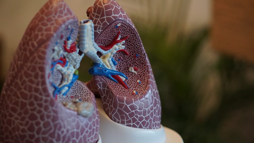
Anatomical model of lungs
Photo by Robina Weermeijer on Unsplash

A study published in the New England Journal of Medicine have found that dual bronchodilators do little to help people who do not have chronic obstructive pulmonary disease (COPD), but who do have respiratory symptoms and a history of smoking.

Millions of people who smoke or used to smoke and have some symptoms of COPD have been prescribed bronchodilators.

“We’ve assumed these medications worked in patients who don’t meet lung function criteria for COPD, but we never checked,” said MeiLan K. Han, MD, a principal investigator and first author of the study. “We now know these existing medications don’t work for these patients.”

According to scientists, the implications are significant. First, they show the importance of diagnosing lung conditions through spirometry, a lung function test Dr Han noted is underutilised in clinical practice. Second, they show the need for new, effective therapies for patients without COPD.

Inhalers have long been the primary go-to treatment for these patients, she explained, because doctors either assume a patient has COPD, or else that their smoking-related symptoms could be helped by the inhalers. But while tobacco smoking causes a large spectrum of lung damage, the study showed bronchodilator therapy only helps patients with enough lung damage that would result in abnormal spirometry readings.

In the 12-week, randomised, double-blinded study, which was part of the Redefining Therapy in Early COPD for the Pulmonary Trials Cooperative (RETHINC), researchers enrolled 535 adults with symptoms of COPD, ages 40–80. Participants used an inhaler twice daily that contained either medication or a placebo.

By the end of the trial, some adults in the medication and placebo groups saw slight respiratory improvements, eg coughed less, produced less phlegm, or felt less winded, which was assessed through the St. George’s Respiratory Questionnaire. However, the researchers found no significant differences between those receiving medication or placebo. They reported 56% (128 of 227) of participants who received the medication saw respiratory symptom improvements, compared to 59% (144 of 244) of those who took the placebo.

According to Dr Han, these data underscore the need to change the standard practice, which is not doing spirometry and just treating patients with the same COPD medications and expecting to see improvement.

Antonello Punturieri, MD, PhD, program director of NHLBI’s Chronic Obstructive Pulmonary Disease/Environment Program, said spirometry testing should be used for any patient who shows signs of COPD, airflow obstruction, or who has a history of cigarette smoking. Though spirometry readings are used during about one-third of medical visits related to COPD, roughly half of patients who would meet criteria for COPD go undiagnosed.

Promoting smoking cessation a primary way to prevent COPD or COPD-like symptoms, the study noted. About one in four current or former smokers without COPD have reported having shortness of breath. In addition to encouraging smoking cessation, doctors can help patients who do not meet lung-function criteria of COPD by working with them to address any other underlying issues, such as overweight and obesity, heart failure, or other lung issues.

“In the meantime, research should be focused on finding new treatments for them,” Dr Han explained. “The next question is, can we develop more targeted therapies for these patients who are on the milder end of the spectrum?”

“Because cough and mucus production show up prominently among these patients, we believe therapies that target mucus production in the airways may be effective,” said Prescott G. Woodruff, MD, a principal investigator and senior author of the study.

Some of these therapies are already in development, and data from other studies may offer insight into the biological causes of excessive airway mucus, which could help point to additional therapies.

Source: NIH/National Heart, Lung and Blood Institute