Tag: 6/4/22

Community-acquired Antimicrobial Resistant UTIs can be More Deadly
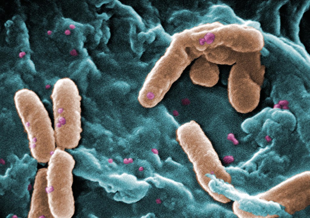
Pseudomonas
Scanning Electron Micrograph of Pseudomonas aeruginosa. Credit: CDC/Janice Carr.

A study from Australia’s scientific organisation CSIRO has revealed that antimicrobial resistant (AMR) bacteria in urinary tract infections are more lethal, especially Enterobacteriaceae. The findings are published online in Open Forum Infectious Diseases.

Antimicrobial resistance (AMR) bacteria can be passed between humans: through hospital transmission and community transmission. While hospital acquired resistance is well researched, there are few studies focusing on the burden of community transmission.

To address this, the study analysed data from 21 268 patients across 134 Queensland hospitals who acquired their infections in the community. The researchers found that patients were almost two and a half (2.43) times more likely to die from community acquired drug-resistant UTIs caused by Pseudomonas aeruginosa and more than three (3.28) times more likely to die from community acquired drug-resistant blood stream infections caused by Enterobacteriaceae than those with drug-sensitive infections. The high prevalence of UTIs make them a major contributor to antibiotic use, said CSIRO research scientist, Dr Teresa Wozniak.

“Our study found patients who contracted drug-resistant UTIs in the community were more than twice as likely to die from the infection in hospital than those without resistant bacteria,” Dr Wozniak said. “Without effective antibiotics, many standard medical procedures and life-saving surgeries will becoming increasingly life-threatening. “Tracking the burden of drug-resistant infections in the community is critical to understanding how far antimicrobial resistance is spreading and how best to mitigate it.”

The study’s findings will provide further guidance for managing AMR in the community, such as developing AMR stewardship programs that draw on data from the population being treated.

CEO of CSIRO’s Australian e-Health Research Centre, Dr David Hansen, said the magnitude of the AMR problem needs to be understood to mitigate it. “Tracking community resistance is difficult because it involves not just one pathogen or disease but multiple strains of bacteria,” Dr Hansen said. “Until now we haven’t been using the best data to support decision making in our fight against AMR. Data on community acquired resistance is an important contribution to solving the puzzle. “Digital health has an important role in using big data sets to describe patterns of disease and drive important population health outcomes.”

Source: CSIRO

Long-term Symptoms of Concussion Tied to Vestibular Nerve Damage

Rugby players
Photo by Olga Guryanova

A clinical study from Lund University in Sweden has shown that long-term problems such as dizziness and difficulty focusing after a concussion originate in an injury to the vestibular nerve. The researchers published their findings in the Journal of Neurotrauma.

Concussion resulting from a blow to the head is a hazard in many sports. In American football, where players who have suffered from repeated concussions have developed dementia, severe depression and cognitive impairment.

Concussion usually has only temporary symptoms, but an increasing number of athletes experience long-term problems that make it difficult to work, go to school or play sports. The symptoms are aggravated by activity or impressions and include headaches, depression, anxiety, nausea, difficulty focusing and problems with balance.

“It has been unclear what causes the symptoms, and it is difficult for healthcare professionals to help these athletes. We wanted to investigate this further to find out what really causes the symptoms,” said Professor Niklas Marklund, one of the study’s researchers.

The study included 21 healthy athletes without previous trauma to the head, and 21 athletes who all suffered from sports-related concussions and who had experienced persisting symptoms for more than six months. The researchers used a 7-Tesla MRI, to study the athletes’ brains to understand more about what caused the symptoms. They discovered impaired function of the balance organs in the inner ear of 13 athletes in the group with long-term problems. In the group of healthy athletes three people had similar findings.

“The test results show that the injury is located to the vestibular nerve, which is connected to the semicircular canals in a cavity inside the skull, and which is directly adjacent to the cochlea in the ear. These injuries lead to the inward nerve impulses not working properly, and the brain therefore does not receive important information about body movements and sensory impressions required to maintain a good balance,” said Anna Gard, doctoral student at Lund University and first author of the study.

Concussion often results from the head rotating too fast.

“We have not examined athletes with short-term problems after blows to the head, so we cannot say anything about them. This study applies to athletes with prolonged symptoms after concussion. The rotation of the head that occurs in connection with a concussion could lead to a stretch of the vestibular nerve, which then leads to impaired function. Now that we have more knowledge about where the problems are located, it is easier to find possible therapies that could help these athletes,” concluded Prof Marklund.

Source: Lund University

Scientists Discover the Neurological Basis of Food Cravings in Pregnancy

Photo by Andriyko Podilnyk on Unsplash

By examining mice, which get pregnancy cravings similar to humans, scientists have identified the neurological basis of food craving during pregnancy.

During pregnancy, the mother’s body undergoes a series of physiological and behavioural changes to create an environment facilitating the embryo’s development. Frequent consumption of tasty, high calorie foods driven by the cravings contributes to weight gain and obesity in pregnancy, with possible negative consequences for the baby’s health.

“There are many myths and popular beliefs regarding these cravings, although the neuronal mechanisms that cause them are not widely known,” noted study leader March Claret, at the University of Barcelona and leader of the study published in the journal Nature Metabolism.

The researchers found that the brains of pregnant female mice undergoes changes in the functional connections of the brain reward circuits, as well as the taste and sensorimotor centres. Mice, like pregnant women, are also more sensitive to sweet food, and develop binge-eating behaviours towards high calorie foods. “The alteration of these structures made us explore the mesolimbic pathway, one of the signal transmission pathways of dopaminergic neurons. Dopamine is a key neurotransmitter in motivational behaviours,” notes Claret, member of the Department of Medicine of the UB and the Diabetes and Associated Metabolic Diseases Networking Biomedical Research Centre (CIBERDEM).

The team saw that dopamine levels and dopamine receptor (D2R) activity increased in the nucleus accumbens, a brain region involved in the reward circuit. “This finding suggests that the pregnancy induces a full reorganisation of the mesolimbic neural circuits through the D2R neurons,” noted study leader Roberta Haddad-Tóvolli. “These neuronal cells – and their alteration – would be responsible for the cravings, since food anxiety, typical during pregnancy, disappeared after blocking their activity.”

The team demonstrated that persistent cravings have consequences for the offspring, affecting the metabolism and development of neural circuits that regulate food intake, leading to weight gain, anxiety and eating disorders. “These results are shocking, since many of the studies are focused on the analysis of how the mother’s permanent habits – such as obesity, malnutrition, or chronic stress – affect the health of the baby. However, this study indicates that short but recurrent behaviours, such as cravings, are enough to increase the psychological and metabolic vulnerability of the offspring,” concluded Claret.

The conclusions of the study could contribute to the improvement of nutritional guidelines for pregnant women in order to ensure a proper prenatal nutrition and prevent the development of diseases.

Source: University of Barcelona

Aggressive Warming during Surgery Does not Reduce Complications

Photo by Anna Shvets on Pexels

When patients kept at a body temperature of 37C with aggressive warming during surgery, there was no reduction cardiac complications compared to patients kept at 35.5C, finds a large new study reported in The Lancet. No differences was seen in number of infections or required blood transfusions in patients kept at cooler body temperatures.

An unintentional drop in body temperature is a normal side effect during surgery, due mostly to anaesthetic medications’ interference with the body’s temperature regulation processes. In Western countries, nursing staff typically use forced-air heaters to keep patients warm during surgery, with a target temperature of 36C. This trial, one of the largest to date, sought to determine whether even greater warming, to 37C, would reduce the risk of cardiac complications, a major cause of mortality in the first 30 days after surgery.

Results showed no significant differences between groups for the trial’s primary endpoint, a composite of troponin elevation due to ischemia (an indicator of heart injury), non-fatal cardiac arrest or death from any cause within 30 days after surgery. Researchers also reported no differences for any of the trial’s secondary endpoints.

“This trial tells us that there is no benefit to aggressively warming patients to 37 C during surgery. It is simply unnecessary, and it doesn’t improve any substantive outcomes,” said Daniel I. Sessler, MD, Michael Cudahy professor and chair of the Department of Outcomes Research at Cleveland Clinic and the trial’s lead author. “Also, the results show that 36C should not be considered the threshold for defining mild hypothermia since there was no harm at 35.5C.”

The researchers enrolled 5050 patients, mostly in Chinese centres. Participants had various major noncardiac surgical procedures, with a minimum duration of two hours and an average duration of four hours. Half of the patients were randomised to routine care, with a target body temperature of 35.5C, and the other half randomised to aggressive warming, with a target body temperature of 37C.

For patients assigned to routine care, nursing staff put a warming cover in position but did not activate it until the patient’s body temperature decreased to less than 35.5C, resulting in an average group body temperature of 35.6C. With the more aggressive warming protocol, nurses covered patients with a heated blanket for 30 minutes before surgery and then used two forced-air heaters to keep patients warmed to a mean of 37.1 C during surgery.

In addition to seeing no benefit in terms of the composite primary endpoint, the trial reported no significant differences between groups in terms of serious wound infections, length of hospitalization, hospital re-admissions or the need for blood transfusions. The investigators were surprised that rates of wound infections and transfusions were similar to previous studies, which suggested that both were more common in patients maintained at lower body temperatures.

While most patients were enrolled in China, Dr Sessler said, the results should still be generalisable to patients and health care settings in other countries.

“This study shows that it is reasonable to keep patients warm, but we saw no evidence whatsoever that it makes a difference if they’re just above or just below 36C,” Dr Sessler said. “Surgical patients should still be warmed, but there’s no need to be super-aggressive about the warming.”

Less serious or non-medical outcomes, such as patient comfort or shivering was not assessed. Dr Sessler said that patients maintained at a lower body temperature may shiver or feel cold after surgery, but both are temporary and unlikely to have a meaningful health impact.

Source: American College of Cardiology

Key Factors in Hospitalisation after Breast Reconstruction Surgery

Photo by Jafar Ahmed on Unsplash

Factors such as anaemia and anticoagulants have more impact on hospitalisation time after breast reconstruction than “common” risk factors according to a new study published in the Journal of Clinical Medicine.

The study investigated the impact of different factors on postoperative blood loss and drainage fluid volume, two factors which can lengthen hospitalisation time of patients after breast reconstruction after breast cancer surgery. The findings of the study allow for an improved risk assessment and planning of reconstructive breast surgery to offer patients personalised and improved treatment.

Partial or total mastectomy is often necessary in breast cancer surgery, and reconstructive breast surgery lessens the psychological stress on the patient. Fast wound healing after surgical breast reconstruction is crucial to not delay subsequent cancer treatments. Factors influencing the length of hospital stay (LOS) or wound healing are therefore particularly significant in cancer treatment. This study identified previously unrecognised risk factors.

Blood loss and drainage fluid volumes after breast reconstruction due to breast cancer were recorded, parameters which are closely linked to the healing process and LOS .Lower loss equals earlier patient discharge and early start of subsequent treatment. “We analysed factors that might affect blood loss and drainage fluid volumes after surgery – but can be identified before the surgery,” explained lead author Dr Tonatiuh Flores, plastic surgeon. “These factors included age, body mass index and smoking status – factors that are known to have a strong impact on the course of disease.” Additionally, haemoglobin levels and possible antithrombotic prophylaxis were reviewed – two parameters that are particularly significant in oncological treatment.

Surprising results emerged from the evaluation of a total of 257 breast reconstructions in 195 patients. Professor Konstantin Bergmeister, senior author of the study explained that “the classic risk factors did not significantly influence postoperative blood loss and drainage fluid output. Haemoglobin levels and anticoagulant concentration, however, did.” The analysis revealed a close relation between low haemoglobin values or anaemia and fluid loss after reconstructive breast surgery. Co-author Prof. Klaus Schroegendorfer, elaborated on this: “Especially breast cancer patients often show perioperative anaemia, caused by the frequently required neoadjuvant chemotherapy which can affect blood values, in particular haemoglobin.”

There were similar findings regarding low molecular heparin used in cancer patients as antithrombotic prophylaxis. Patients receiving heparin tended to have increased drainage fluid output after surgery, though the effect was not as strong as with perioperative anaemia.

The study authors recommend that, to cut LOS and continue the necessary cancer treatment after reconstructive breast surgery in cancer patients as early as possible, patients should preoperatively be screened for anaemia and administration of low molecular heparin should be adapted to the patients’ risk. In correlation to the results, follow-up treatment can be improved, patients can be discharged earlier and cancer treatment can be continued.

Source: Karl Landsteiner University of Health Sciences