Tag: 5/3/25

Our HIV Response Will Collapse Without US Funding – Unless We Act Urgently

Reassurances that state clinics will pick up the lost services are empty

Sign outside a Wits RHI clinic in Johannesburg. Photo: Ihsaan Haffejee

By Francois Venter

South Africa faces its worst health crisis in 20 years. Worse than COVID, and one that will overshadow diabetes as a major killer, while pouring petrol on a dwindling TB fire. But it is preventable if our government steps up urgently.

Nearly eight-million people have HIV in South Africa; they need life-long antiretroviral medicines to stay healthy.

The near-total removal of US government funding last week, a programme called PEPFAR, will see every important measure of the HIV programme worsen, including hospitalisations, new infections in adults and children, and death. Unless government meaningfully steps in to continue funding the network of highly efficient organisations that currently fill key gaps in national care, an epidemic that was tantalisingly close to coming under control will again be out of our reach. Millions of people in South Africa will become infected with HIV and hundreds of thousands more will die in the next ten years. 2025 will end much more like 2004, when we started our HIV treatment programme.

Many fail to recognize the danger. Commentators, public health officials, and government spokespeople have downplayed the US financial contributions to the HIV response, suggesting services can be absorbed within current services. The funding cuts amount to approximately 17% of the entire budget for HIV and largely go to salaries for health staff. On the face of it, this indeed seems replaceable. So why are the consequences so deadly?

To understand the impact, one must recognize how US funding has supported HIV care. The money is largely allocated to a network of non-government organisations through a competitive, focused, and rigorously monitored program in four key areas:

  1. Active case finding: The best way to prevent new cases of HIV is find everyone with the disease early on, and get them on treatment. These organisations deploy people in high-risk areas, to test for HIV and screen for TB, and shepherd people who test positive to treatment programmes. People are almost always healthy when they start treatment, and remain healthy, with greatly reduced time to transmit the virus, and much less chance of ever “burdening” the health sector with an opportunistic infection. They are hugely cost-effective.
  2. Tracing people who disappear from care: Patients on antiretrovirals fall out of care for many reasons, ranging from changing their address, to life chaos such as losing their job or mental illness. Or they are simply mixed up in the filing dysfunction within clinics. The US supported programmes helped finding people ‘lost from care’, maintaining systems able to track who has not come back, and how to contact them, often spending considerable time cleaning redundant records as people move between facilities.
  3. Vulnerable population programmes: Services include those for sex workers, LGBTQ+ people, adolescents, people who use drugs, and victims of gender violence. These programs are for people who need tailored services beyond the straightforward HIV care offered in state clinics. They are often discriminated against in routine services and also at significant risk of contracting HIV.
  4. Supporting parts of the health system: This includes technical positions supporting medicine supply lines, laboratories and large information systems, as well as organisations doing advocacy or monitoring the quality of services. All of this keeps the health system ticking over.

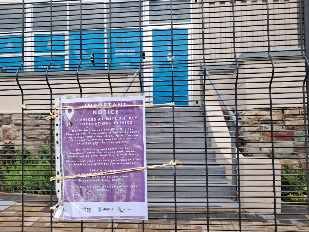
In central Johannesburg, where I work, HIV testing services have collapsed. The people who fell out of programmes are not coming back. HIV prevention and TB screening have largely stopped.

Reassurances that state clinics will pick up testing are empty – the staff do not exist, and testing has not resumed. State clinics do not trace people who fall out of care for any illness, let alone for HIV. The data systems maintained by PEPFAR-supported organisations are now gone.

What happens now? The first hard sign that things are failing will be a large drop in the number of people starting treatment, versus what happened in the same month one year ago. The next metric to watch will be hospitalisations for tuberculosis and other infections associated with untreated HIV infection. This will happen towards the end of the year, as immune systems fail. Not long after, death rates will rise. We will see that in death certificates among younger people – the parents and younger adults.

Unfortunately, much of this information will not be available to the health department for at least a year or two, because among the staff laid off in this crisis are the data collectors for the programmes that tracked vital metrics.

The above should come as no surprise, especially to the public health commentators and health department, which is why it is so surprising to hear how certain they are that the PEPFAR programme can easily be absorbed into the state services. The timing of this crisis could not be worse, with huge budget holes in provincial health departments.

Why should this be a priority? After starting the HIV programme in 2004, we spent the next few years muddling through how to deliver antiretrovirals to millions of people in primary care, before we realised we also needed to diagnose them earlier. In 2004, the average CD4 count (a measure of immune strength) at initiation of treatment was about 80 cells/ul, devastatingly low – normal is > 500 cells/ul. A quarter were ill with TB.

This CD4 count average took years to go up, but only by pushing testing into clinic queues, communities, and special services for key populations, not waiting till they were sick. Recently, the average initiation CD4 count was about 400 cells/ul, stopping years of transmission, with most people healthy, and only a small number with TB.

There are many reasons to criticise the relationship between PEPFAR and the health department. It suited both parties to have a symbiotic relationship that meant each got on with their job and ticked their respective output boxes, but neither had to tussle with the messiness of trying to move the PEPFAR deliverables into the health department. As we move forward, learning from these fragilities to plan for the future of the HIV care programme, and for other diseases, will be critical.

Since the suspension of funding, many people have said, “We don’t hear much about HIV anymore”. That is because when the system works well, you don’t hear about it. Some things are far better compared to 2004:

  • We have a government not in denial about HIV being a problem nor encouraging pseudoscience or crackpots.
  • Our frontline health workers, in over 3000 clinics, have vast experience initiating and maintaining antiretrovirals.
  • Antiretrovirals are cheaper, more potent, more durable, and safer.
  • Treatment protocols are simpler.
  • New infection rates are way down.
  • Government delivery systems have improved.
  • Data systems suggest that the majority of ‘lost’ patients are in care, often simply in another clinic.

A sensible emergency plan would do this:

  • Fund existing programmes for a limited time, understanding that the level of reach and expertise is impossible for the health department to replicate at short notice.
  • Couple this with a plan to make posts more sustainable over the next year or two.
  • Learn from the PEPFAR programme that rigorously held organisations accountable, so that provinces can similarly be answerable for their HIV metrics.
  • Ask hard questions why single patient identifiers, and government information systems, that could easily be linked to laboratory, pharmacy and radiology databases, are still not integrated within the public systems, as they are throughout the private health system.
  • Accept that certain key functions and clinics may best be sited outside of the health department.

This will not save the large and valuable research programmes, which need other help. Much of the rest of Africa needs a Marshall Plan to rescue their entire HIV service, as they are almost totally dependent on US government funding.

But ideas like the above will preserve the current South African HIV response and allow us to imagine interventions that could end the disease as a threat for future generations.

No one disputes we need a move away from donor-assisted health programmes. But the scale and immense urgency of this oncoming emergency needs to be understood. We need a plan and a budget, and fast. Or we will have an overwhelmed hospital system and busy funeral services again.

Professor Francois Venter works for Ezintsha, a policy and research unit at Wits. He has been involved in the HIV programme since 2001, and ran several large PEPFAR programmes till 2012. Venter and his unit do not receive funding from PEPFAR, USAID or CDC. Thank you to several experts for supplying analysis and ideas for the initial draft of the article.

Published by GroundUp and Spotlight

Republished from GroundUp under a Creative Commons Attribution-NoDerivatives 4.0 International License.

Read the original article.

Scientists Crack the Puzzle of How Retinoic Acid Works Against Neuroblastoma

Photo by Anna Shvets

Neuroblastoma is a solid tumour that occurs in children. When high-risk, the disease has a poor prognosis. Decades ago, adding the drug retinoic acid to neuroblastoma treatment increased survival by 10–15%. However, this effect was only evident in post-chemotherapy consolidation after bulky primary tumours had largely been eliminated. Why retinoic acid is effective in this setting but not against primary tumours, has been speculated about for nearly 50 years. 

St. Jude Children’s Research Hospital scientists resolved the mystery in a new study, showing retinoic acid uses a novel mechanism to kill metastasised neuroblastoma. The drug “hijacks” a normal developmental pathway to trigger cancer cell death. The findings, which have implications for future combination therapy approaches, appears in Nature Communications

“We’ve come up with an explanation for a decades-long contradiction about why retinoic acid works in post-chemotherapy consolidation but has little impact on primary neuroblastoma tumours,” said senior co-corresponding author Paul Geeleher, PhD. “Retinoic acid’s activity heavily depends on the cellular microenvironment.” 

The cellular microenvironment is the soup of chemicals, proteins and other signals that surround a cell, and which is unique to that part of the body. For example, the bone marrow microenvironment contains signals to grow blood cells and restructure bone. Metastasised neuroblastoma cells often migrate to bone marrow, where the bone morphogenetic protein (BMP) pathway signalling is highly active. The researchers showed that BMP signaling makes neuroblastoma cells much more vulnerable to retinoic acid. 

“Unexpectedly, we found that cells expressing genes from the BMP signaling pathway were very sensitive to retinoic acid,” said co-first and co-corresponding author Min Pan, PhD, St. Jude Department of Computational Biology. “However, since the bone marrow microenvironment causes neuroblastoma cells there to have higher BMP activity, it neatly explained why retinoic acid is very effective at treating those cells during consolidation therapy, but not the primary tumours during up-front treatment.” 

Hijacking development to drive metastatic neuroblastoma cell death 

Using gene editing technology, the scientists uncovered the relationship between BMP signaling and retinoic acid. They assembled a group of neuroblastoma cell lines susceptible to retinoic acid, then cut out genes to find which were responsible for the drug’s activity. Genes in the BMP pathway had the largest effect while providing a plausible explanation for retinoic acid’s varying outcomes in patients.

“We found that, in neuroblastoma, BMP signaling works with retinoic acid signaling in the same way as during development,” said co-first author Yinwen Zhang, PhD, who characterised how transcription factors, the proteins that bind DNA to regulate gene expression, led to different results in highly retinoic acid-sensitive or insensitive neuroblastoma cells. “If there are a lot of BMP-signaling pathway transcription factors already on DNA, then retinoic acid signaling combines with it to promote downstream cell death–related gene expression. This occurs both in normal embryonic development and neuroblastoma cells in certain microenvironments.” 

“We are the first to uncover such an example of ‘hijacking’ a normal embryonic developmental process preserved in cancer that we can exploit therapeutically,” Geeleher said. “Now, we can look for similar processes in other diseases to design less toxic and more effective treatment strategies.” 

Source: St. Jude Children’s Research Hospital

Gut Microbes also Feed on Sugar to Produce Crucial Short-chain Fatty Acids

Source: CC0

Gut microbes that were thought to feed exclusively on dietary fibre also get fed sugar from our guts, from which they produce short-chain fatty acids that are crucial to many body functions. The Kobe University discovery of this symbiotic relationship also points the way to developing novel therapeutics.

Gut microbes produce many substances that our body needs but cannot produce itself. Among them are short-chain fatty acids that are the primary energy source for the cells lining our guts but have other important roles, too, and that are thought to be produced by bacteria who feed on undigested fibre. However, in a previous study, the Kobe University endocrinologist Ogawa Wataru found that people who take the diabetes drug metformin excrete the sugar glucose to the inside of their guts. He says: “If glucose is indeed excreted into the gut, it is conceivable that this could affect the symbiotic relationship between the gut microbiome and the host.”

Ogawa and his team set out to learn more about the details of the glucose excretion and its relationship with the gut microbiota. “We had to develop unprecedented bioimaging methods and establish novel analytical techniques for the products of the gut microbial metabolism,” he says. They used their new methods to not only see where and how much glucose enters the guts, but also used mouse experiments to find out how the sugar is transformed after that. In addition, they also checked how the diabetes drug metformin influences these results both in humans and in mice.

The Kobe University team now published their results in the journal Communications Medicine. They found that, first, glucose is excreted in the jejunum and is transported from there inside the gut to the large intestine and the rectum. “It was surprising to find that even individuals not taking metformin exhibited a certain level of glucose excretion into the intestine. This finding suggests that intestinal glucose excretion is a universal physiological phenomenon in animals, with metformin acting to enhance this process,” Ogawa explains. In both humans and mice, irrespective of whether they were diabetic or not, metformin increased the excretion by a factor of almost four.

And second, on the way down, the glucose gets transformed into short-chain fatty acids. Ogawa says: “The production of short-chain fatty acids from the excreted glucose is a huge discovery. While these compounds are traditionally thought to be produced through the fermentation of indigestible dietary fibres by gut microbiota, this newly identified mechanism highlights a novel symbiotic relationship between the host and its microbiota.”

Ogawa and his team are now conducting further studies with the aim of understanding how metformin and other diabetes drugs affect glucose excretion, the gut microbiome and their metabolic products. He says: “Intestinal glucose excretion represents a previously unrecognised physiological phenomenon. Understanding the underlying molecular mechanisms and how drugs interfere with this process could lead to the development of novel therapeutics aimed at the regulation of gut microbiota and their metabolites.”

Source: Kobe University

Brown Fat Could Help Maintain Exercise Capacity in Aging

Photo by Barbara Olsen on Pexels

Rutgers Health researchers have made discoveries about brown fat that may open a new path to helping people stay physically fit as they age. A team from Rutgers New Jersey Medical School found that mice lacking a specific gene developed an unusually potent form of brown fat tissue that expanded lifespan and increased exercise capacity by roughly 30%. The team is working on a drug that could mimic these effects in humans.

“Exercise capacity diminishes as you get older, and to have a technique that could enhance exercise performance would be very beneficial for healthful aging,” said Stephen Vatner, university professor and director of the Cardiovascular Research Institute in the medical school’s Department of Cell Biology and Molecular Medicine and senior author of the study in Aging Cell. “This mouse model performs exercise better than their normal littermates.”

Unlike white fat, which stores energy, brown fat burns calories and helps regulate body temperature. This study revealed brown fat also plays a crucial role in exercise capacity by improving blood flow to muscles during physical activity.

The genetically modified mice produced unusually high amounts of active brown fat and showed about 30% better exercise performance than normal mice, both in speed and time to exhaustion.

The discovery emerged from broader research into healthy aging. The modified mice, which lack a protein called RGS14, live about 20% longer than normal mice, with females living longer than males – similar to the pattern seen in humans. Even at advanced ages, they maintain a healthier appearance, avoiding the typical signs of aging, such as loss of hair and graying that appear in normal elderly mice. Their brown adipose tissue also protects them from obesity, glucose intolerance, cardiovascular disorders, cancer and Alzheimer’s disease, in addition to reduced exercise tolerance.

To test whether the brown fat – rather than some other result from the missing genes –accounted for the benefits, the researchers transplanted the brown fat to normal mice. They noted that the recipients gained similar benefits within days. Transplants using regular brown fat from normal mice, by contrast, took eight weeks to produce much milder improvements.

The discovery could eventually improve human lifespans – the total time when people enjoy good mental and physical health.

“With all the medical advances, aging and longevity have increased in humans, but unfortunately, healthful aging hasn’t,” Vatner said. “There are a lot of diseases associated with aging – obesity, diabetes, myocardial ischemia, heart failure, cancer – and what we have to do is find new drugs based on models of healthful aging.”

Rather than develop a treatment that addresses aging broadly, which poses regulatory challenges, Vatner said his team plans to test for specific benefits such as improved exercise capacity and metabolism. This approach builds on their previous success in developing a drug based on a different mouse healthful longevity model.

“We’re working with some people to develop this agent, and hopefully, in another year or so, we’ll have a drug that we can test,” Vatner said.

In the meantime, techniques such as deliberate cold exposure can increase brown fat naturally. Studies have found such efforts to produce short-term benefits that range from enhanced immune system function to improved metabolic health, but Vatner said none of the studies have run long enough to find any effect on healthful aging.

He added that most people would prefer to increase brown fat levels by taking pills rather than ice baths and is optimistic about translating the newest finding into an effective medication.

Source: Rutgers University

Researchers Debunk Common Beliefs About ‘Cycle Syncing’ and Muscles

Photo by John Arano on Unsplash

New research from McMaster kinesiologists is challenging the internet belief that timing resistance training to specific phases of the menstrual cycle boosts the body’s ability to build muscle and strength.

The researchers have shown that exercising at various points in the cycle had no impact — positive or negative — on the synthesis of new muscle proteins, a process essential to building and maintaining muscle.

The results, published in the print edition of the Journal of Physiology, debunk the popularly touted practice of cycle syncing, or tailoring workouts to align with the way hormones change throughout a woman’s menstrual cycle.

“Our findings conflict with the popular notion that there is some kind of hormonal advantage to performing different exercises in each phase,” explains Lauren Colenso-Semple, lead author of the study and a former graduate student in the Department of Kinesiology, who conducted the work while at McMaster.

“We saw no differences, regardless of cycle timing.”

For the study, researchers monitored the menstrual cycles of participants — all healthy young women — for three months to confirm their cycles were normal. Contrary to popular belief, only a small percentage of women — about 12 per cent — have a consistent 28-day cycle and ovulate regularly on Day 14 or the “textbook” menstrual cycle.

Participants then ingested a tracer molecule, a benign substance designed to track and monitor muscle protein levels. They performed heavy resistance exercise during two distinct phases of their menstrual cycles: the follicular phase, when estrogen levels are at their peak; and the luteal phase, characterized by peak progesterone levels.

Researchers observed no effect of either menstrual cycle phase on the production of muscle proteins.

Cycle syncing has been made popular by internet influencers to coordinate workouts, certain diets and lifestyle behaviours with the menstrual cycle.

There are fitness apps for tracking cycles, and social media channels are rife with advice and recommendations.

Proponents routinely cite a handful of scientific studies on animals as evidence that fluctuations in ovarian hormones can affect how human muscles respond to exercise, but this study shows that not to be correct.

“Our work shows that women who want to lift weights and recondition their muscles should feel free to do so in any phase of their cycle. There is no physiological difference in response to the exercise,” says Stuart Phillips, the Canada Research Chair in Skeletal Muscle Health at McMaster who supervised the study.

“It is important to tailor your training to how you feel.”

Scientists highlight the need for further research, particularly studies that focus on women’s health. This includes investigating how training, in relation to the menstrual cycle, affects women and how both oral and non-oral contraceptives influence their responses to exercise.

This article was first published on Brighter World. Read the original article.

Research Draws a Potential Association Between Tattoos and Cancer Risk

Photo by Benjamin Lehman on Unsplash

Tattoo ink does not just stay under the skin – some of it makes its way into the lymph nodes. Researchers from the Department of Public Health and the Department of Clinical Research at the University of Southern Denmark (SDU), together with the University of Helsinki, have investigated whether this could have health consequences. Using data from Danish twin pairs, they found that tattooed individuals are more frequently diagnosed with skin and lymphoma cancers compared to those without tattoos.

Ink particles in the body may affect the immune system

When tattoo ink penetrates the skin, some of it is absorbed into the lymph nodes, a key part of the immune system. The researchers are particularly concerned that tattoo ink may trigger chronic inflammation in the lymph nodes, which over time could lead to abnormal cell growth and an increased risk of cancer.

“We can see that ink particles accumulate in the lymph nodes, and we suspect that the body perceives them as foreign substances,” explains Henrik Frederiksen, consultant in haematology at Odense University Hospital and clinical professor at SDU.

“This may mean that the immune system is constantly trying to respond to the ink, and we do not yet know whether this persistent strain could weaken the function of the lymph nodes or have other health consequences.”

Studying this link is challenging because cancer can take years to develop. This means that exposure in youth may not lead to illness until decades later, making it difficult to measure a direct effect.

Twin data provides a unique opportunity to study the link

The study is based on data from the Danish Twin Tattoo Cohort, where researchers have information from more than 5900 Danish twins. By analysing tattoo patterns alongside cancer diagnoses, they found a higher occurrence of both skin and lymphoma cancers in tattooed individuals.

“The unique aspect of our approach is that we can compare twin pairs where one has cancer, but they otherwise share many genetic and environmental factors,” says Jacob von Bornemann Hjelmborg, professor of biostatistics at SDU.

“This provides us with a stronger method for investigating whether tattoos themselves may influence cancer risk.”

The size of tattoos matters

The results show that the link between tattoos and cancer is most evident in those with large tattoos – defined as bigger than a palm. 

For lymphoma, the rate is nearly three times higher for the group of individuals with large tattoos compared to those without tattoos.  This rate (more specifically, ‘hazard rate’) accounts for age, the timing of the tattoo, and how long the individuals have been followed in the study. 

“This suggests that the bigger the tattoo and the longer it has been there, the more ink accumulates in the lymph nodes. The extent of the impact on the immune system should be further investigated so that we can better understand the mechanisms at play,” says Signe Bedsted Clemmensen, assistant professor of biostatistics at SDU.

Another study from the Danish Twin Tattoo Cohort shows that tattoos are becoming increasingly common. Researchers estimate that four in ten women and three in ten men will have tattoos by the age of 25.

The link to lymphoma has also been observed in an independent Swedish study from 2024.

Are some ink colours worse than others?

Previous research has suggested that certain pigments in tattoo ink may be more problematic than others.

“In our study, we do not see a clear link between cancer occurrence and specific ink colours, but this does not mean that colour is irrelevant. We know from other studies that ink can contain potentially harmful substances, and for example, red ink more often causes allergic reactions. This is an area we would like to explore further,” says Signe Bedsted Clemmensen.

What are the next steps?

The researchers now plan to investigate how ink particles affect the function of lymph nodes at a molecular level and whether certain types of lymphoma are more linked to tattoos than others.

“We want to gain a better understanding of the biological mechanisms – what happens in the lymph nodes when they are exposed to ink particles over decades? This can help us assess whether there is a real health risk and what we might do to reduce it,” concludes Signe Bedsted Clemmensen.

Source: University of Southern Denmark Faculty of Health Sciences