Tag: 4/9/24

White Matter may Aid Recovery from Spinal Cord Injuries

View of the spinal cord. Credit: Scientific Animations CC4.0

Injuries, infection and inflammatory diseases that damage the spinal cord can lead to intractable pain and disability but some degree of recovery may be possible. The question is, how best to stimulate the regrowth and healing of damaged nerves.

At the Vanderbilt University Institute of Imaging Science (VUIIS), scientists are focusing on a previously understudied part of the brain and spinal cord – white matter, which is made up of axons that relay signals. Their discoveries could lead to treatments that restore nerve activity through the targeted delivery of electromagnetic stimuli or drugs.

In a recent paper published in the Proceedings of the National Academy of SciencesAnirban Sengupta, PhD, John Gore, PhD, and their colleagues report the detection of signals from white matter in the spinal cord in response to a stimulus that are as robust as grey matter signals.

“In the spinal cord, the white matter signal is quite large and detectable, unlike in the brain, where it has less amplitude than the grey matter (signal),” said Sengupta, research instructor in Radiology and Radiological Sciences at Vanderbilt University Medical Center.

“This may be due to the larger volume of white matter in the spinal cord compared to the brain,” he added. Alternatively, the signal could represent “an intrinsic demand” in metabolism within the white matter, reflecting its critical role in supporting grey matter.

For several years, Gore, who directs the VUIIS, and his colleagues have used functional magnetic resonance imaging (fMRI) to detect blood oxygenation-level dependent (BOLD) signals, a key marker of nervous system activity, in white matter.

Last year, they reported that when participants undergoing fMRI perform a task, like wiggling their fingers, BOLD signals increase in white matter throughout the brain.

The current study monitored changes in BOLD signals in the white matter of the spinal cord at rest and in response to a vibrotactile stimulus applied to the fingers in an animal model. In response to stimulation, white matter activity was higher in “tracts” of ascending fibres that carry the signal from the spine to the brain.

This result is consistent with white matter’s known neurobiological function, the researchers noted. White matter contains non-neuronal glial cells that do not produce electrical impulses, but which regulate blood flow and neurotransmitters, the signaling molecules that transmit signals between nerve cells.

Much remains to be learned about the function of white matter in the spinal cord. But the findings from this research may help in improved understanding of diseases that affect white matter in the spinal cord, including multiple sclerosis, Sengupta said.

“We will be able to see how activity in the white matter changes in different stages of the disease,” he said. Researchers also may be able to monitor the effectiveness of therapeutic interventions, including neuromodulation, in promoting recovery following spinal cord injury.

Source: Vanderbilt University Medical Center

Inflammation Leaves a Long-lasting Impression on Intestinal Stem Cells

Irritable bowel syndrome. Credit: Scientific Animations CC4.0

Researchers at Baylor College of Medicine, the University of Michigan and collaborating institutions have discovered that inflammation in the gut leaves long-term marks on intestinal stem cells (ISCs) that reduce their ability to heal the intestine, even after inflammation has receded. This is important because it affects ISCs’ response to future challenges. The study appeared in Cell Stem Cell.

“We study graft-vs-host disease (GVHD), a major cause of mortality after bone marrow transplantation, a potentially curative therapy for many blood diseases. One of our goals is to better understand GVHD and identify strategies to control it,” said corresponding author Dr Pavan Reddy, professor and director of the Dan L Duncan Comprehensive Cancer Center at Baylor and previously at the University of Michigan.

“GVHD is an inflammatory reaction in which immune T cells from the bone marrow transplant donor attack the host gut cells, mainly ISCs,” said first author Dr Dongchang Zhao, in Reddy’s lab.

Although many ISCs perish during GVHD, survivors remain. However, it’s not known whether they are fully functional or can return to their full functionality after the resolution of GVHD, which has fundamental implications for host resilience and repair.

“In the current study, we investigated the consequences of inflammation on ISCs in well-defined clinically relevant models of GVHD,” Reddy said.

“Using cellular and animal models, we found that exposure to inflammation drove ISCs to change their metabolism in ways that resulted in the accumulation of succinate, a product of cellular processes, which in turn reprogramed the epigenome,” Zhao said.

The epigenome is a system of chemical marks on the DNA that regulates the genes expressed by the cell. Inflammation-led epigenome reprogramming changed the expression of genes involved in cell reproduction. Overall, reprogrammed ISCs were less able of regenerating, a first step toward healing the intestine.

“We then investigated whether ISCs would be able to recuperate their regenerative ability after inflammation had resolved,” Reddy said. “We found that ISCs had not overcome their initial exposure to inflammation. Despite mitigating GVHD inflammation for 28 days, ISCs retained a reduced regenerative capacity that led to poor recovery and increased mortality from challenges, such as non-lethal radiation exposure, in animal models. More research is on the way to design strategies to help ISCs ‘forget’ their encounter with inflammation and enhance their resilience against immune attacks.”

Source: Baylor College of Medicine

Finerenone Reduces Worsening Heart Failure and Cardiovascular Death in Clinical Trial

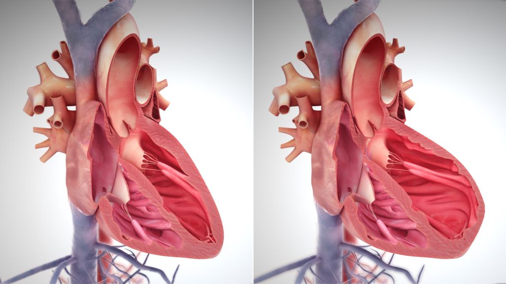
Right side heart failure. Credit: Scientific Animations CC4.0

Finerenone reduced the composite of total first and recurrent heart failure (HF) events (hospitalisations for HF or urgent HF visits) and cardiovascular death in patients with HF and mildly reduced or preserved ejection fraction, according to an international clinical trial led by investigators from Brigham and Women’s Hospital.

Heart failure events and cardiovascular death were less common in the finerenone group than in the placebo group. Overall, the rate of serious adverse events was similar across the groups, but rates of hyperkalaemia were higher for the group taking finerenone. Results were presented at the European Society of Cardiology Congress 2024 and published simultaneously in the New England Journal of Medicine.

“We saw benefit regardless of the ejection fraction and even in patients who were on other approved therapies,” said trial principal investigator and corresponding author Scott Solomon, MD, the director of the Clinical Trials Outcomes Center at Mass General Brigham and the Edward D. Frohlich Distinguished Chair at Brigham and Women’s Hospital. “This drug represents a new drug class that may become a pillar of therapy for this disease.”

HF is the progressive decline in the heart’s ability to fill with and pump blood. It affects over 60 million people worldwide. Approximately half of all people living with HF have mildly reduced or preserved left ventricular ejection fraction, a condition with limited treatment options. These findings suggest that the non-steroidal mineralocorticoid receptor antagonist finerenone could represent a new therapeutic option for patients.

The FINEARTS-HF trial, funded by Bayer, assigned 6000 patients to receive either finerenone or placebo in addition to their existing therapies. The trial’s limitations include few Black patients, although the percentage of Black patients was proportional to their regional population. “Our group continues to study novel therapies for heart failure,” Solomon said. “There’s huge residual risk in these patients and so more room for new therapies.“

Source: Brigham and Women’s Hospital

fMRI Discovers Where Love Resides in the Brain

The image represents a statistical average of how different types of love light up different regions of the brain. Photo: Pärttyli Rinne et al 2024, Aalto University.

We use the word ‘love’ in a bewildering range of contexts, from sexual adoration to parental love or the love of nature. Now, more comprehensive imaging of the brain may shed light on why we use the same word for such a diverse collection of human experiences.

“You see your newborn child for the first time. The baby is soft, healthy and hearty – your life’s greatest wonder. You feel love for the little one.”

The above statement was one of many simple scenarios presented to 55 parents, self-described as being in a loving relationship. Researchers from Aalto University utilised functional magnetic resonance imaging (fMRI) to measure brain activity while subjects mulled brief stories related to six different types of love.

“We now provide a more comprehensive picture of the brain activity associated with different types of love than previous research,” says Pärttyli Rinne, the philosopher and researcher who coordinated the study. “The activation pattern of love is generated in social situations in the basal ganglia, the midline of the forehead, the precuneus and the temporoparietal junction at the sides of the back of the head.”

Love for one’s children generated the most intense brain activity, closely followed by romantic love.

“In parental love, there was activation deep in the brain’s reward system in the striatum area while imagining love, and this was not seen for any other kind of love,” says Rinne. Love for romantic partners, friends, strangers, pets and nature were also part of the study, which was published in the journal Cerebral Cortex.

According to the research, brain activity is influenced not only by the closeness of the object of love, but also by whether it is a human being, another species or nature.

Unsurprisingly, compassionate love for strangers was less rewarding and caused less brain activation than love in close relationships. Meanwhile, love of nature activated the reward system and visual areas of the brain, but not the social brain areas.

Pet-owners identifiable by brain activity

The biggest surprise for the researchers was that the brain areas associated with love between people ended up being very similar, with differences lying primarily in the intensity of activation. All types of interpersonal love activated areas of the brain associated with social cognition, in contrast to love for pets or nature – with one exception.

Subjects’ brain responses to a statement like the following, on average, revealed whether or not they shared their life with a furry friend:

“You are home lolling on the couch and your pet cat pads over to you. The cat curls up next to you and purrs sleepily. You love your pet.”

“When looking at love for pets and the brain activity associated with it, brain areas associated with sociality statistically reveal whether or not the person is a pet owner. When it comes to the pet owners, these areas are more activated than with non-pet owners,” says Rinne.

Love activations were controlled for in the study with neutral stories in which very little happened. For example, looking out the bus window or absent-mindedly brushing your teeth. After hearing a professional actor’s rendition of each ‘love story’, participants were asked to imagine each emotion for 10 seconds.

This is not the first effort at finding love for Rinne and his team, which includes researchers Juha Lahnakoski, Heini Saarimäki, Mikke Tavast, Mikko Sams and Linda Henriksson. They have undertaken several studies seeking to deepen our scientific knowledge of human emotions. The group released research mapping subjects’ bodily experiences of love a year ago, with the earlier study also linking the strongest physical experiences of love with close interpersonal relationships.

Not only can understanding the neural mechanisms of love help guide philosophical discussions about the nature of love, consciousness, and human connection, but also, the researchers hope that their work will enhance mental health interventions in conditions like attachment disorders, depression or relationship issues.

Source: Aalto University