Tag: 30/5/22

By Now, Nearly All South Africans Have COVID Antibodies

South African flag with COVID theme
Image by Quicknews

The latest COVID seroprevalence survey shows that nearly every adult in South Africa has either been vaccinated or had COVID. For many, it’s both.

The study analysed blood from over 3000 blood donors. It was conducted by the South African National Blood Service, which is responsible for blood donations in eight provinces, and the Western Cape Blood Service.

The researchers estimated that by March 2022, before the fifth wave which appears to have peaked in the last few weeks, 98% of adults had some detectable antibodies, whether from COVID or from vaccination. This means that only 2% had neither been vaccinated nor been infected.

Only 10% had been vaccinated but not infected by COVID.

Read the study

(Note: The study has been published as a preprint and has not been peer-reviewed.)

What the survey tested for

Blood samples were collected and tested from 3395 consenting donors from all provinces in mid-March 2022. While blood donors are not precisely representative of the population, the researchers have argued that the study is representative enough.

This is the first time the blood services researchers have been able to look for two types of antibodies.

One test indicates if a sample has antibodies to the nucleocapsid proteins (anti-nucleocapsid antibodies). These antibodies develop if someone is infected, but won’t develop after a person receives a vaccine only (at least not those vaccines currently available in South Africa).

The other test indicates if the sample has antibodies to the spike protein (anti-spike antibodies). These antibodies develop when someone has been infected or has been vaccinated (or both).

Using these two tests together, researchers can, for the first time, evaluate the proportion of the population that has been vaccinated and not infected.

Results

After weighting the results to reflect national demographics, the researchers found that a mere 2% of the population had neither anti-spike nor anti-nucleocapsid antibodies. These are people who have likely never had COVID nor been vaccinated.

10% had only anti-spike antibodies. These are people who were likely vaccinated, but never infected.

The researchers noted that there is “an increasing incidence of reinfection” with the omicron wave.

Blood service survey is the best we have

The blood services have been regularly testing blood samples from donors throughout the pandemic, looking at the presence of anti-nucleocapsid antibodies.

While other surveys might be more representative of the population than the blood donor ones, these have been infrequently published or published long after the survey was conducted. By contrast the blood donor surveys are relatively affordable and quick to publish. Also, as far as we are aware, it is the only survey repeatedly testing the same group of people, so that comparisons across time are possible.

Past blood surveys

The blood services’ survey from samples taken in May 2021 estimated that 47% of the adult population had previously been infected.

The next survey of blood samples was taken in November 2021 after the delta wave. This was just before the omicron wave. The researchers estimated that about 70% of people had been infected.

The latest survey indicates that about 87% of people have been infected.

The previous surveys found that levels of infection differed by province. Now these differences have “largely disappeared as prevalence appears to have saturated”.

Differences across race

There are significant differences in rates of infection when different races are compared.

The November survey showed that about 80% of black donors and 40% of white donors had been infected with COVID.

In the latest survey the proportion of white and Asian donors that only have anti-spike antibodies (indicating vaccination but no infection) was higher than black and coloured donors.

The researchers suggest that “white donors are both unusually likely to avail themselves of vaccination, and they are unusually able to avoid exposure, for instance by working predominantly from home, [and] living in smaller family units.”

Article by By James Stent. Republished from GroundUp under a Creative Commons Attribution-NoDerivatives 4.0 International License.

Source: GroundUp

Vitamin D Doesn’t Prevent the Development of Type 2 Diabetes

Vitamin D pills
Photo by Michele Blackwell on Unsplash

Daily vitamin D supplements do not seem to prevent the development of type 2 diabetes in people already at high risk of the condition, according to a Japanese trial published by The BMJ.

While no clinically meaningful effect was seen in high risk adults, the results hinted that there may be a benefit for people with insufficient insulin secretion.

Type 2 diabetes affects around 480 million people worldwide, and is predicted to increase to 700 million by 2045. Another half a billion people have impaired glucose tolerance or pre-diabetes (higher than normal blood sugar levels that, if left untreated, can develop into type 2 diabetes).

Weight loss and exercise can lower the risk of progression to type 2 diabetes, but are difficult to sustain, so new strategies are needed to tackle the problem.

An association between vitamin D deficiency and an increased risk of future diabetes has been shown in some studies, but trials of vitamin D supplements for preventing type 2 diabetes show inconsistent results.

To address this knowledge gap, researchers therefore set out to assess whether eldecalcitol (an active form of vitamin D used to treat osteoporosis in Japan) could reduce the risk of developing type 2 diabetes among people with impaired glucose tolerance.

The study involved 1256 Japanese adults with impaired glucose tolerance with an average age of 61 years; 46% were women, and 59% had a family history of type 2 diabetes.

Participants were randomly assigned to receive either a standard daily dose of eldecalcitol (630 participants) or placebo (626 participants) and were assessed for diabetes every three months over a three-year follow-up period.

During this period, the researchers found no meaningful differences between groups in those who developed diabetes (12.5% in the eldecalcitol group and 14% in the placebo group) or whose blood sugar levels returned to normal (23% in the eldecalcitol group and 20% in the placebo group).

However, after adjusting for 11 potentially influential factors, including age, sex, blood pressure, body mass index, and family history of diabetes, the results suggested that eldecalcitol might prevent type 2 diabetes in pre-diabetic patients with insufficient insulin secretion. But this finding remains unclear and the researchers say further work is needed before any firm conclusions can be made.

They did, however, find a significant increase in both lower back and hip bone mineral densities among those taking eldecalcitol compared with placebo.

No significant difference in serious adverse events was seen between the two groups.

The researchers acknowledge some uncertainties, such as whether the dose of eldecalcitol used was appropriate for preventing diabetes, and whether the results apply to all ethnicities. Nevertheless, this was a large trial with regular follow-up and high adherence to treatment, suggesting that the findings are robust.

As such, they say: “Although treatment with eldecalcitol did not significantly reduce the incidence of diabetes among people with pre-diabetes, the results suggested the potential for a beneficial effect of eldecalcitol on people with insufficient insulin secretion.” And they call for further research to determine whether vitamin D is beneficial to people with pre-diabetes.

This new trial was well conducted and results are consistent with two other recent trials, said Tatiana Christides at Queen Mary University of London in a linked editorial.

However, several questions remain, she writes, including whether vitamin D supplementation may be more effective for particular populations, and whether longer duration of treatment or younger age at initiation might be more beneficial.

Until further data are available from high quality randomized trials, she suggests healthcare professionals “should continue to discuss with patients the musculoskeletal health benefits of vitamin D and support them to achieve and maintain lifestyle changes that, although challenging to sustain, are known to decrease development of type 2 diabetes.”

Source: News-Medical.Net

MRI Unveils Secrets of Brains under Anaesthesia

Depiction of a human brain
Image by Fakurian Design on Unsplash

A study published in eLife reveals how the brains of humans and other primates under anaesthesia differ from mammals such as mice, with the visual cortex in primates being isolated from certain effects.

Anaesthesia still holds mysteries for modern science. Electroencephalography (EEG) studies show that, during anaesthesia, the brain is put into a deep sleep-like state in which periods of rhythmic electrical activity alternate with periods of complete inactivity. This state is called burst-suppression. Until now, it was unclear where exactly this state happens in the brain and which brain areas are involved.

Shedding light on the phenomenon would help better understand how the brain functions under anaesthesia. To this end, researchers used functional magnetic resonance imaging (fMRI) to study the precise spatial distribution of synchronously working brain regions in anaesthetised humans, long-tailed macaques, common marmosets and rats. They were able to show for the first time that the areas where burst-suppression is evident differ significantly in primates and rodents. While in rats large parts of the cerebral cortex synchronously show the burst-suppression pattern, in primates individual sensory regions, such as the visual cortex, are excluded from it.

“Our brain can be thought of as a full soccer stadium when we are awake,” explained Nikoloz Sirmpilatze, lead author of the study. “Our active neurons are like tens of thousands of spectators all talking at once. Under anaesthesia, however, neuronal activity is synchronised. You can measure this activity using EEG as uniform waves, as if all the spectators in the stadium were singing the same song. In deep anaesthesia, this song is repeatedly interrupted by periods of silence. This is called burst-suppression. The deeper the anaesthesia, the shorter the phases of uniform activity, the bursts, and the longer the periodically recurring inactive phases, the so-called suppressions.”

The phenomenon is caused by many different anaesthetics, some of which vary in their mechanisms of action. And burst-suppression is also detectable in coma patients. However, it is not known whether this condition is a protective reaction of the brain or a sign of impaired functioning. It has also been unclear where in the brain burst-suppression occurs and which brain areas are involved, as localisation by EEG alone is not possible.

To answer this question, the researchers fMRI. In the first part of the study, the researchers established a system to evaluate fMRI data in humans, monkeys and rodents in a standardised manner using the same method. To do this, they used simultaneously-measured EEG and fMRI data from anaesthetised patients that had been generated in a previous study. “We first looked to see whether the burst-suppression detected in the EEG was also visible in the fMRI data and whether it showed a certain pattern,” says Nikoloz Sirmpilatze. “Based on that, we developed a new algorithm that allowed detecting burst-suppression events in the experimental animals using fMRI, without additional EEG measurement.”

The researchers then performed fMRI measurements in anaesthetised long-tailed macaques, common marmosets and rats. In all animals, they were able to detect and precisely localise burst-suppression as a function of anesthetic concentration. The spatial distribution of burst-suppression showed that in both humans and monkey species, certain sensory areas, such as the visual cortex, were excluded from it. In contrast, in the rats, the entire cerebral cortex was affected by burst-suppression.

“At the moment, we can only speculate about the reasons,” said Nikoloz Sirmpilatze, who was awarded the German Primate Center’s 2021 PhD Thesis Award for his work. “Primates orient themselves mainly through their sense of sight. Therefore, the visual cortex is a highly specialised region that differs from other brain areas by special cell types and structures. In rats, this is not the case. In future studies, we will investigate what exactly happens in these regions during anaesthesia to ultimately understand why burst-suppression is not detectable there with fMRI.”

Susann Boretius, senior author of the study adds: “The study not only raises the question of the extent to which rodents are suitable models for many areas of human brain research, especially when it comes to anaesthesia, but the results also have many implications for neuroscience and the evolution of neural networks in general.”

Source: Deutsches Primatenzentrum (DPZ)/German Primate Center

An Anti-HIV Drug for Memory Recall in Older Adults?

Old man
Photo by Kindel Media on Pexels

The human brain usually stores memories in groups so that the recollection of one significant memory triggers the recall of others connected by time. With ageing, the brain gradually loses this ability to link related memories.

Now, researchers have discovered a key molecular mechanism behind this memory linking, and also identified a way to restore this brain function in middle-aged mice. They also found an anti-HIV drug that can do this.

Published in Nature, the findings suggest a new method for strengthening human memory in middle age and a possible early intervention for dementia.

“Our memories are a huge part of who we are,” explained Professor Alcino Silva. “The ability to link related experiences teaches how to stay safe and operate successfully in the world.”

The team from UCLA focused on a gene called CCR5 that encodes the CCR5 receptor – the same one that HIV hitches a ride on to infect brain cells, resulting in memory loss in AIDS patients.

In previous work, Prof Silva’s lab showed that CCR5 expression reduced memory recall.

In the current study, Prof Silva and his colleagues discovered a central mechanism underlying mice’s ability to link their memories of two different cages. Using a tiny microscope, the researchers observed neurons firing and creating new memories in the brains of the mice.

They found that boosting CCR5 gene expression in the brains of middle-aged mice interfered with memory linking, with animals forgetting the connection between the two cages.

Mice with the CCR5 gene knocked out were able to link memories that normal mice could not.

Proof Silva had previously studied the anti-HIV drug maraviroc, which inhibits the entry of HIV into human cells. His lab discovered that maraviroc also suppressed CCR5 in the brains of mice.

“When we gave maraviroc to older mice, the drug duplicated the effect of genetically deleting CCR5 from their DNA,” said Prof Silva. “The older animals were able to link memories again.”

The finding suggests that maraviroc could be used off-label to help restore middle-aged memory loss, as well as reverse the cognitive deficits caused by HIV infection.

“Our next step will be to organise a clinical trial to test maraviroc’s influence on early memory loss with the goal of early intervention,” said Prof Silva. “Once we fully understand how memory declines, we possess the potential to slow down the process.”

All of this raises a question: what’s the purpose of a gene that interferes with the brain’s ability to link memories?

“Life would be impossible if we remembered everything,” said Prof Silva. “We suspect that CCR5 enables the brain to connect meaningful experiences by filtering out less significant details.”

Source: University of California – Los Angeles Health Sciences

Tissue-sparing Radiotherapy for Lung Cancer Brain Metastasis is Effective

MRI or CT machine
Photo by Mart Production on Pexels

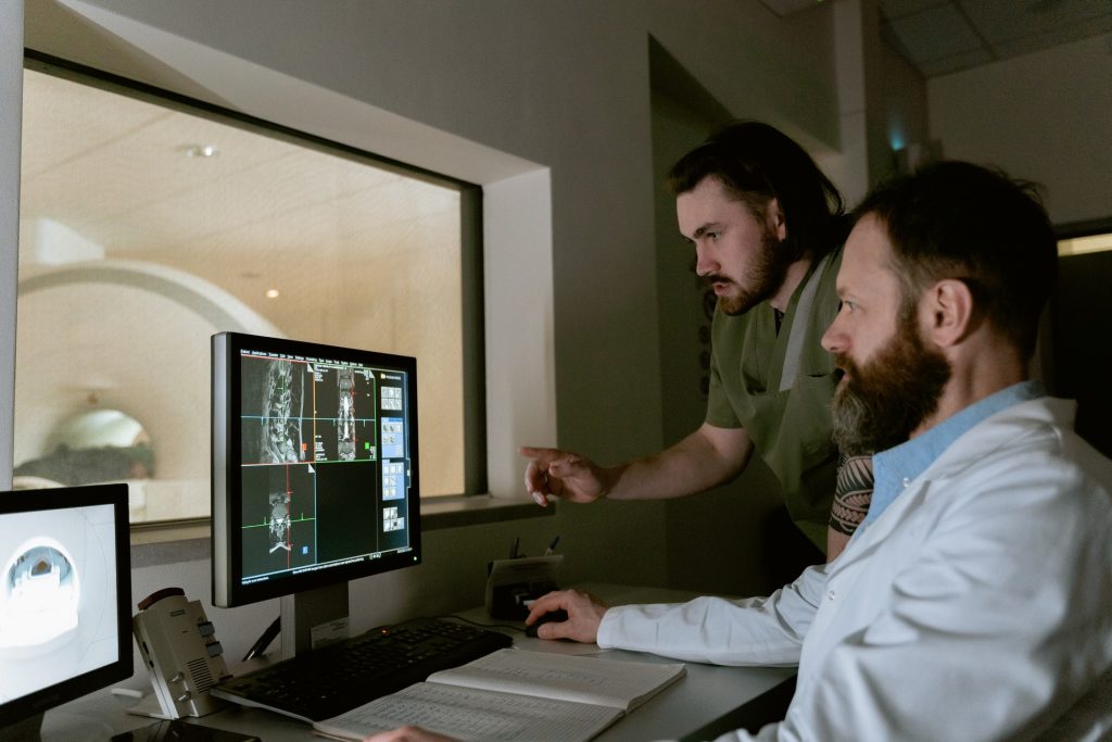
A new study appearing in The Lancet Oncology suggests that a targeted radiation therapy is as effective as standard care for patients with lung cancer brain metastasis.

The findings suggests that patients could benefit from this targeted approach as it is known to have have fewer negative cognitive consequences.

In non-small-cell (NSLC) lung cancer, about 57% of patients present with metastatic disease, and 20% present with brain metastases. Brain metastasis is currently treated with whole brain radiation therapy, which targets the entire brain. While this approach treats even microscopic tumours, it results in memory problems and decreases cognitive function. The alternative, stereotactic radiosurgery, spares healthy brain tissue by precisely targeting the tumour, has been shown to have less severe cognitive consequences but has not yet been studied in patients with small cell lung cancer that has metastasised to the brain.

“For many years, it made sense to treat these patients with whole brain radiation because their survival was quite poor,” said Karolina Gaebe, a research student in Dr Sunit Das’s lab, who led the study.

“For them, long-term consequences of the treatment were not as crucial as reducing the impact of disease in the short-term. But now, as treatments for their lung cancer have improved, these patients are surviving much longer.”

The researchers set out to learn more after noticing patients with longer survival times were also living with severe cognitive impairments due to the treatments for their brain metastases. They wanted to understand whether a more targeted brain radiation regimen might be as beneficial for these patients, as has been demonstrated for most other cancer types.

As a first step, they undertook this meta-analysis, reviewing current literature to examine survival and brain outcomes following stereotactic radiosurgery for patients with small cell lung cancer that had spread to the brain. The team analysed data from 31 studies and included 18 130 patients, the largest cohort of small cell lung cancer patients with brain metastases to be studied so far.

The next steps are to conduct a large clinical trial to investigate cognitive outcome differences between stereotactic radiosurgery and whole brain radiation therapy for such patients.

“Because this is a meta-analysis, we can’t use this as absolute evidence that all patients should be treated in this way,” Dr Das said. “But essentially, this means that we need to challenge our standing worldwide paradigms for treating patients with this disease and revisit the idea that these patients should receive whole brain radiation therapy.”

Source: EurekAlert!