Tag: 30/1/23

High-fat Diets Overload the Ability to Moderate Calorie Intake

Regularly eating a high fat/calorie diet could reduce the brain’s ability to regulate calorie intake, according to a study published in The Journal of Physiology. Rat studies revealed a signalling pathway which causes a quick response to high fat/high calorie intake, reducing food and calorie intake. But continuously eating a high fat/calorie diet seems to disrupt this signalling pathway, sabotaging this short-term protection.

Senior author Dr Kirsteen Browning said, “Calorie intake seems to be regulated in the short-term by astrocytes. We found that a brief exposure (three to five days) of high fat/calorie diet has the greatest effect on astrocytes, triggering the normal signalling pathway to control the stomach. Over time, astrocytes seem to desensitise to the high fat food. Around 10–14 days of eating high fat/calorie diet, astrocytes seem to fail to react and the brain’s ability to regulate calorie intake seems to be lost. This disrupts the signalling to the stomach and delays how it empties.”

Astrocytes initially react when high fat/calorie food is ingested, triggering the release of gliotransmitters, chemicals (including glutamate and ATP) that excite nerve cells and enable normal signalling pathways to stimulate neurons that control stomach function. This ensures the stomach contracts correctly to fill and empty in response to food passing through the digestive system. When astrocytes are inhibited, the cascade is disrupted. The decrease in signalling chemicals leads to a delay in digestion because the stomach doesn’t fill and empty appropriately.

The vigorous investigation used behavioural observation to monitor food intake in rats which were fed a control or high fat/calorie diet for one, three, five or 14 days. This was combined with pharmacological and specialist genetic approaches (both in vivo and in vitro) to target distinct neural circuits, which enabled the researchers to specifically inhibit astrocytes in a particular region of the brainstem. In this way, they assessed the response of individual neurons.

Human studies will need to be carried out to confirm if the same mechanism occurs in humans. If this is the case, further testing will be required to assess if the mechanism could be safely targeted without disrupting other neural pathways.

The researchers have plans to further explore the mechanism. Dr Browning said, “We have yet to find out whether the loss of astrocyte activity and the signalling mechanism is the cause of overeating or that it occurs in response to the overeating. We are eager to find out whether it is possible to reactivate the brain’s apparent lost ability to regulate calorie intake. If this is the case, it could lead to interventions to help restore calorie regulation in humans.”

Source: The Physiological Society

Actin Filaments Act as Pipelines for Metastatic Factors

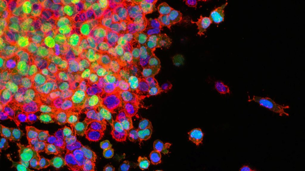
Lung cancer cells in the process of metastasising. Source: National Cancer Institute on Unsplash

When cancer cells metastasise, they have to break connections with neighbouring cells and migrate to other tissues. Both processes are promoted by signalling molecules released by the cancer cells, which thereby increase the malignancy of tumours. Researchers found that the release of these ‘prometastatic’ factors is influenced by the cellular skeleton – specifically, actin filaments. The study was published in Advanced Science.

Actin’s multiple role functions in cancer propagation

Actin filaments are part of the cell skeleton and essential for stability and motility. They form a network that dynamically builds up and gets broken down by the addition or detachment of building blocks at the filaments’ ends. These processes are precisely regulated by other molecules, such as formins. The dynamics of the actin network enable the movement of cells, for example during development or wound closure, but also that of spreading cancer cells. Actin also plays a role in the transport of substances within the cell. However, this is less well understood than that of other intracellular transport mechanisms.

The research team led by Prof Dr Robert Grosse and Dr Carsten Schwan from the University of Freiburg, now found that the actin network also enables the release of prometastatic factors, such as ANGPTL4 which is an important prometastatic factor that promotes the formation of metastases in various types of cancer. For their study, they used high-resolution microscopy to track the movement of individual transport vesicles within living cancer cells.

“We observed that ANGPTL4-loaded vesicles are conveyed to the periphery of the cell by means of dynamic and localised polymerisation of actin filaments,” says Grosse, who is a member of the Cluster of Excellence CIBSS – Centre for Integrative Biological Signalling Studies at the University of Freiburg.

Transportation along actin filaments

Based on microscopic observations and genetic analyses, the scientists conclude that the vesicles’ movement is controlled by the formin-like molecule FMNL2 by initiating polymerisation (ie elongation) of actin filaments directly at the vesicle. “We already knew that increased FMNL2 activity has prometastatic effects in many types of tumours,” says Grosse. “In our current work we could now demonstrate an important underlying process and a connection to the TGFbeta signalling pathway.” According to the scientist, this knowledge could be used for tumour diagnostics or therapy. for example, by developing an antibody that indicates the presence of active FMNL2 or pharmacologically targets active, phosphorylated FMNL2.

Source: University of Freiburg

Momelotinib Trumps Standard Care in Treating Myelofibrosis

Photo by Louise Reed on Unsplash

A Phase III trial testing the targeted therapy momelotinib showed that patients with myelofibrosis had clinically significant improvement in disease-related symptoms, including anaemia and spleen enlargement.

The findings, published in The Lancet, support the use of momelotinib over the standard therapy (danazol) in treating myelofibrosis patients that were resistant, refractory or intolerant to firstline therapy, especially symptomatic patients and those with anemia.

“Current options for managing anaemia in our myelofibrosis patients provide only modest and temporary benefits, so we are excited about these findings,” said study lead Srdan Verstovsek, MD, PhD, professor of Leukemia at University of Texas. “The trial results suggest that momelotinib is safe, well-tolerated and can improve one of the most common and debilitating clinical problems for this patient population.”

Myelofibrosis is an uncommon bone marrow cancer that is part of a group of diseases known as myeloproliferative neoplasms. A hallmark of the disease is dysregulated JAK signalling, which disrupts blood cell production and leads to symptoms including an enlarged spleen and anaemia. Chronic anaemia in these patients is associated with poor prognoses.

Currently approved JAK inhibitors can improve spleen responses and other disease-related symptoms, but they also can worsen anaemia. In this trial, momelotinib improved anaemia and reduced transfusion dependency in myelofibrosis patients previously treated with a JAK inhibitor. Momelotinib, a potent ACVR1/ALK2 and JAK1/2 inhibitor, can be administered and maintained at full dose because it does not suppress bone marrow activity like other JAK inhibitors.  

The randomised Phase III MOMENTUM trial was designed to compare the clinical benefits of momelotinib to danazol, a synthetic androgen currently used to treat anaemia in symptomatic myelofibrosis patients.

The trial enrolled 195 adult patients (63% male, 37% female) from 107 research sites across 21 countries. Trial participants were randomised (2:1) to receive momelotinib plus placebo or danazol plus placebo. A significantly greater proportion of patients who received momelotinib saw benefits in their disease symptoms (25%) compared to those receiving danazol (9%).

Patients treated with momelotinib also experienced a significant reduction in their spleen size, with 25% responding after 24 weeks of therapy. Additionally, these patients required fewer blood transfusions compared to those receiving danazol.

The safety profile of momelotinib was comparable to previous clinical trials. The most common non-haematological side effects experienced by trial participants in the momelotinib group included diarrhoea, nausea, weakness and itching or irritated skin.

“If approved, momelotinib could offer an effective option for patients with myelofibrosis to improve anemia, splenomegaly and other disease-related symptoms over other approved medications so far,” Verstovsek said. “Momelotinib may also be an ideal partner for combinations with other investigational agents in development to further control myelofibrosis symptoms.”

Patient follow-up is ongoing and long-term survival continues to be monitored.

Source: University of Texas MD Anderson Cancer Center

Periods of Hypoglycaemia Worsen Progression of Diabetic Retinopathy

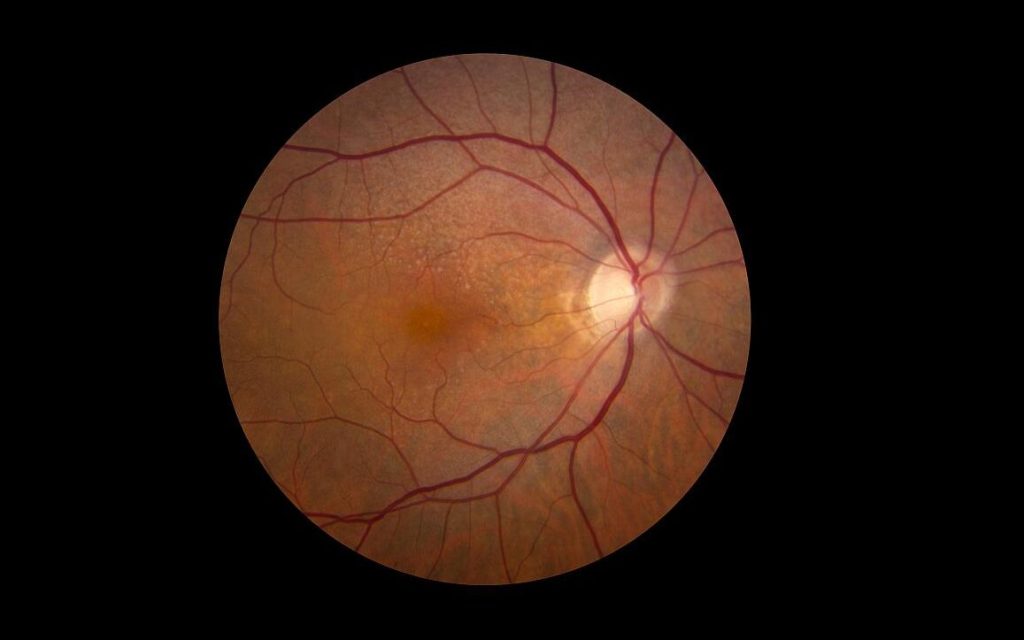
Credit: National Eye Institute

People with diabetes who experience periods of hypoglycaemia, a common event in those new to blood sugar management, are more likely to have worsening diabetic eye disease. Now, researchers say they have linked such low blood sugar levels with a molecular pathway that is activated in hypoxic cells in the eye.

The research, involving human and mouse eye cells and intact retinas grown in a low glucose environment in the laboratory, as well as mice with low glucose levels, was published in Cell Reports.

“Temporary episodes of low glucose happen once or twice a day in people with insulin-dependent diabetes and often among people newly diagnosed with the condition,” says Akrit Sodhi, MD, PhD, Johns Hopkins Medicine professor. Low glucose levels can also occur during sleep in people with non-insulin dependent diabetes. “Our results show that these periodic low glucose levels cause an increase in certain retinal cell proteins, resulting in an overgrowth of blood vessels and worsening diabetic eye disease,” adds Sodhi.

Up to a third of diabetic patients will develop diabetic retinopathy, which is characterised by the overgrowth of abnormal blood vessels in the retina.

Sodhi says the current study suggests that people with diabetic retinopathy may be particularly vulnerable to periods of low glucose, and keeping glucose levels stable should be an important part of glucose control.

For the study, the researchers analysed protein levels in human and mouse retinal cells and intact retinas grown in an environment of low glucose in the laboratory, as well as in mice that had occasional low blood sugar.

In human and mouse retinal cells, low glucose levels triggered a cascade of molecular changes that can lead to blood vessel overgrowth. First, the researchers saw that low glucose caused a decrease in retinal cells’ ability to break down glucose for energy.

When the researchers focused on Müller glial cells, which are supportive cells for neurons in the retina and rely primarily on glucose for energy production, they found that the cells increased the expression of the GLUT1 gene, which makes a protein that transports glucose into cells.

The researchers found that, in response to low glucose, the cells increased levels of a transcription factor, hypoxia-inducible factor (HIF)-1α. This turned on the cellular machinery, including GLUT1, needed to improve their ability to utilise available glucose, preserving the limited oxygen available for energy production by retinal neurons.

However, in hypoxic environments, as occurs in the retinas of patients with diabetic eye disease, this normal, physiologic response to low glucose triggered a flood of HIF-1α protein into the nucleus.

This resulted in an increase in the production of proteins such as VEGF and ANGPTL4, which cause the growth of abnormal, leaky blood vessels – the key culprit of vision loss in people with diabetic eye disease.

The researchers plan to study whether low glucose levels in people with diabetes may impact similar molecular pathways in other organs, such as the kidney and brain.

Sodhi says the HIF-1α pathway may serve as an effective target for developing new treatments for diabetic eye disease.

Source: Johns Hopkins Medicine

Inpatient Blood Draws are Often Performed During Sleep Hours

Blood sample being drawn
Photo by Hush Naidoo Jade Photography on Unsplash

The sleep of hospitalised patients may be often interrupted due to non-urgent blood draws, according to findings from a Yale study published in JAMA. This may be exacerbating sleep deprivation, and putting them at greater risk for health events later on.

In an analysis of more than 5 million non-urgent blood draws collected at Yale New Haven Hospital from 2016 to 2019, a team of researchers found that a high proportion of them occurred during a three-hour window in the early morning.

“We found that nearly four in 10 of total daily blood draws were performed between 4am and 7am,” said César Caraballo-Cordovez, MD, a postdoctoral associate at Yale Center for Outcomes Research and Evaluation (CORE) and co-lead author of the study. “Importantly, we found that this occurred across patients with different sociodemographic characteristics, including older individuals who are at highest risk of adverse health events from sleep deprivation.”

Although early morning blood draws are often considered necessary to inform decisions during morning medical rounds, the authors suggest that sleep interruptions may increase the risk of delirium and other adverse events. “Patients who were recently hospitalised experience a period of generalised risk for myriad adverse health events, a condition named posthospital syndrome,” added Dr Caraballo-Cordovez. “The stress that patients experience during the hospitalisation – including stress from sleep deprivation – is a key contributor to this period of increased risk.”

“This is not an issue at just one hospital,” said Harlan M. Krumholz, MD, SM, professor of medicine and public health at Yale and CORE director. “Our findings reflect an aspect of how inpatient hospital care is being delivered in modern medicine. A more patient-centered care would limit nonurgent tests during sleep hours. However, these early morning blood draws are often considered necessary to make decisions during rounds.”

“We need to re-design our process to protect patients’ sleep, but major changes in our practice must be informed by solid studies that demonstrate the efficacy of strategies to do so without untoward effects,” added Krumholz.

Source: Yale School of Medicine